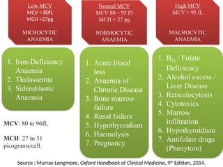
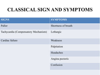
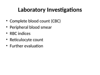
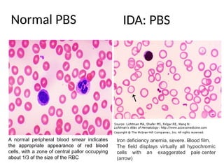
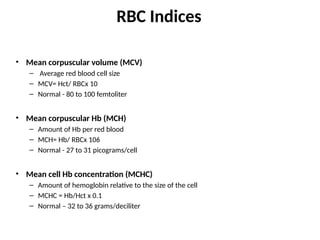
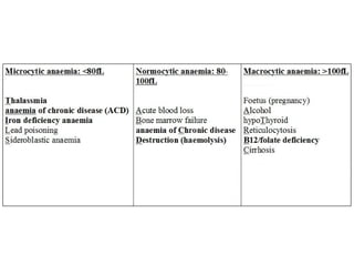
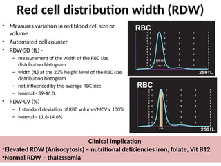
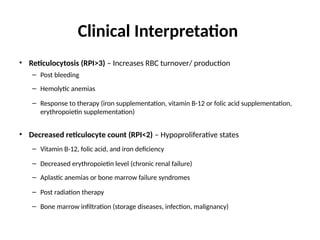
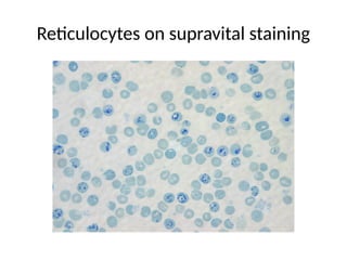
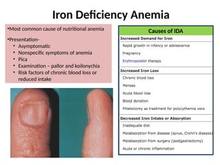
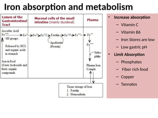
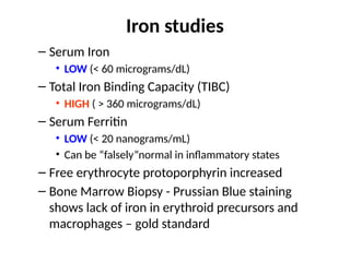
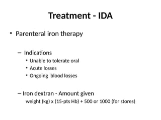
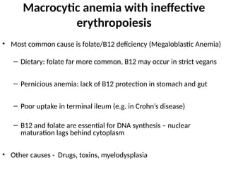
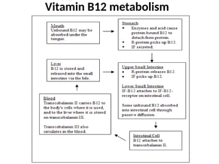
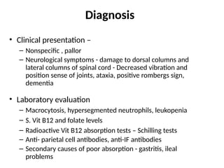
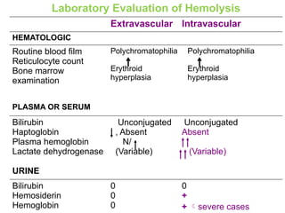
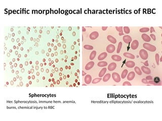
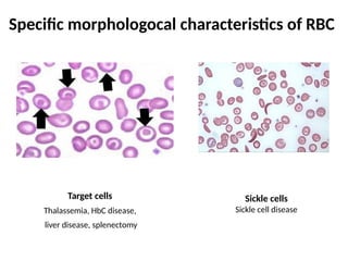
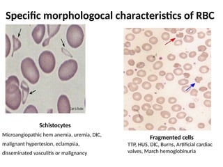
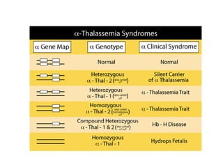
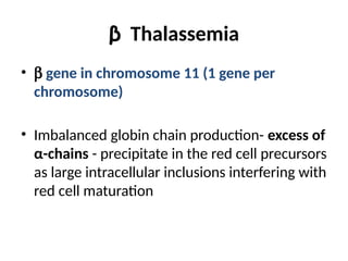
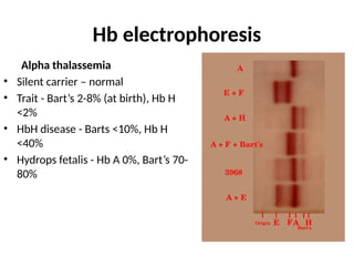
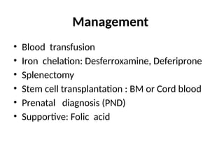
Anemia
Dr. V R
M.B.B.S., M.D.
Clinical Biochemistry
Objectives
• Definition
• Classification and Causes
• Laboratory investigations
• Overview of Common anemias
Definition
• Reduction of total circulating red cell mass
below normal limits for age, sex and
pregnancy status – reduction in oxygen
carrying capacity and hence tissue hypoxia
• Red cell mass - hematocrit / hemoglobin
concentration / isotopic dilution methods
Robbins & Cotran Pathologic Basis of Disease 9th
ed
Anaemia
Definition
WHO : ANAEMIA is a condition in which the number of
red blood cells or their oxygen carrying capacity is
insufficient to meet physiologic needs, which vary by
age, sex, altitude, smoking and pregnancy status.
OXFORD : ANAEMIA is defined as low heamoglobin
(Hb) concentration, and may be due either to a low red
cell mass or increased plasma volume.
Anemia is a laboratory diagnosis
Age/ Sex Hemoglobin (g/dl) Hematocrit (%)
At birth 17 52
Childhood 12 36
Adolescent 13 40
Adult man 16 (+/- 2) 47 (+/- 6)
Adult woman
(menstruating)
13 (+/- 2) 40 (+/- 6)
Adult woman
(postmenopausal)
14 (+/- 2) 42 (+/- 6)
Pregnant woman 12 (+/- 2) 37 (+/- 6)
WHO criteria for anemia – Hb < 13 g/dl for men, <12 g/dl for non
pregnant women and <11 g/dl for pregnant women and children.
Harrison’s Principles of Internal Medicine, 18th
ed.
COMPOSITION OF BLOOD
1. Cellular Portion (45% of
total blood volume)
-Erythrocytes (RBCs)
-Leukocyes (WBCs)
-Thrombocytes
2. Fluid portion (Plasma, 55%
of total blood volume)
Erythrocytes
Biconcave Disk
Flattened
Flexible
Semi-permeable
membrane
Contains Antigen
(ABO & Rh)  Blood
type
Anuclear
No Mitochondria
Erythrocytes
Life Span
100-120 days
FUNCTIONS
Transport O2
to tissues
Transport CO2
from tissues
Fate
Destroyed by
macrophage cells
(liver, spleen,
bone marrow)
33% of RBC cell
mass consist of
hemoglobin
Normal Erythrocytes
Normal Value
Male : 4.32 – 5.72 X 10¹²
cells/L
Female : 3.90 – 5.03 X 10¹²
cells/L
Source :
myoclinic.org/test-procedures/complete-
blood-count/basics/results/prc-20014088
Classification
Classification
of Anemia
Cause
Blood loss
Increased RBC
destuction
(Hemolysis)
Decreased RBC
production
Morphology
Microcytic and
Hypochromic
Normocytic
and
Normochromic
Macrocytic
Robbins & Cotran Pathologic Basis of Disease 9th
ed
Types of Anemia according to Morphology
Based on Mean Cell Volume (MCV)
- Normal MCV : 76-96 fL (femtolitres)
Low MCV
MICROCYTIC
ANAEMIA
Normal MCV
NORMOCYTIC
ANAEMIA
High MCV
MACROCYTIC
ANAEMIA
Varying MCV
HAEMOLYTIC ANAEMIA
Low MCV
MCV < 80fL
MCH <27pg
MICROCYTIC
ANAEMIA
Normal MCV
MCV 80 – 95 Fl
MCH > 27 pg
NORMOCYTIC
ANAEMIA
High MCV
MCV > 95 fL
MACROCYTIC
ANAEMIA
1. Iron-Deficiency
Anaemia
2. Thalassemia
3. Sideroblastic
Anaemia
1. Acute blood
loss
2. Anaemia of
Chronic Disease
3. Bone marrow
failure
4. Renal failure
5. Hypothyroidism
6. Haemolysis
7. Pregnancy
1. B12 / Folate
Deficiency
2. Alcohol excess /
Liver Disease
3. Reticulocytosis
4. Cytotoxics
5. Marrow
infiltration
6. Hypothyroidism
7. Antifolate drugs
(Phenytoin)
Source : Murray Longmore, Oxford Handbook of Clinical Medicine, 9th
Edition, 2014.
MCV: 80 to 96fL
MCH: 27 to 31
picograms/cell.
Etiology
Blood loss
• Acute
• Chronic
Hemolysis
• Intracorpuscular defects
• RBC memberane defecrs –
hereditary spherocytosis/
elliptocytosis
• Enzyme defects – G6PD or PK
deficiency
• Hemoglobinopathies –
thalassemias, sickle cell
disease
• Extracorpuscular defects
• Antibody mediated damage -
transfusion reactions,
autoimmune disorders, drug
induced
• Infections – malaria
• Mechanical trauma – prosthetic
valves
• Toxic – snake venom, lead
• Microangiopathic hemolytic
anemia – HUS, DIC, TTP
• Sequestration - Hypersplenism
Decreased RBC
production
• Genetic defects
• Stem cell depletion – Fanconi
anemia
• Erythroblast maturation -
thalassemias
• Nutritional deficiency
• Affecting DNA synthesis – B12 and
folate def
• Affecting HB synthesis – IDA
• EPO deficiency – anemia of chronic
disease, Renal failure
• Inflammation mediated - anemia of
chronic disease
• Marrow infiltration – malignancy,
granulomatous diseases
• Immune mediated injury to
progenitors – aplastic anemias, pure
red cell dyscrasias
Robbins & Cotran Pathologic Basis of Disease 9th
ed
CLASSICAL SIGN AND SYMPTOMS
SIGNS SYMPTOMS
Pallor Shortness of breath
Tachycardia (Compensatory Mechanism) Lethargic
Cardiac failure Weakness
Palpitation
Headaches
Angina pectoris
Confusion
Clinical presentation
• History
– Fatigue, decreased exercise tolerance, SOB, palpitations, lightheadedness
– Paresthesias, numbness
– Pica – IDA
– Acute or chronic blood loss
– h/o transfusions – thalassemia
– Family history - genetic causes
– Vegetarian diet – B12 deficiency
– Malabsorption – nutritional deficiency
– Drug intake
• Examination
– Pallor
– Jaundice – hemolytic anemia
– Tachycardia, bounding pulses, Systolic flow murmur
– Koilonychia - IDA
– Glossitis, Angular cheilosis – nutritional anemias
– Decreased vibratory sense/ joint position sense / ataxia/ positive Romberg
sign - B12/folate deficiency
Laboratory Investigations
• Complete blood count (CBC)
• Peripheral blood smear
• RBC indices
• Reticulocyte count
• Further evaluation
Complete blood count
• Evaluate
– Hb concentration (g/dl)
– Hct - % of blood volume
occupied by RBCs
– RBC count
– TLC and DLC
– Platelet count
Clinical implications
•Aplastic anemia – associated leukopenia and/or thrombocytopenia
•Leukocytosis – infections, leukemoid reactions, leukemias
Peripheral blood smear
• Manual RBC, WBC and platelet counts
• RBC morphology
– Microcytic, normocytic, macrocytic
– Specific abnormalities
– Staining – hypochromic, normochromic,
polychromasia (correlates with reticulocytosis)
• WBC morphology
– Hypersegmented neutrophils in megaloblastic
anemia
– Blasts in leukemias
RBC morphology Cause
Target cells (small normally stained
center surrounded by a hypochromic
ring)
Thalassemia, post splenectomy, IDA
Spherocytes Hereditary spherocytosis,
hypersplenism, other extracorpuscular
hemolytic anemias
Acanthocytes (spur cells) Liver diseases
Burr cells Uremia
Schistocytes/ fragmented cells HUS, TTP, DIC
Tear drop cells Myeloproliferative disorders
Nucleated RBC Postsplenectomy, intense marrow
stimulation
Basophilic stippling (aggregates of
rRNA)
Lead poisoning, Thalassemia
Howell-jolly bodies (nuclear remnants) Postsplenectomy
Heinz bodies (denatured Hb) Thalassemia, asplenia
Normal PBS
Iron deficiency anemia, severe. Blood film.
The field displays virtually all hypochromic
cells with an exaggerated pale center
(arrow)
A normal peripheral blood smear indicates
the appropriate appearance of red blood
cells, with a zone of central pallor occupying
about 1/3 of the size of the RBC
IDA: PBS
RBC Indices
• Mean corpuscular volume (MCV)
– Average red blood cell size
– MCV= Hct/ RBCx 10
– Normal - 80 to 100 femtoliter
• Mean corpuscular Hb (MCH)
– Amount of Hb per red blood
– MCH= Hb/ RBCx 106
– Normal - 27 to 31 picograms/cell
• Mean cell Hb concentration (MCHC)
– Amount of hemoglobin relative to the size of the cell
– MCHC = Hb/Hct x 0.1
– Normal – 32 to 36 grams/deciliter
Red cell distribution width (RDW)
• Measures variation in red blood cell size or
volume
• Automated cell counter
• RDW-SD (fL) -
– measurement of the width of the RBC size
distribution histogram
– width (fL) at the 20% height level of the RBC size
distribution histogram
– not influenced by the average RBC size
– Normal - 39-46 fL
• RDW-CV (%)
– 1 standard deviation of RBC volume/MCV x 100%
– Normal - 11.6-14.6%
Clinical implication
•Elevated RDW (Anisocytosis) – nutritional deficiencies iron, folate, Vit B12
•Normal RDW – thalassemia
Reticulocyte count
• Immature RBCs
• Indicator of bone marrow activity / effective erythropoesis
• PBS stained by supravital staining (Romanowsky staining) or Automated counter by
combination of laser excitation, detectors and flouroscent dyes
• Normal range - 0.5-1.5%
• Corrected Retic count
– Adjusted for the patient's hematocrit
– At lower hematocrit reticulocytes released earlier from marrow
– Corrected retic = Patients retic. x (Patients Hct/45)
• Reticulocyte index (RPI) = corrected retic. count/Maturation time
• Absolute reticulocyte count = retic x RBC number.
Clinical Interpretation
• Reticulocytosis (RPI>3) – Increases RBC turnover/ production
– Post bleeding
– Hemolytic anemias
– Response to therapy (iron supplementation, vitamin B-12 or folic acid supplementation,
erythropoietin supplementation)
• Decreased reticulocyte count (RPI<2) – Hypoproliferative states
– Vitamin B-12, folic acid, and iron deficiency
– Decreased erythropoietin level (chronic renal failure)
– Aplastic anemias or bone marrow failure syndromes
– Post radiation therapy
– Bone marrow infiltration (storage diseases, infection, malignancy)
Reticulocytes on supravital staining
Overview of common anemias
Iron Deficiency Anemia
•Most common cause of nutritional anemia
•Presentation-
• Asymptomatic
• Nonspecific symptoms of anemia
• Pica
• Examination – pallor and koilonychia
• Risk factors of chronic blood loss or
reduced intake
Causes of IDA
Iron absorption and metabolism
• Source of Iron
– Jaggery, Beef, chicken,
fortified breakfast cereal,
beans, whole wheat
grains, spinach
• Daily iron absorption –
– 5-10% from vegetarian
diet, 20% from
nonvegetarian diet
• RDA for Iron
Iron absorption and metabolism
• Increase absorption
– Vitamin C
– Vitamin B6
– Iron Stores are low
– Low gastric pH
• Limit Absorption
– Phosphates
– Fiber rich food
– Copper
– Tannates
IDA - Diagnosis
• Peripheral smear shows microcytic, hypochromic red cells with marked
anisopoikilocytosis
• Target cells, pencil shaped cells
• Low retic count, high RDW
Iron studies
– Serum Iron
• LOW (< 60 micrograms/dL)
– Total Iron Binding Capacity (TIBC)
• HIGH ( > 360 micrograms/dL)
– Serum Ferritin
• LOW (< 20 nanograms/mL)
• Can be “falsely”normal in inflammatory states
– Free erythrocyte protoporphyrin increased
– Bone Marrow Biopsy - Prussian Blue staining
shows lack of iron in erythroid precursors and
macrophages – gold standard
Further evaluation
• Endoscopy – upper or lower GI bleeding in males in
pstmenopausal females
• Stool for occult blood
• Stool for ova (hookworm infestation)
• Meckels scan for meckels diverticulum in children
Treatment - IDA
• Oral iron therapy
– 300 mg elemental iron per day (3-4 divided doses)
– Empty stomach preferred
– Vitamin C can facilitate iron absorption
– Sustained therapy for 6-12 months after correction of anemia – stores
replenished
– Response – increase in retic count by 4-7 days, peak at 1.5 weeks
• Failure of oral therapy
– Incorrect diagnosis (eg, thalassemia)
– Anemia of chronic disease?
– Poor compliance
– Poor absorption
Treatment - IDA
• Parenteral iron therapy
– Indications
• Unable to tolerate oral
• Acute losses
• Ongoing blood losses
– Iron dextran - Amount given
weight (kg) x (15-pts Hb) + 500 or 1000 (for stores)
Anemia of Chronic disease (ACD)
Hypoproliferative anemias
Pathophysiology – Inadequate EPO or inadequate response of marrow to EPO
Soluble Transferrin Receptor: Elevated in cases of iron deficiency
Ferritin: Elevated in anemia of chronic disease
Treatment - ACD
• Treat the underlying cause
• Consider co-existent iron deficiency as well
• If underlying disease state requires consider
EPO injection
Macrocytic anemia with ineffective
erythropoiesis
• Most common cause is folate/B12 deficiency (Megaloblastic Anemia)
– Dietary: folate far more common, B12 may occur in strict vegans
– Pernicious anemia: lack of B12 protection in stomach and gut
– Poor uptake in terminal ileum (e.g. in Crohn’s disease)
– B12 and folate are essential for DNA synthesis – nuclear
maturation lags behind cytoplasm
• Other causes - Drugs, toxins, myelodysplasia
Vitamin B12 metabolism
Diagnosis
• Clinical presentation –
– Nonspecific , pallor
– Neurological symptoms - damage to dorsal columns and
lateral columns of spinal cord - Decreased vibration and
position sense of joints, ataxia, positive rombergs sign,
dementia
• Laboratory evaluation
– Macrocytosis, hypersegmented neutrophils, leukopenia
– S. Vit B12 and folate levels
– Radioactive Vit B12 absorption tests – Schilling tests
– Anti- parietal cell antibodies, anti-IF antibodies
– Secondary causes of poor absorption - gastritis, ileal
problems
Treatment– supplementation
• Do NOT correct folate levels unless B12 is OK
– Correction of folate deficiency will correct hematologic abnormalities
without correcting neurological abnormalities
– Check B12 and correct first
– Oral folic acid 5-15 mg per day
• B12 usually 1000 mg I.M. weekly upto 6 doses followed by
maintanence 3 monthly
– B12 stores take a long time to deplete; missed doses are not usually a
problem
– Oral supplementation is gaining support; usually effective in pernicious
anemia (1-2 mg PO QD)
• Reticulocyte count should respond in 1 wk
Hemolytic anemias
• Clinical suspision
– Pallor with jaundice
– Dark urine
– Hepatosplenomegaly
– Gallstones
– Hemolytic facies
– Leg ulcers
– Crises – aplastic or hemolytic
Laboratory evidence of hemolysis
• Normocytic, normochromic, leukoytosis,
thrombocytosis
• Increased indirect bilirubin - sensitive but not
specific
• Reticulocytosis, polychromasia
• Increased LDH (LDH1) - sensitive but not specific.
• Decreased haptoglobin - specific but not sensitive.
• Hemoglobinemia
• Hemoglobinuria
Laboratory Evaluation of Hemolysis
Extravascular Intravascular
HEMATOLOGIC
Routine blood film
Reticulocyte count
Bone marrow
examination
Polychromatophilia
Erythroid
hyperplasia
Polychromatophilia
Erythroid
hyperplasia
PLASMA OR SERUM
Bilirubin
Haptoglobin
Plasma hemoglobin
Lactate dehydrogenase
Unconjugated
, Absent
N/
(Variable)
Unconjugated
Absent
(Variable)
URINE
Bilirubin
Hemosiderin
Hemoglobin
0
0
0
0
+
+ severe cases
Specific morphologocal characteristics of RBC
Spherocytes
Her. Spherocytosis, immune hem. anemia,
burns, chemical injury to RBC
Elliptocytes
Hereditary elliptocytosis/ ovalocytosis
Specific morphologocal characteristics of RBC
Target cells
Thalassemia, HbC disease,
liver disease, splenectomy
Sickle cells
Sickle cell disease
Specific morphologocal characteristics of RBC
Schistocytes
Microangiopathic hem anemia, uremia, DIC,
malignant hypertesion, eclampsia,
disseminated vasculitis or malignancy
Fragmented cells
TTP, HUS, DIC, Burns, Artificial cardiac
valves, March hemoglobinuria
Specific morphologocal characteristics of RBC
Heinz bodies
Unstable (denatured) Hb,
G6PD deficiency and oxidant
stress
Howel-jolly bodies
(nuclear remnants)
Post-splenectomy
Specific morphologocal characteristics of RBC
Stomatocytes
Hereditary stomatocytosis, alcoholism
Burr cells
Uremia
Acanthocytes
Liver disease, abetalipoproteinemia
Special Lab. Examinations
• Coombs antiglobulin test - immune hemolysis
• Osmotic fragility test - spherocytosis
• Autohemolysis- G6PD,PK, spherocytosis
• Membrane protein analysis- membrane defects
• Red cell sickling- sickle cell anemia
• Hemoglobin electrophoresis and HbA2, Hb F ,
HHb,etc - Hemoglobinopathies and thalassemias
• Red cell enzyme assays- RBC enzyme defects
Treatment
• Hereditary spherocytosis
– Folic Acid 5mg weekly, prophylaxis life long
– Splenectomy
– Cholecystectomy for gall stones
– Blood transfusion in Acute severe hemolytic crisis
• G6PD deficiency
– Removal of offending drugs
– Transfusion if required
• Immune hemolytic anemias
– Correct the underlying cause
– Prednisolone 1mg/kg po until Hb reaches 10mg/dl
then taper slowly and stop
– Transfusion: for life threatening problems
– If no response to steroids  Spleenectomy or
Immunosuppressive: Azathioprine,
Cyclophosphamide
Haemoglobinopathies
• Quantitative or qualitative defects in
production of globin chains
• Qualitative defects
– Sickle cell disease - point mutation at codon 6
of b-globin gene; Glutamic acid-->Valine
• Quantitative defect
– Thalassemia
Thalassemias
• Autosomal resessive
• Globin chain affected
– a-thalassemia
– b-thalassemia
• Pathophysiology related to increased/ excess
production of other chains forming polymers –
ineffective erythropoesis and reduced RBC
survival – marrow hyperplasia
Alpha Thalassemia
• a globin – chromosome 16, each chromosome has 2 genes
• Absence of α chains will result in increase/ excess of g chains
during fetal life and excess β chains later in life - Hb Bart's (g4) or
HbH (β4) (very high affinity for O2)
• 4 clinival syndromes
– Silent Carrier State
– Alpha Thalassemia Trait (Alpha Thalassemia Minor)
– Hemoglobin H Disease
– Bart's Hydrops Fetalis Syndrome
β Thalassemia
• b gene in chromosome 11 (1 gene per
chromosome)
• Imbalanced globin chain production- excess of
α-chains - precipitate in the red cell precursors
as large intracellular inclusions interfering with
red cell maturation
3 types of b Thalassemia mutations:
o b++
Thalassemia: production of b chain is mildly
reduced
o b+
Thalassemia: The production of b chain is
more reduced than b++
But NOT ABSENT
o b0
Thalassemia: ABSENCE of b chain production.
Silent b Thal
(
mildest form
) b++
/bN
Thal. Trait
(
mild hypochromic, microcytic
hemolytic anemia
)
b++
/ b
++
b+
/ bN
bo
/ bN
Thal. intermedia
b++
/ b
++
b++
/ bo
b+
/ b
+
b+
/ bo
bo
/ bo
Thal. Major
(
severe life long
transfusion-dependent
hemolytic anemia
)
There is
overlap in
presentation
4
types of b Thalassemia syndromes
Clinical presentation
• Hemolytic facies
• Mild pallor to hydrops fetalis
• Hepatosplenomegaly
• Hair on end appearance
• Short stature
• Features of iron overload
– Skin pigmentation
– Endocrine defects and delayed puberty
– Liver cirrhosis
– Heart failure
Laboratory diagnosis
• Microcytic and hypochromic anemia
• PBS - Anisopoikilocytosis , HbH, Barts Hb,
inclusion bodies, tear drop and fragmented
cells, nucleated RBC
HbH Barts Hb
Hb electrophoresis
Alpha thalassemia
• Silent carrier – normal
• Trait - Bart’s 2-8% (at birth), Hb H
<2%
• HbH disease - Barts <10%, Hb H
<40%
• Hydrops fetalis - Hb A 0%, Bart’s 70-
80%
Hb electrophoresis
• Beta thalassemia
Management
• Blood transfusion
• Iron chelation: Desferroxamine, Deferiprone
• Splenectomy
• Stem cell transplantation : BM or Cord blood
• Prenatal diagnosis (PND)
• Supportive: Folic acid

Anemia classification, basic lab diagnosis.pptx

  • 1.
    Anemia Dr. V R M.B.B.S.,M.D. Clinical Biochemistry
  • 2.
    Objectives • Definition • Classificationand Causes • Laboratory investigations • Overview of Common anemias
  • 3.
    Definition • Reduction oftotal circulating red cell mass below normal limits for age, sex and pregnancy status – reduction in oxygen carrying capacity and hence tissue hypoxia • Red cell mass - hematocrit / hemoglobin concentration / isotopic dilution methods Robbins & Cotran Pathologic Basis of Disease 9th ed
  • 4.
    Anaemia Definition WHO : ANAEMIAis a condition in which the number of red blood cells or their oxygen carrying capacity is insufficient to meet physiologic needs, which vary by age, sex, altitude, smoking and pregnancy status. OXFORD : ANAEMIA is defined as low heamoglobin (Hb) concentration, and may be due either to a low red cell mass or increased plasma volume.
  • 5.
    Anemia is alaboratory diagnosis Age/ Sex Hemoglobin (g/dl) Hematocrit (%) At birth 17 52 Childhood 12 36 Adolescent 13 40 Adult man 16 (+/- 2) 47 (+/- 6) Adult woman (menstruating) 13 (+/- 2) 40 (+/- 6) Adult woman (postmenopausal) 14 (+/- 2) 42 (+/- 6) Pregnant woman 12 (+/- 2) 37 (+/- 6) WHO criteria for anemia – Hb < 13 g/dl for men, <12 g/dl for non pregnant women and <11 g/dl for pregnant women and children. Harrison’s Principles of Internal Medicine, 18th ed.
  • 6.
    COMPOSITION OF BLOOD 1.Cellular Portion (45% of total blood volume) -Erythrocytes (RBCs) -Leukocyes (WBCs) -Thrombocytes 2. Fluid portion (Plasma, 55% of total blood volume)
  • 7.
  • 8.
    Erythrocytes Life Span 100-120 days FUNCTIONS TransportO2 to tissues Transport CO2 from tissues Fate Destroyed by macrophage cells (liver, spleen, bone marrow) 33% of RBC cell mass consist of hemoglobin
  • 9.
    Normal Erythrocytes Normal Value Male: 4.32 – 5.72 X 10¹² cells/L Female : 3.90 – 5.03 X 10¹² cells/L Source : myoclinic.org/test-procedures/complete- blood-count/basics/results/prc-20014088
  • 10.
    Classification Classification of Anemia Cause Blood loss IncreasedRBC destuction (Hemolysis) Decreased RBC production Morphology Microcytic and Hypochromic Normocytic and Normochromic Macrocytic Robbins & Cotran Pathologic Basis of Disease 9th ed
  • 11.
    Types of Anemiaaccording to Morphology Based on Mean Cell Volume (MCV) - Normal MCV : 76-96 fL (femtolitres) Low MCV MICROCYTIC ANAEMIA Normal MCV NORMOCYTIC ANAEMIA High MCV MACROCYTIC ANAEMIA Varying MCV HAEMOLYTIC ANAEMIA
  • 12.
    Low MCV MCV <80fL MCH <27pg MICROCYTIC ANAEMIA Normal MCV MCV 80 – 95 Fl MCH > 27 pg NORMOCYTIC ANAEMIA High MCV MCV > 95 fL MACROCYTIC ANAEMIA 1. Iron-Deficiency Anaemia 2. Thalassemia 3. Sideroblastic Anaemia 1. Acute blood loss 2. Anaemia of Chronic Disease 3. Bone marrow failure 4. Renal failure 5. Hypothyroidism 6. Haemolysis 7. Pregnancy 1. B12 / Folate Deficiency 2. Alcohol excess / Liver Disease 3. Reticulocytosis 4. Cytotoxics 5. Marrow infiltration 6. Hypothyroidism 7. Antifolate drugs (Phenytoin) Source : Murray Longmore, Oxford Handbook of Clinical Medicine, 9th Edition, 2014. MCV: 80 to 96fL MCH: 27 to 31 picograms/cell.
  • 13.
    Etiology Blood loss • Acute •Chronic Hemolysis • Intracorpuscular defects • RBC memberane defecrs – hereditary spherocytosis/ elliptocytosis • Enzyme defects – G6PD or PK deficiency • Hemoglobinopathies – thalassemias, sickle cell disease • Extracorpuscular defects • Antibody mediated damage - transfusion reactions, autoimmune disorders, drug induced • Infections – malaria • Mechanical trauma – prosthetic valves • Toxic – snake venom, lead • Microangiopathic hemolytic anemia – HUS, DIC, TTP • Sequestration - Hypersplenism Decreased RBC production • Genetic defects • Stem cell depletion – Fanconi anemia • Erythroblast maturation - thalassemias • Nutritional deficiency • Affecting DNA synthesis – B12 and folate def • Affecting HB synthesis – IDA • EPO deficiency – anemia of chronic disease, Renal failure • Inflammation mediated - anemia of chronic disease • Marrow infiltration – malignancy, granulomatous diseases • Immune mediated injury to progenitors – aplastic anemias, pure red cell dyscrasias Robbins & Cotran Pathologic Basis of Disease 9th ed
  • 14.
    CLASSICAL SIGN ANDSYMPTOMS SIGNS SYMPTOMS Pallor Shortness of breath Tachycardia (Compensatory Mechanism) Lethargic Cardiac failure Weakness Palpitation Headaches Angina pectoris Confusion
  • 15.
    Clinical presentation • History –Fatigue, decreased exercise tolerance, SOB, palpitations, lightheadedness – Paresthesias, numbness – Pica – IDA – Acute or chronic blood loss – h/o transfusions – thalassemia – Family history - genetic causes – Vegetarian diet – B12 deficiency – Malabsorption – nutritional deficiency – Drug intake • Examination – Pallor – Jaundice – hemolytic anemia – Tachycardia, bounding pulses, Systolic flow murmur – Koilonychia - IDA – Glossitis, Angular cheilosis – nutritional anemias – Decreased vibratory sense/ joint position sense / ataxia/ positive Romberg sign - B12/folate deficiency
  • 16.
    Laboratory Investigations • Completeblood count (CBC) • Peripheral blood smear • RBC indices • Reticulocyte count • Further evaluation
  • 17.
    Complete blood count •Evaluate – Hb concentration (g/dl) – Hct - % of blood volume occupied by RBCs – RBC count – TLC and DLC – Platelet count Clinical implications •Aplastic anemia – associated leukopenia and/or thrombocytopenia •Leukocytosis – infections, leukemoid reactions, leukemias
  • 18.
    Peripheral blood smear •Manual RBC, WBC and platelet counts • RBC morphology – Microcytic, normocytic, macrocytic – Specific abnormalities – Staining – hypochromic, normochromic, polychromasia (correlates with reticulocytosis) • WBC morphology – Hypersegmented neutrophils in megaloblastic anemia – Blasts in leukemias
  • 19.
    RBC morphology Cause Targetcells (small normally stained center surrounded by a hypochromic ring) Thalassemia, post splenectomy, IDA Spherocytes Hereditary spherocytosis, hypersplenism, other extracorpuscular hemolytic anemias Acanthocytes (spur cells) Liver diseases Burr cells Uremia Schistocytes/ fragmented cells HUS, TTP, DIC Tear drop cells Myeloproliferative disorders Nucleated RBC Postsplenectomy, intense marrow stimulation Basophilic stippling (aggregates of rRNA) Lead poisoning, Thalassemia Howell-jolly bodies (nuclear remnants) Postsplenectomy Heinz bodies (denatured Hb) Thalassemia, asplenia
  • 20.
    Normal PBS Iron deficiencyanemia, severe. Blood film. The field displays virtually all hypochromic cells with an exaggerated pale center (arrow) A normal peripheral blood smear indicates the appropriate appearance of red blood cells, with a zone of central pallor occupying about 1/3 of the size of the RBC IDA: PBS
  • 21.
    RBC Indices • Meancorpuscular volume (MCV) – Average red blood cell size – MCV= Hct/ RBCx 10 – Normal - 80 to 100 femtoliter • Mean corpuscular Hb (MCH) – Amount of Hb per red blood – MCH= Hb/ RBCx 106 – Normal - 27 to 31 picograms/cell • Mean cell Hb concentration (MCHC) – Amount of hemoglobin relative to the size of the cell – MCHC = Hb/Hct x 0.1 – Normal – 32 to 36 grams/deciliter
  • 23.
    Red cell distributionwidth (RDW) • Measures variation in red blood cell size or volume • Automated cell counter • RDW-SD (fL) - – measurement of the width of the RBC size distribution histogram – width (fL) at the 20% height level of the RBC size distribution histogram – not influenced by the average RBC size – Normal - 39-46 fL • RDW-CV (%) – 1 standard deviation of RBC volume/MCV x 100% – Normal - 11.6-14.6% Clinical implication •Elevated RDW (Anisocytosis) – nutritional deficiencies iron, folate, Vit B12 •Normal RDW – thalassemia
  • 24.
    Reticulocyte count • ImmatureRBCs • Indicator of bone marrow activity / effective erythropoesis • PBS stained by supravital staining (Romanowsky staining) or Automated counter by combination of laser excitation, detectors and flouroscent dyes • Normal range - 0.5-1.5% • Corrected Retic count – Adjusted for the patient's hematocrit – At lower hematocrit reticulocytes released earlier from marrow – Corrected retic = Patients retic. x (Patients Hct/45) • Reticulocyte index (RPI) = corrected retic. count/Maturation time • Absolute reticulocyte count = retic x RBC number.
  • 25.
    Clinical Interpretation • Reticulocytosis(RPI>3) – Increases RBC turnover/ production – Post bleeding – Hemolytic anemias – Response to therapy (iron supplementation, vitamin B-12 or folic acid supplementation, erythropoietin supplementation) • Decreased reticulocyte count (RPI<2) – Hypoproliferative states – Vitamin B-12, folic acid, and iron deficiency – Decreased erythropoietin level (chronic renal failure) – Aplastic anemias or bone marrow failure syndromes – Post radiation therapy – Bone marrow infiltration (storage diseases, infection, malignancy)
  • 26.
  • 27.
  • 28.
    Iron Deficiency Anemia •Mostcommon cause of nutritional anemia •Presentation- • Asymptomatic • Nonspecific symptoms of anemia • Pica • Examination – pallor and koilonychia • Risk factors of chronic blood loss or reduced intake Causes of IDA
  • 29.
    Iron absorption andmetabolism • Source of Iron – Jaggery, Beef, chicken, fortified breakfast cereal, beans, whole wheat grains, spinach • Daily iron absorption – – 5-10% from vegetarian diet, 20% from nonvegetarian diet • RDA for Iron
  • 30.
    Iron absorption andmetabolism • Increase absorption – Vitamin C – Vitamin B6 – Iron Stores are low – Low gastric pH • Limit Absorption – Phosphates – Fiber rich food – Copper – Tannates
  • 31.
    IDA - Diagnosis •Peripheral smear shows microcytic, hypochromic red cells with marked anisopoikilocytosis • Target cells, pencil shaped cells • Low retic count, high RDW
  • 32.
    Iron studies – SerumIron • LOW (< 60 micrograms/dL) – Total Iron Binding Capacity (TIBC) • HIGH ( > 360 micrograms/dL) – Serum Ferritin • LOW (< 20 nanograms/mL) • Can be “falsely”normal in inflammatory states – Free erythrocyte protoporphyrin increased – Bone Marrow Biopsy - Prussian Blue staining shows lack of iron in erythroid precursors and macrophages – gold standard
  • 33.
    Further evaluation • Endoscopy– upper or lower GI bleeding in males in pstmenopausal females • Stool for occult blood • Stool for ova (hookworm infestation) • Meckels scan for meckels diverticulum in children
  • 34.
    Treatment - IDA •Oral iron therapy – 300 mg elemental iron per day (3-4 divided doses) – Empty stomach preferred – Vitamin C can facilitate iron absorption – Sustained therapy for 6-12 months after correction of anemia – stores replenished – Response – increase in retic count by 4-7 days, peak at 1.5 weeks • Failure of oral therapy – Incorrect diagnosis (eg, thalassemia) – Anemia of chronic disease? – Poor compliance – Poor absorption
  • 35.
    Treatment - IDA •Parenteral iron therapy – Indications • Unable to tolerate oral • Acute losses • Ongoing blood losses – Iron dextran - Amount given weight (kg) x (15-pts Hb) + 500 or 1000 (for stores)
  • 36.
    Anemia of Chronicdisease (ACD) Hypoproliferative anemias Pathophysiology – Inadequate EPO or inadequate response of marrow to EPO Soluble Transferrin Receptor: Elevated in cases of iron deficiency Ferritin: Elevated in anemia of chronic disease
  • 37.
    Treatment - ACD •Treat the underlying cause • Consider co-existent iron deficiency as well • If underlying disease state requires consider EPO injection
  • 38.
    Macrocytic anemia withineffective erythropoiesis • Most common cause is folate/B12 deficiency (Megaloblastic Anemia) – Dietary: folate far more common, B12 may occur in strict vegans – Pernicious anemia: lack of B12 protection in stomach and gut – Poor uptake in terminal ileum (e.g. in Crohn’s disease) – B12 and folate are essential for DNA synthesis – nuclear maturation lags behind cytoplasm • Other causes - Drugs, toxins, myelodysplasia
  • 39.
  • 40.
    Diagnosis • Clinical presentation– – Nonspecific , pallor – Neurological symptoms - damage to dorsal columns and lateral columns of spinal cord - Decreased vibration and position sense of joints, ataxia, positive rombergs sign, dementia • Laboratory evaluation – Macrocytosis, hypersegmented neutrophils, leukopenia – S. Vit B12 and folate levels – Radioactive Vit B12 absorption tests – Schilling tests – Anti- parietal cell antibodies, anti-IF antibodies – Secondary causes of poor absorption - gastritis, ileal problems
  • 41.
    Treatment– supplementation • DoNOT correct folate levels unless B12 is OK – Correction of folate deficiency will correct hematologic abnormalities without correcting neurological abnormalities – Check B12 and correct first – Oral folic acid 5-15 mg per day • B12 usually 1000 mg I.M. weekly upto 6 doses followed by maintanence 3 monthly – B12 stores take a long time to deplete; missed doses are not usually a problem – Oral supplementation is gaining support; usually effective in pernicious anemia (1-2 mg PO QD) • Reticulocyte count should respond in 1 wk
  • 42.
    Hemolytic anemias • Clinicalsuspision – Pallor with jaundice – Dark urine – Hepatosplenomegaly – Gallstones – Hemolytic facies – Leg ulcers – Crises – aplastic or hemolytic
  • 43.
    Laboratory evidence ofhemolysis • Normocytic, normochromic, leukoytosis, thrombocytosis • Increased indirect bilirubin - sensitive but not specific • Reticulocytosis, polychromasia • Increased LDH (LDH1) - sensitive but not specific. • Decreased haptoglobin - specific but not sensitive. • Hemoglobinemia • Hemoglobinuria
  • 44.
    Laboratory Evaluation ofHemolysis Extravascular Intravascular HEMATOLOGIC Routine blood film Reticulocyte count Bone marrow examination Polychromatophilia Erythroid hyperplasia Polychromatophilia Erythroid hyperplasia PLASMA OR SERUM Bilirubin Haptoglobin Plasma hemoglobin Lactate dehydrogenase Unconjugated , Absent N/ (Variable) Unconjugated Absent (Variable) URINE Bilirubin Hemosiderin Hemoglobin 0 0 0 0 + + severe cases
  • 45.
    Specific morphologocal characteristicsof RBC Spherocytes Her. Spherocytosis, immune hem. anemia, burns, chemical injury to RBC Elliptocytes Hereditary elliptocytosis/ ovalocytosis
  • 46.
  • 47.
    Specific morphologocal characteristicsof RBC Target cells Thalassemia, HbC disease, liver disease, splenectomy Sickle cells Sickle cell disease
  • 48.
    Specific morphologocal characteristicsof RBC Schistocytes Microangiopathic hem anemia, uremia, DIC, malignant hypertesion, eclampsia, disseminated vasculitis or malignancy Fragmented cells TTP, HUS, DIC, Burns, Artificial cardiac valves, March hemoglobinuria
  • 49.
    Specific morphologocal characteristicsof RBC Heinz bodies Unstable (denatured) Hb, G6PD deficiency and oxidant stress Howel-jolly bodies (nuclear remnants) Post-splenectomy
  • 50.
    Specific morphologocal characteristicsof RBC Stomatocytes Hereditary stomatocytosis, alcoholism Burr cells Uremia Acanthocytes Liver disease, abetalipoproteinemia
  • 51.
    Special Lab. Examinations •Coombs antiglobulin test - immune hemolysis • Osmotic fragility test - spherocytosis • Autohemolysis- G6PD,PK, spherocytosis • Membrane protein analysis- membrane defects • Red cell sickling- sickle cell anemia • Hemoglobin electrophoresis and HbA2, Hb F , HHb,etc - Hemoglobinopathies and thalassemias • Red cell enzyme assays- RBC enzyme defects
  • 52.
    Treatment • Hereditary spherocytosis –Folic Acid 5mg weekly, prophylaxis life long – Splenectomy – Cholecystectomy for gall stones – Blood transfusion in Acute severe hemolytic crisis • G6PD deficiency – Removal of offending drugs – Transfusion if required
  • 53.
    • Immune hemolyticanemias – Correct the underlying cause – Prednisolone 1mg/kg po until Hb reaches 10mg/dl then taper slowly and stop – Transfusion: for life threatening problems – If no response to steroids  Spleenectomy or Immunosuppressive: Azathioprine, Cyclophosphamide
  • 54.
    Haemoglobinopathies • Quantitative orqualitative defects in production of globin chains • Qualitative defects – Sickle cell disease - point mutation at codon 6 of b-globin gene; Glutamic acid-->Valine • Quantitative defect – Thalassemia
  • 55.
    Thalassemias • Autosomal resessive •Globin chain affected – a-thalassemia – b-thalassemia • Pathophysiology related to increased/ excess production of other chains forming polymers – ineffective erythropoesis and reduced RBC survival – marrow hyperplasia
  • 56.
    Alpha Thalassemia • aglobin – chromosome 16, each chromosome has 2 genes • Absence of α chains will result in increase/ excess of g chains during fetal life and excess β chains later in life - Hb Bart's (g4) or HbH (β4) (very high affinity for O2) • 4 clinival syndromes – Silent Carrier State – Alpha Thalassemia Trait (Alpha Thalassemia Minor) – Hemoglobin H Disease – Bart's Hydrops Fetalis Syndrome
  • 58.
    β Thalassemia • bgene in chromosome 11 (1 gene per chromosome) • Imbalanced globin chain production- excess of α-chains - precipitate in the red cell precursors as large intracellular inclusions interfering with red cell maturation
  • 59.
    3 types ofb Thalassemia mutations: o b++ Thalassemia: production of b chain is mildly reduced o b+ Thalassemia: The production of b chain is more reduced than b++ But NOT ABSENT o b0 Thalassemia: ABSENCE of b chain production.
  • 60.
    Silent b Thal ( mildestform ) b++ /bN Thal. Trait ( mild hypochromic, microcytic hemolytic anemia ) b++ / b ++ b+ / bN bo / bN Thal. intermedia b++ / b ++ b++ / bo b+ / b + b+ / bo bo / bo Thal. Major ( severe life long transfusion-dependent hemolytic anemia ) There is overlap in presentation 4 types of b Thalassemia syndromes
  • 62.
    Clinical presentation • Hemolyticfacies • Mild pallor to hydrops fetalis • Hepatosplenomegaly • Hair on end appearance • Short stature • Features of iron overload – Skin pigmentation – Endocrine defects and delayed puberty – Liver cirrhosis – Heart failure
  • 63.
    Laboratory diagnosis • Microcyticand hypochromic anemia • PBS - Anisopoikilocytosis , HbH, Barts Hb, inclusion bodies, tear drop and fragmented cells, nucleated RBC HbH Barts Hb
  • 64.
    Hb electrophoresis Alpha thalassemia •Silent carrier – normal • Trait - Bart’s 2-8% (at birth), Hb H <2% • HbH disease - Barts <10%, Hb H <40% • Hydrops fetalis - Hb A 0%, Bart’s 70- 80%
  • 65.
  • 66.
    Management • Blood transfusion •Iron chelation: Desferroxamine, Deferiprone • Splenectomy • Stem cell transplantation : BM or Cord blood • Prenatal diagnosis (PND) • Supportive: Folic acid