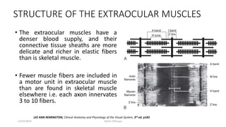
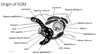
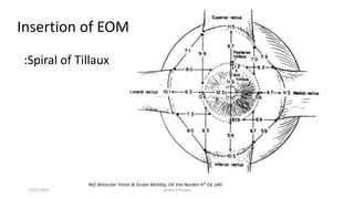
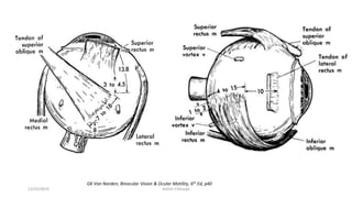
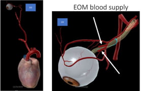
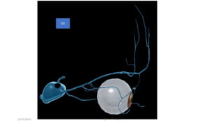
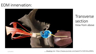
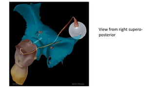
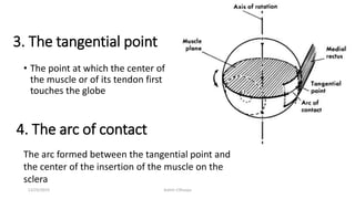
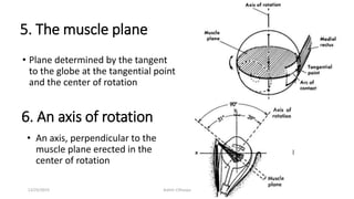
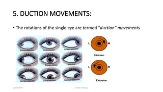
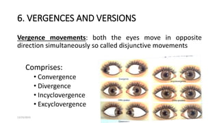
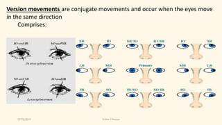
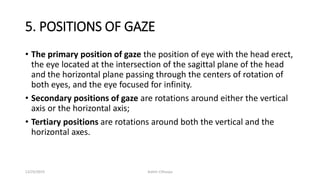
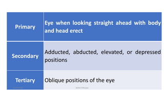
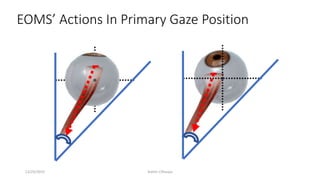
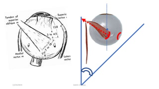
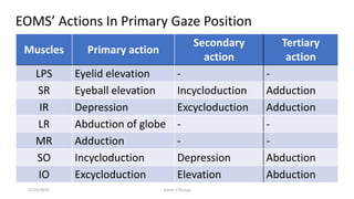
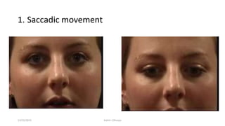
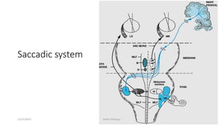
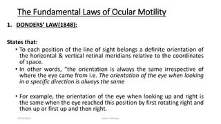
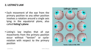
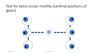
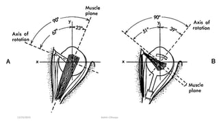
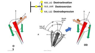
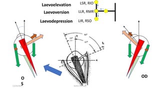
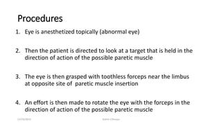
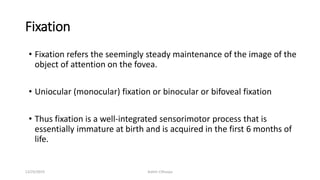
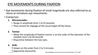
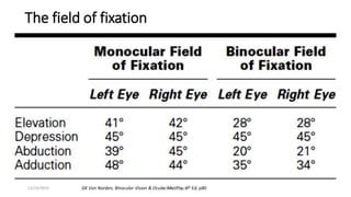
Extraocular muscle actions &
motility
Presenter: Ashim Chhusya
B.Optometry, Tilganga Institute of
Ophthalmology
12/25/2019 Ashim Chhusya
STRUCTURE OF THE EXTRAOCULAR MUSCLES
• The extraocular muscles have a
denser blood supply, and their
connective tissue sheaths are more
delicate and richer in elastic fibers
than is skeletal muscle.
• Fewer muscle fibers are included in
a motor unit in extraocular muscle
than are found in skeletal muscle
elsewhere i.e. each axon innervates
3 to 10 fibers.
LEE ANN REMINGTON; Clinical Anatomy and Physiology of the Visual System, 3rd ed. p183
12/25/2019 Ashim Chhusya
• This dense innervation provides for precise fine motor control of the
extraocular muscles resulting in high velocity ocular movements,
necessary in saccades, (up to 1000 degrees per second) and very
accurate pursuits (velocities of 100 degrees per second) and fixations.
• The extraocular muscles have a range of fiber sizes, with the fibers
closer to the surface generally having smaller diameters (5 to 15 μm)
and those deeper within the muscle generally having larger
diameters (10 to 40 μm).
12/25/2019 Ashim Chhusya
Origin & Insertion of EOMs
12/25/2019 Ashim Chhusya
Origin of EOM
12/25/2019 Ashim Chhusya
Insertion of EOM
Ref: Binocular Vision & Ocular Motility, GK Von Norden 6th Ed, p40
:Spiral of Tillaux
12/25/2019 Ashim Chhusya
GK Von Norden; Binocular Vision & Ocular Motility, 6th Ed, p40
12/25/2019 Ashim Chhusya
Origin & insertion of EOM
LPS:
Origin : Lesser wings of sphenoid bone
Insertion: Tarsus, pretarsal tissues & skin, lateral & medial palpebral
ligaments
LR:
origin on the common tendinous ring and the spina recti lateralis, a
prominence on the greater wing of the sphenoid bone
Insertion approximately 6.9 mm from the limbus (parallels that of the
medial rectus) and is, and the length of the tendon is approximately 8.8
mm.12/25/2019 Ashim Chhusya
SR:
• Origin on the superior part of the common tendinous ring and the sheath of
the optic nerve
• Insertion of the superior rectus is approximately 7.7 mm from the limbus
MR
• Origin: the upper and the lower parts of the common ring tendon and from
the sheath of the optic nerve
• Insertion of the medial rectus is about 5.5 mm from the limbus, and the
tendon is approximately 3.7 mm long
12/25/2019 Ashim Chhusya
Clinical pearl: Retrobulbar Optic Neuritis
• An inflammation affecting the sheaths of the optic nerve.
• Pain with extreme eye movement can be one of the early presenting
signs.
• The optic nerve sheath is supplied with a dense sensory nerve
network and because of the close association of muscle sheath and
optic nerve sheath, eye movement can cause stretching of the optic
nerve sheath, resulting in a sensation of pain
12/25/2019 Ashim Chhusya
INFERIOR RECTUS MUSCLE:
• Origin on the lower limb of the tendinous ring
• Insertion is about 6.5 mm from the limbus
SUPERIOR OBLIQUE MUSCLE
• Origin on the lesser wing of the sphenoid bone, supero-medial to the optic
canal near the fronto-ethmoid suture
• Courses forward and passes through the trochlea, a u-shaped piece of
cartilage attached to the orbital plate of the frontal bone
• Insertion of the superior oblique muscle attaches in the super-posterior
lateral aspect of the globe
INFERIOR OBLIQUE MUSCLE
• Origin on the maxillary bone just posterior to the inferior medial orbital rim
and lateral to the nasolacrimal canal.
• Insertion of the inferior oblique is on the posterior portion of the globe on
the lateral side, mostly inferior, lying just outer to the macular area
12/25/2019 Ashim Chhusya
EOM blood supply
OD
OD
12/25/2019 Ashim Chhusya
OD
12/25/2019 Ashim Chhusya
EOM innervation:
Reading site: https://www.youtube.com/watch?v=5OD-BnuOMHs
Transverse
section
View from above
12/25/2019 Ashim Chhusya
View from right supero-
posterior
12/25/2019 Ashim Chhusya
Terminologies in ocular movements
1. Center of Rotation
• A center point of globe around which the
eye performs rotary movements
• The approximate geometric center of the
eye
• In primary position the center of rotation is
located
• About 13.5 mm (in myopes, 14.5 mm)
behind the apex of the cornea on the
line of sight
• 1.3 mm behind the equatorial plane.
GK Von Norden; Binocular Vision & Ocular Motility, 6th Ed, p5212/25/2019 Ashim Chhusya
2. Fick’s axes
• All eye movement can be described as rotations
around one or more axes called FICK’S AXES.
• Comprises:
The x-axis The horizontal or transverse axis and runs from nasal to
temporal
The y-axis The sagittal axis running from the anterior pole to the
Posterior pole
The z-axis Is the vertical axis and runs from superior to inferior
12/25/2019 Ashim Chhusya
3. The tangential point
• The point at which the center of
the muscle or of its tendon first
touches the globe
4. The arc of contact
The arc formed between the tangential point and
the center of the insertion of the muscle on the
sclera
12/25/2019 Ashim Chhusya
5. The muscle plane
• Plane determined by the tangent
to the globe at the tangential point
and the center of rotation
6. An axis of rotation
• An axis, perpendicular to the
muscle plane erected in the
center of rotation
12/25/2019 Ashim Chhusya
5. DUCTION MOVEMENTS:
• The rotations of the single eye are termed “duction” movements
12/25/2019 Ashim Chhusya
6. VERGENCES AND VERSIONS
Vergence movements: both the eyes move in opposite
direction simultaneously so called disjunctive movements
Comprises:
• Convergence
• Divergence
• Incyclovergence
• Excyclovergence
12/25/2019 Ashim Chhusya
Version movements are conjugate movements and occur when the eyes move
in the same direction
Comprises:
12/25/2019 Ashim Chhusya
5. POSITIONS OF GAZE
• The primary position of gaze the position of eye with the head erect,
the eye located at the intersection of the sagittal plane of the head
and the horizontal plane passing through the centers of rotation of
both eyes, and the eye focused for infinity.
• Secondary positions of gaze are rotations around either the vertical
axis or the horizontal axis;
• Tertiary positions are rotations around both the vertical and the
horizontal axes.
12/25/2019 Ashim Chhusya
Primary
Eye when looking straight ahead with body
and head erect
Secondary
Adducted, abducted, elevated, or depressed
positions
Tertiary Oblique positions of the eye
12/25/2019 Ashim Chhusya
EOMS’ Actions In Primary Gaze Position
12/25/2019 Ashim Chhusya
Muscles Primary action
Secondary
action
Tertiary
action
LPS Eyelid elevation - -
SR Eyeball elevation Incycloduction Adduction
IR Depression Excycloduction Adduction
LR Abduction of globe - -
MR Adduction - -
SO Incycloduction Depression Abduction
IO Excycloduction Elevation Abduction
EOMS’ Actions In Primary Gaze Position
12/25/2019 Ashim Chhusya
The vertical gaze center: Rostral interstitial
Nucleus Of Medial Longitudinal Fasciculus
(riMLF) & INC
at midbrain (thalamo-mesencephalic junction)
The horizontal gaze center:-
PPRF in Pons adler’s physio.p235
Ipsilateral trochlear
nuclei adler’s physio.p235
B/L oculomotor
nuclei
The ipsilateral
abducens nucleus
adler’s physio.p235
Contralateral oculomotor nucleus
Ipsilateral LR Contralateral yoke muscle
Yoke muscle
What are the centers for making Eye Movements?
=2 centers
12/25/2019 Ashim Chhusya
Parinaud's Syndrome
Parinaud's syndrome, is a group of abnormalities
of eye movement and pupil dysfunction caused
by lesions that compresses the vertical gaze
center at riMLF at thalamo-mesencephalic
junction resulting an inability to move the eyes
up (most commonly) and down.
It is named after Henri Parinaud, French
ophthalmologist and neurologist
12/25/2019 Ashim Chhusya
How does eyes makes movements for gazing?
1. Saccadic movement
2. Smooth pursuit movement
3. Vergence movement
4. Vestibulocular movement
12/25/2019 Ashim Chhusya
1. Saccadic movement
12/25/2019 Ashim Chhusya
Saccadic system
12/25/2019 Ashim Chhusya
Test for saccadic movement
a. To each side of the head for horizontal movement
b. Above & below the head for vertical movement
• The movement, speed & accuracy of either eye is compared & any
asymmetry between the eyes is noted
• The patient is asked to look from one
target to another, one held on the
primary position and the other:
12/25/2019 Ashim Chhusya
Smooth Pursuit System
Dorsal lateral pontine premotor nuclei
(DLPN)
Ref: https://www.ncbi.nlm.nih.gov/pmc/articles/PMC3164106/
12/25/2019 Ashim Chhusya
Test for smooth pursuit movement
• Smooth pursuit movements are tested monocularly & binocularly.
• Version SPM test is done using a spotlight to observe the movement from
the primary position into the eight cardinal positions of gaze with the
head erect & immobile.
• Ductions are tested to compare with versions & the following is noted:
• Symmetry of eyes’ movement
• Whether the movement is smooth or jerky
• If, presence of Nystagmus
• Effect of fatigue
• Torsional movement
• Abnormal head movements
12/25/2019 Ashim Chhusya
Vergence system
Supraoculomotor area (SOA) in the midbrain
Cortical neural signals
Stimuli: disparity between images on the retina, retinal blur (an
unfocused image), proximity of objects, the change in size of an
image
Cortical processing: occipital lobe, the parietal lobe and frontal eye
fields
Evidence that an area located anteriorly to the saccade
and smooth pursuit zone in the frontal eye fields is linked
with depth perception suggests that this area could be
involved with the initiation of vergence eye movement
https://www.ncbi.nlm.nih.gov/pmc/articles/PMC5122972/
3rd & 6th CN nuclei12/25/2019 Ashim Chhusya
Vestibulo-ocular
reflex movement
Head rotation
12/25/2019 Ashim Chhusya
Vestibulocular reflex test
1. Doll’s head maneuver
• Patients head is moved quickly by the examiner to the right & left for
horizontal movement & up-down for vertical movement.
• A normal response is of the eyes is moving in the opposite direction to the
head movement
2. Induced vestibular nystagmus
• Warm water: slow phase to opposite side & fast phase to same side
• Cold water: slow phase to the same side & fast phase to opposite side
• Vertical caloric nystagmus: by stimulating both ears
12/25/2019 Ashim Chhusya
The Fundamental Laws of Ocular Motility
1. DONDERS’ LAW(1848):
States that:
• To each position of the line of sight belongs a definite orientation of
the horizontal & vertical retinal meridians relative to the coordinates
of space.
• In other words, “the orientation is always the same irrespective of
where the eye came from i.e. The orientation of the eye when looking
in a specific direction is always the same
• For example, the orientation of the eye when looking up and right is
the same when the eye reached this position by first rotating right and
then up or first up and then right.
12/25/2019 Ashim Chhusya
2. LISTING’S LAW
• Each movement of the eye from the
primary position to any other position
involves a rotation around a single axis
lying in the equatorial plane, also
called listing’s plane
• Listing’s law implies that all eye
movements from the primary position
occur without ‘‘torsion’’ or cyclo-
rotation with respect to the primary
position
12/25/2019 Ashim Chhusya
Does Listing's Law hold for other types of eye
movements?
a) Vestibular ocular reflex (VOR):
Function: to keep the image of the world stationary on the retina
when the head rotates
It does this by rotating the eyes in the opposite direction of the head.
If the head tilts to the side (torsionally) the eye counter-rolls, i.e. it
moves out of Listing's plane.
Thus the VOR does not obey Listing's law.
http://schorlab.berkeley.edu/vilis/othereye.htm
12/25/2019 Ashim Chhusya
b) Saccades & Pursuit
Function: to keep the fovea on a small moving target, e.g. tracking a
tennis ball.
As in saccades, pointing the fovea at this moving ball is a 2D problem
and Listing's law used to resolve which of many eye position to choose.
Thus listing law is obeyed.
http://schorlab.berkeley.edu/vilis/othereye.htm
12/25/2019 Ashim Chhusya
c) Vergence
Function: to align a near target on the fovea of each eye.
Listing's law is violated during vergence between targets at different
distances.
Listing's law holds at a constant distance.
http://schorlab.berkeley.edu/vilis/othereye.htm
12/25/2019 Ashim Chhusya
3. Herring's Law of Equal Innervation
• It states “whenever an impulse for the performance of an eye
movement is sent out, corresponding muscles of each eye receive
equal innervations to contract or relax.
• Also called the law of motor correspondence of the eyes
• Herring's law applies only to extraocular muscles
• For example: rt. LR & lt. MR in dextroversion.
12/25/2019 Ashim Chhusya
4. Sherrington’s Law of Reciprocal Innervation
• “Whenever an agonist receives an impulse to contract, an equivalent
inhibitory impulse is sent to its antagonist, which relaxes and actually
lengthens”
• The finely graded interplay between opposing eye muscles makes
movements of the globe smooth and steady
• Sherrington’s law is not limited to the extraocular muscles and
applies to all striated muscles of the body
12/25/2019 Ashim Chhusya
Test for extra-ocular motility (cardinal positions of
gazes)
12/25/2019 Ashim Chhusya
RIR, LSO
RSR, LIO
RLR, LMR
ODO
S
Dextroelevation
Dextrodepression
Dextroversion
12/25/2019 Ashim Chhusya
LIR, RSO
LSR, RIO
LLR, RMR
ODO
S
Laevoelevation
Laevodepression
Laevoversion
23⁰
67⁰
90⁰
12/25/2019 Ashim Chhusya
RSR LIO
RLR LMR
RIR LSO
LSRRIO
LLR
RM
R
RSO LIR
LeftRight
12/25/2019 Ashim Chhusya
How to test Extra-ocular Motility?
Have the patient look in the six cardinal positions of gaze.
• With monocularly to test ductions.
• with both eyes open to assess versions
12/25/2019 Ashim Chhusya
Recording of ocular Motility
Ocular movements are graded on a 9-point scale of −4 through to +4.
-4 No movement of the eye(s) past midline (marked).
-3 75% deficit of movement (moderate).
-2 50% deficit of movement (small).
-1 25% deficit of movement (slight).
0 Full normal movement
# Limitations of movement
Fiona J. Rowe; Clinical Orthoptics 3rd ed, p42412/25/2019 Ashim Chhusya
-1
-1
-2
0
0
00
0
0
0
0
0
R L
12/25/2019 Ashim Chhusya
Force Duction Test
• Also known as traction test
• Done in patients with impaired extraocular
motility / strabismic
• It helps to differentiate whether the deviation is due to paralysis of EOM or
due to mechanical restriction of the ocular movement.
12/25/2019 Ashim Chhusya
Procedures
1. Eye is anesthetized topically (abnormal eye)
2. Then the patient is directed to look at a target that is held in the
direction of action of the possible paretic muscle
3. The eye is then grasped with toothless forceps near the limbus
at opposite site of paretic muscle insertion
4. An effort is then made to rotate the eye with the forceps in the
direction of action of the possible paretic muscle
12/25/2019 Ashim Chhusya
Results:
• If the eye can be rotated freely with the forceps – FDT is recorded as
negative i.e. the impaired extraocular motility is not due to
restriction but the muscle paresis or palsy.
• FDT is positive in cases of squint (incomitant) due to mechanical
restriction
12/25/2019 Ashim Chhusya
Fixation
• Fixation refers the seemingly steady maintenance of the image of the
object of attention on the fovea.
• Uniocular (monocular) fixation or binocular or bifoveal fixation
• Thus fixation is a well-integrated sensorimotor process that is
essentially immature at birth and is acquired in the first 6 months of
life.
12/25/2019 Ashim Chhusya
EYE MOVEMENTS DURING FIXATION
• Eye movements during fixation of small magnitude are also referred to as
micro or miniature eye movements.
• Comprises:
1. Microsaccades:
• Range in amplitude from 1 to 23 seconds,
• They correct for slippage of the visual target off the fovea.
2. Tremor:
• When the amplitude of fixation tremor is on the order of the diameter of the
smallest cone 5 to 50 seconds
• Disjunctive between the two eyes.
3. Drift:
 Slower on the order from 2 to 5 minutes
 Apparently disjunctive GK Von Norden; Binocular Vision & Ocular Motility, 6th Ed, p8012/25/2019 Ashim Chhusya
The field of fixation
GK Von Norden; Binocular Vision & Ocular Motility, 6th Ed, p8012/25/2019 Ashim Chhusya
Any Query?
Thank YOU ^_^
12/25/2019 Ashim Chhusya

Extraocular muscle actions & motility

  • 1.
    Extraocular muscle actions& motility Presenter: Ashim Chhusya B.Optometry, Tilganga Institute of Ophthalmology 12/25/2019 Ashim Chhusya
  • 2.
    STRUCTURE OF THEEXTRAOCULAR MUSCLES • The extraocular muscles have a denser blood supply, and their connective tissue sheaths are more delicate and richer in elastic fibers than is skeletal muscle. • Fewer muscle fibers are included in a motor unit in extraocular muscle than are found in skeletal muscle elsewhere i.e. each axon innervates 3 to 10 fibers. LEE ANN REMINGTON; Clinical Anatomy and Physiology of the Visual System, 3rd ed. p183 12/25/2019 Ashim Chhusya
  • 3.
    • This denseinnervation provides for precise fine motor control of the extraocular muscles resulting in high velocity ocular movements, necessary in saccades, (up to 1000 degrees per second) and very accurate pursuits (velocities of 100 degrees per second) and fixations. • The extraocular muscles have a range of fiber sizes, with the fibers closer to the surface generally having smaller diameters (5 to 15 μm) and those deeper within the muscle generally having larger diameters (10 to 40 μm). 12/25/2019 Ashim Chhusya
  • 4.
    Origin & Insertionof EOMs 12/25/2019 Ashim Chhusya
  • 5.
  • 6.
  • 7.
  • 8.
    Insertion of EOM Ref:Binocular Vision & Ocular Motility, GK Von Norden 6th Ed, p40 :Spiral of Tillaux 12/25/2019 Ashim Chhusya
  • 9.
    GK Von Norden;Binocular Vision & Ocular Motility, 6th Ed, p40 12/25/2019 Ashim Chhusya
  • 10.
    Origin & insertionof EOM LPS: Origin : Lesser wings of sphenoid bone Insertion: Tarsus, pretarsal tissues & skin, lateral & medial palpebral ligaments LR: origin on the common tendinous ring and the spina recti lateralis, a prominence on the greater wing of the sphenoid bone Insertion approximately 6.9 mm from the limbus (parallels that of the medial rectus) and is, and the length of the tendon is approximately 8.8 mm.12/25/2019 Ashim Chhusya
  • 11.
    SR: • Origin onthe superior part of the common tendinous ring and the sheath of the optic nerve • Insertion of the superior rectus is approximately 7.7 mm from the limbus MR • Origin: the upper and the lower parts of the common ring tendon and from the sheath of the optic nerve • Insertion of the medial rectus is about 5.5 mm from the limbus, and the tendon is approximately 3.7 mm long 12/25/2019 Ashim Chhusya
  • 12.
    Clinical pearl: RetrobulbarOptic Neuritis • An inflammation affecting the sheaths of the optic nerve. • Pain with extreme eye movement can be one of the early presenting signs. • The optic nerve sheath is supplied with a dense sensory nerve network and because of the close association of muscle sheath and optic nerve sheath, eye movement can cause stretching of the optic nerve sheath, resulting in a sensation of pain 12/25/2019 Ashim Chhusya
  • 13.
    INFERIOR RECTUS MUSCLE: •Origin on the lower limb of the tendinous ring • Insertion is about 6.5 mm from the limbus SUPERIOR OBLIQUE MUSCLE • Origin on the lesser wing of the sphenoid bone, supero-medial to the optic canal near the fronto-ethmoid suture • Courses forward and passes through the trochlea, a u-shaped piece of cartilage attached to the orbital plate of the frontal bone • Insertion of the superior oblique muscle attaches in the super-posterior lateral aspect of the globe INFERIOR OBLIQUE MUSCLE • Origin on the maxillary bone just posterior to the inferior medial orbital rim and lateral to the nasolacrimal canal. • Insertion of the inferior oblique is on the posterior portion of the globe on the lateral side, mostly inferior, lying just outer to the macular area 12/25/2019 Ashim Chhusya
  • 14.
  • 15.
  • 16.
    EOM innervation: Reading site:https://www.youtube.com/watch?v=5OD-BnuOMHs Transverse section View from above 12/25/2019 Ashim Chhusya
  • 17.
    View from rightsupero- posterior 12/25/2019 Ashim Chhusya
  • 18.
  • 19.
    Terminologies in ocularmovements 1. Center of Rotation • A center point of globe around which the eye performs rotary movements • The approximate geometric center of the eye • In primary position the center of rotation is located • About 13.5 mm (in myopes, 14.5 mm) behind the apex of the cornea on the line of sight • 1.3 mm behind the equatorial plane. GK Von Norden; Binocular Vision & Ocular Motility, 6th Ed, p5212/25/2019 Ashim Chhusya
  • 20.
    2. Fick’s axes •All eye movement can be described as rotations around one or more axes called FICK’S AXES. • Comprises: The x-axis The horizontal or transverse axis and runs from nasal to temporal The y-axis The sagittal axis running from the anterior pole to the Posterior pole The z-axis Is the vertical axis and runs from superior to inferior 12/25/2019 Ashim Chhusya
  • 21.
  • 22.
    3. The tangentialpoint • The point at which the center of the muscle or of its tendon first touches the globe 4. The arc of contact The arc formed between the tangential point and the center of the insertion of the muscle on the sclera 12/25/2019 Ashim Chhusya
  • 23.
    5. The muscleplane • Plane determined by the tangent to the globe at the tangential point and the center of rotation 6. An axis of rotation • An axis, perpendicular to the muscle plane erected in the center of rotation 12/25/2019 Ashim Chhusya
  • 24.
    5. DUCTION MOVEMENTS: •The rotations of the single eye are termed “duction” movements 12/25/2019 Ashim Chhusya
  • 25.
    6. VERGENCES ANDVERSIONS Vergence movements: both the eyes move in opposite direction simultaneously so called disjunctive movements Comprises: • Convergence • Divergence • Incyclovergence • Excyclovergence 12/25/2019 Ashim Chhusya
  • 26.
    Version movements areconjugate movements and occur when the eyes move in the same direction Comprises: 12/25/2019 Ashim Chhusya
  • 27.
    5. POSITIONS OFGAZE • The primary position of gaze the position of eye with the head erect, the eye located at the intersection of the sagittal plane of the head and the horizontal plane passing through the centers of rotation of both eyes, and the eye focused for infinity. • Secondary positions of gaze are rotations around either the vertical axis or the horizontal axis; • Tertiary positions are rotations around both the vertical and the horizontal axes. 12/25/2019 Ashim Chhusya
  • 28.
    Primary Eye when lookingstraight ahead with body and head erect Secondary Adducted, abducted, elevated, or depressed positions Tertiary Oblique positions of the eye 12/25/2019 Ashim Chhusya
  • 29.
    EOMS’ Actions InPrimary Gaze Position 12/25/2019 Ashim Chhusya
  • 30.
  • 31.
    Muscles Primary action Secondary action Tertiary action LPSEyelid elevation - - SR Eyeball elevation Incycloduction Adduction IR Depression Excycloduction Adduction LR Abduction of globe - - MR Adduction - - SO Incycloduction Depression Abduction IO Excycloduction Elevation Abduction EOMS’ Actions In Primary Gaze Position 12/25/2019 Ashim Chhusya
  • 32.
    The vertical gazecenter: Rostral interstitial Nucleus Of Medial Longitudinal Fasciculus (riMLF) & INC at midbrain (thalamo-mesencephalic junction) The horizontal gaze center:- PPRF in Pons adler’s physio.p235 Ipsilateral trochlear nuclei adler’s physio.p235 B/L oculomotor nuclei The ipsilateral abducens nucleus adler’s physio.p235 Contralateral oculomotor nucleus Ipsilateral LR Contralateral yoke muscle Yoke muscle What are the centers for making Eye Movements? =2 centers 12/25/2019 Ashim Chhusya
  • 33.
    Parinaud's Syndrome Parinaud's syndrome,is a group of abnormalities of eye movement and pupil dysfunction caused by lesions that compresses the vertical gaze center at riMLF at thalamo-mesencephalic junction resulting an inability to move the eyes up (most commonly) and down. It is named after Henri Parinaud, French ophthalmologist and neurologist 12/25/2019 Ashim Chhusya
  • 34.
    How does eyesmakes movements for gazing? 1. Saccadic movement 2. Smooth pursuit movement 3. Vergence movement 4. Vestibulocular movement 12/25/2019 Ashim Chhusya
  • 35.
  • 36.
  • 37.
    Test for saccadicmovement a. To each side of the head for horizontal movement b. Above & below the head for vertical movement • The movement, speed & accuracy of either eye is compared & any asymmetry between the eyes is noted • The patient is asked to look from one target to another, one held on the primary position and the other: 12/25/2019 Ashim Chhusya
  • 38.
    Smooth Pursuit System Dorsallateral pontine premotor nuclei (DLPN) Ref: https://www.ncbi.nlm.nih.gov/pmc/articles/PMC3164106/ 12/25/2019 Ashim Chhusya
  • 39.
    Test for smoothpursuit movement • Smooth pursuit movements are tested monocularly & binocularly. • Version SPM test is done using a spotlight to observe the movement from the primary position into the eight cardinal positions of gaze with the head erect & immobile. • Ductions are tested to compare with versions & the following is noted: • Symmetry of eyes’ movement • Whether the movement is smooth or jerky • If, presence of Nystagmus • Effect of fatigue • Torsional movement • Abnormal head movements 12/25/2019 Ashim Chhusya
  • 40.
    Vergence system Supraoculomotor area(SOA) in the midbrain Cortical neural signals Stimuli: disparity between images on the retina, retinal blur (an unfocused image), proximity of objects, the change in size of an image Cortical processing: occipital lobe, the parietal lobe and frontal eye fields Evidence that an area located anteriorly to the saccade and smooth pursuit zone in the frontal eye fields is linked with depth perception suggests that this area could be involved with the initiation of vergence eye movement https://www.ncbi.nlm.nih.gov/pmc/articles/PMC5122972/ 3rd & 6th CN nuclei12/25/2019 Ashim Chhusya
  • 41.
  • 42.
    Vestibulocular reflex test 1.Doll’s head maneuver • Patients head is moved quickly by the examiner to the right & left for horizontal movement & up-down for vertical movement. • A normal response is of the eyes is moving in the opposite direction to the head movement 2. Induced vestibular nystagmus • Warm water: slow phase to opposite side & fast phase to same side • Cold water: slow phase to the same side & fast phase to opposite side • Vertical caloric nystagmus: by stimulating both ears 12/25/2019 Ashim Chhusya
  • 43.
    The Fundamental Lawsof Ocular Motility 1. DONDERS’ LAW(1848): States that: • To each position of the line of sight belongs a definite orientation of the horizontal & vertical retinal meridians relative to the coordinates of space. • In other words, “the orientation is always the same irrespective of where the eye came from i.e. The orientation of the eye when looking in a specific direction is always the same • For example, the orientation of the eye when looking up and right is the same when the eye reached this position by first rotating right and then up or first up and then right. 12/25/2019 Ashim Chhusya
  • 44.
    2. LISTING’S LAW •Each movement of the eye from the primary position to any other position involves a rotation around a single axis lying in the equatorial plane, also called listing’s plane • Listing’s law implies that all eye movements from the primary position occur without ‘‘torsion’’ or cyclo- rotation with respect to the primary position 12/25/2019 Ashim Chhusya
  • 45.
  • 46.
    Does Listing's Lawhold for other types of eye movements? a) Vestibular ocular reflex (VOR): Function: to keep the image of the world stationary on the retina when the head rotates It does this by rotating the eyes in the opposite direction of the head. If the head tilts to the side (torsionally) the eye counter-rolls, i.e. it moves out of Listing's plane. Thus the VOR does not obey Listing's law. http://schorlab.berkeley.edu/vilis/othereye.htm 12/25/2019 Ashim Chhusya
  • 47.
    b) Saccades &Pursuit Function: to keep the fovea on a small moving target, e.g. tracking a tennis ball. As in saccades, pointing the fovea at this moving ball is a 2D problem and Listing's law used to resolve which of many eye position to choose. Thus listing law is obeyed. http://schorlab.berkeley.edu/vilis/othereye.htm 12/25/2019 Ashim Chhusya
  • 48.
    c) Vergence Function: toalign a near target on the fovea of each eye. Listing's law is violated during vergence between targets at different distances. Listing's law holds at a constant distance. http://schorlab.berkeley.edu/vilis/othereye.htm 12/25/2019 Ashim Chhusya
  • 49.
    3. Herring's Lawof Equal Innervation • It states “whenever an impulse for the performance of an eye movement is sent out, corresponding muscles of each eye receive equal innervations to contract or relax. • Also called the law of motor correspondence of the eyes • Herring's law applies only to extraocular muscles • For example: rt. LR & lt. MR in dextroversion. 12/25/2019 Ashim Chhusya
  • 50.
    4. Sherrington’s Lawof Reciprocal Innervation • “Whenever an agonist receives an impulse to contract, an equivalent inhibitory impulse is sent to its antagonist, which relaxes and actually lengthens” • The finely graded interplay between opposing eye muscles makes movements of the globe smooth and steady • Sherrington’s law is not limited to the extraocular muscles and applies to all striated muscles of the body 12/25/2019 Ashim Chhusya
  • 51.
    Test for extra-ocularmotility (cardinal positions of gazes) 12/25/2019 Ashim Chhusya
  • 52.
  • 53.
    RIR, LSO RSR, LIO RLR,LMR ODO S Dextroelevation Dextrodepression Dextroversion 12/25/2019 Ashim Chhusya
  • 54.
    LIR, RSO LSR, RIO LLR,RMR ODO S Laevoelevation Laevodepression Laevoversion 23⁰ 67⁰ 90⁰ 12/25/2019 Ashim Chhusya
  • 55.
    RSR LIO RLR LMR RIRLSO LSRRIO LLR RM R RSO LIR LeftRight 12/25/2019 Ashim Chhusya
  • 56.
    How to testExtra-ocular Motility? Have the patient look in the six cardinal positions of gaze. • With monocularly to test ductions. • with both eyes open to assess versions 12/25/2019 Ashim Chhusya
  • 57.
    Recording of ocularMotility Ocular movements are graded on a 9-point scale of −4 through to +4. -4 No movement of the eye(s) past midline (marked). -3 75% deficit of movement (moderate). -2 50% deficit of movement (small). -1 25% deficit of movement (slight). 0 Full normal movement # Limitations of movement Fiona J. Rowe; Clinical Orthoptics 3rd ed, p42412/25/2019 Ashim Chhusya
  • 58.
  • 59.
    Force Duction Test •Also known as traction test • Done in patients with impaired extraocular motility / strabismic • It helps to differentiate whether the deviation is due to paralysis of EOM or due to mechanical restriction of the ocular movement. 12/25/2019 Ashim Chhusya
  • 60.
    Procedures 1. Eye isanesthetized topically (abnormal eye) 2. Then the patient is directed to look at a target that is held in the direction of action of the possible paretic muscle 3. The eye is then grasped with toothless forceps near the limbus at opposite site of paretic muscle insertion 4. An effort is then made to rotate the eye with the forceps in the direction of action of the possible paretic muscle 12/25/2019 Ashim Chhusya
  • 61.
    Results: • If theeye can be rotated freely with the forceps – FDT is recorded as negative i.e. the impaired extraocular motility is not due to restriction but the muscle paresis or palsy. • FDT is positive in cases of squint (incomitant) due to mechanical restriction 12/25/2019 Ashim Chhusya
  • 62.
    Fixation • Fixation refersthe seemingly steady maintenance of the image of the object of attention on the fovea. • Uniocular (monocular) fixation or binocular or bifoveal fixation • Thus fixation is a well-integrated sensorimotor process that is essentially immature at birth and is acquired in the first 6 months of life. 12/25/2019 Ashim Chhusya
  • 63.
    EYE MOVEMENTS DURINGFIXATION • Eye movements during fixation of small magnitude are also referred to as micro or miniature eye movements. • Comprises: 1. Microsaccades: • Range in amplitude from 1 to 23 seconds, • They correct for slippage of the visual target off the fovea. 2. Tremor: • When the amplitude of fixation tremor is on the order of the diameter of the smallest cone 5 to 50 seconds • Disjunctive between the two eyes. 3. Drift:  Slower on the order from 2 to 5 minutes  Apparently disjunctive GK Von Norden; Binocular Vision & Ocular Motility, 6th Ed, p8012/25/2019 Ashim Chhusya
  • 64.
    The field offixation GK Von Norden; Binocular Vision & Ocular Motility, 6th Ed, p8012/25/2019 Ashim Chhusya
  • 65.
    Any Query? Thank YOU^_^ 12/25/2019 Ashim Chhusya