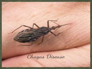
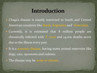
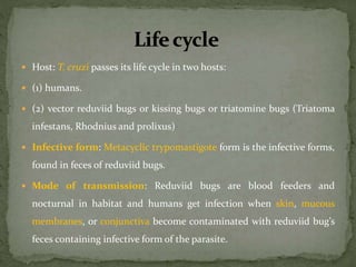
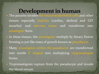
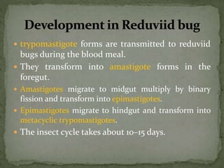
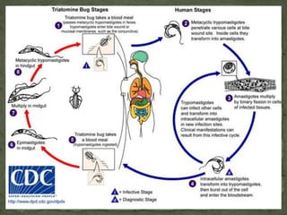
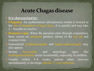
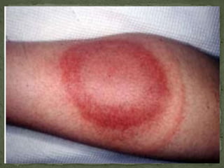
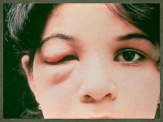
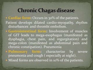
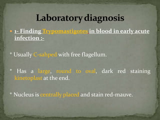
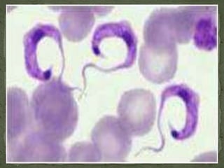
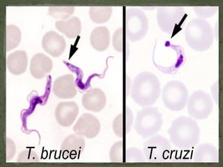
By
Montasir Ghaneim Alzain
M.Sc in Medical Parasitology / UOG
 By the end of this presentation each student
should:
 Know the causative agent of American trypanosoma.
 Understand how the parasite is transmitted and the
life cycle of it.
 Know briefly about the clinical picture of the acute &
chronic forms of the disease.
 Know the methods of laboratory diagnosis.
 American trypanosomasis also called Chaga’s disease, it is
caused by Trypanosoma Cruzi.
 It was first discovered in the vector by Carlos Chagas in
1907 and then shown to cause human disease.
 He named the parasite as T. cruzi after his guide Oswaldo
Cruz.
 The vector of T. cruzi are winged bugs called triatominae
bug (AKA; Reduviidae bug).
 Chaga’s disease is mainly restricted to South and Central
American countries like Brazil, Argentina and Venezuela.
 Currently, it is estimated that 8 million people are
chronically infected with T. cruzi and 14,000 deaths occur
due to the illness every year.
 It is a zoonotic disease, having many animal reservoirs like
dogs, cats, opossums and rodents.
 The disease may be acute or chronic.
 Host: T. cruzi passes its life cycle in two hosts:
 (1) humans.
 (2) vector reduviid bugs or kissing bugs or triatomine bugs (Triatoma
infestans, Rhodnius and prolixus)
 Infective form: Metacyclic trypomastigote form is the infective forms,
found in feces of reduviid bugs.
 Mode of transmission: Reduviid bugs are blood feeders and
nocturnal in habitat and humans get infection when skin, mucous
membranes, or conjunctiva become contaminated with reduviid bug’s
feces containing infective form of the parasite.
 T. cruzi can also be transmitted by the blood
transfusion, organ transplantation, from mother to
fetus.
 The parasite invades the reticuloendothelial cells and other
tissues especially muscles (cardiac, skeletal and GIT
muscles) and nervous tissue and transforms into
amastigote form.
 In these tissues, the amastigotes multiply by binary fission
forming a cyst like mass of growth known as pseudocyst.
 Many amastigotes within the pseudocyst are transformed
into motile C shaped non multiplying trypomastigote
forms.
 Trypomastigotes rupture from the pseudocyst and invade
the blood stream.
 trypomastigote forms are transmitted to reduviid
bugs during the blood meal.
 They transform into amastigote forms in the
foregut.
 Amastigotes migrate to midgut multiply by binary
fission and transform into epimastigotes.
 Epimastigotes migrate to hindgut and transform into
metacyclic trypomastigotes.
 The insect cycle takes about 10–15 days.
 It is characterized by:
 Chagoma: An erythematous subcutaneous nodule is formed at
the site of deposition of bug’s feces. It is painful and may take
2 - 3months to resolve
 Romana’s sign: When the parasites enter through conjunctiva,
there occurs an unilateral painless edema of the eye lid and
conjunctivitis.
 Generalized lymphadenopathy and hepato-splenomegaly may
also appear.
 Severe myocarditis and neurologic signs like
meningoencephalitis occur occasionally, especially in children.
 Usually within 4–8 weeks, patient either recovers
spontaneously or develops chronic T. cruzi infection.
 Chronic Chagas’ disease manifests years or even
decades after the initial infection.
 It occurs due to multiplication of the parasites in
the muscles (skeletal, cardiac and GIT) and
nervous tissue.
 The mechanism of turning from acute to chronic
and immune evasion mechanism is not fully
understood.
 Cardiac form: Occurs in 30% of the patients.
Patient develops dilated cardio-myopathy, rhythm
disturbances and thrombi embolism.
 Gastrointestinal form: Involvement of muscles
of GIT leads to mega-esophagus (manifested as
dysphagia, chest pain, and regurgitation) and
mega-colon (manifested as abdominal pain and
chronic constipation). Pneumonia
 Pulmonary form: characterize by severe
pneumonia and cough ( especially at night).
 Mixed forms are observed in 10% of the patients.
 1- Finding Trypomastigotes in blood in early acute
infection :-
* Usually C-sahped with free flagellum.
* Has a large, round to oval, dark red staining
kinetoplast at the end.
* Nucleus is centrally placed and stain red-mauve.
 2- Blood culture to detect Epimastigotes of
Trypanosoma cruzi:
 Culture technique is used mainly to diagnose chronic
Chaga's disease.
 Blood is inoculated in NNN medium or Yager’s
liver infusion tryptose medium, incubated at 25°C
and observed for the epimastigote forms for up to
30 days before they are considered negative.
 3- Xenodiagnosis:
 Uninfected, laboratory reared triatomine bugs are
starved for 2 weeks and then fed on the patient’s
blood.
 If trypomastigotes are ingested they will multiply and
develop into epimastigotes and metacyclic
trypomastigotes, which can be found in the bug feces.
 4-other techniques are used like:
 Serological tests.
 Molecular diagnosis.
 Animal inoculation.
* Blood of the patients is inoculated intra-peritoneally
into mice.
* Trypomastigotes can be demonstrated from the blood
of mice within 10 days of inoculation.
That’s ALL

American trypanosoma

  • 1.
    By Montasir Ghaneim Alzain M.Scin Medical Parasitology / UOG
  • 2.
     By theend of this presentation each student should:  Know the causative agent of American trypanosoma.  Understand how the parasite is transmitted and the life cycle of it.  Know briefly about the clinical picture of the acute & chronic forms of the disease.  Know the methods of laboratory diagnosis.
  • 3.
     American trypanosomasisalso called Chaga’s disease, it is caused by Trypanosoma Cruzi.  It was first discovered in the vector by Carlos Chagas in 1907 and then shown to cause human disease.  He named the parasite as T. cruzi after his guide Oswaldo Cruz.  The vector of T. cruzi are winged bugs called triatominae bug (AKA; Reduviidae bug).
  • 5.
     Chaga’s diseaseis mainly restricted to South and Central American countries like Brazil, Argentina and Venezuela.  Currently, it is estimated that 8 million people are chronically infected with T. cruzi and 14,000 deaths occur due to the illness every year.  It is a zoonotic disease, having many animal reservoirs like dogs, cats, opossums and rodents.  The disease may be acute or chronic.
  • 6.
     Host: T.cruzi passes its life cycle in two hosts:  (1) humans.  (2) vector reduviid bugs or kissing bugs or triatomine bugs (Triatoma infestans, Rhodnius and prolixus)  Infective form: Metacyclic trypomastigote form is the infective forms, found in feces of reduviid bugs.  Mode of transmission: Reduviid bugs are blood feeders and nocturnal in habitat and humans get infection when skin, mucous membranes, or conjunctiva become contaminated with reduviid bug’s feces containing infective form of the parasite.
  • 7.
     T. cruzican also be transmitted by the blood transfusion, organ transplantation, from mother to fetus.
  • 8.
     The parasiteinvades the reticuloendothelial cells and other tissues especially muscles (cardiac, skeletal and GIT muscles) and nervous tissue and transforms into amastigote form.  In these tissues, the amastigotes multiply by binary fission forming a cyst like mass of growth known as pseudocyst.  Many amastigotes within the pseudocyst are transformed into motile C shaped non multiplying trypomastigote forms.  Trypomastigotes rupture from the pseudocyst and invade the blood stream.
  • 9.
     trypomastigote formsare transmitted to reduviid bugs during the blood meal.  They transform into amastigote forms in the foregut.  Amastigotes migrate to midgut multiply by binary fission and transform into epimastigotes.  Epimastigotes migrate to hindgut and transform into metacyclic trypomastigotes.  The insect cycle takes about 10–15 days.
  • 11.
     It ischaracterized by:  Chagoma: An erythematous subcutaneous nodule is formed at the site of deposition of bug’s feces. It is painful and may take 2 - 3months to resolve  Romana’s sign: When the parasites enter through conjunctiva, there occurs an unilateral painless edema of the eye lid and conjunctivitis.  Generalized lymphadenopathy and hepato-splenomegaly may also appear.  Severe myocarditis and neurologic signs like meningoencephalitis occur occasionally, especially in children.  Usually within 4–8 weeks, patient either recovers spontaneously or develops chronic T. cruzi infection.
  • 14.
     Chronic Chagas’disease manifests years or even decades after the initial infection.  It occurs due to multiplication of the parasites in the muscles (skeletal, cardiac and GIT) and nervous tissue.  The mechanism of turning from acute to chronic and immune evasion mechanism is not fully understood.
  • 15.
     Cardiac form:Occurs in 30% of the patients. Patient develops dilated cardio-myopathy, rhythm disturbances and thrombi embolism.  Gastrointestinal form: Involvement of muscles of GIT leads to mega-esophagus (manifested as dysphagia, chest pain, and regurgitation) and mega-colon (manifested as abdominal pain and chronic constipation). Pneumonia  Pulmonary form: characterize by severe pneumonia and cough ( especially at night).  Mixed forms are observed in 10% of the patients.
  • 16.
     1- FindingTrypomastigotes in blood in early acute infection :- * Usually C-sahped with free flagellum. * Has a large, round to oval, dark red staining kinetoplast at the end. * Nucleus is centrally placed and stain red-mauve.
  • 19.
     2- Bloodculture to detect Epimastigotes of Trypanosoma cruzi:  Culture technique is used mainly to diagnose chronic Chaga's disease.  Blood is inoculated in NNN medium or Yager’s liver infusion tryptose medium, incubated at 25°C and observed for the epimastigote forms for up to 30 days before they are considered negative.
  • 21.
     3- Xenodiagnosis: Uninfected, laboratory reared triatomine bugs are starved for 2 weeks and then fed on the patient’s blood.  If trypomastigotes are ingested they will multiply and develop into epimastigotes and metacyclic trypomastigotes, which can be found in the bug feces.
  • 22.
     4-other techniquesare used like:  Serological tests.  Molecular diagnosis.  Animal inoculation. * Blood of the patients is inoculated intra-peritoneally into mice. * Trypomastigotes can be demonstrated from the blood of mice within 10 days of inoculation.
  • 23.