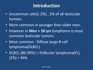
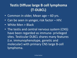
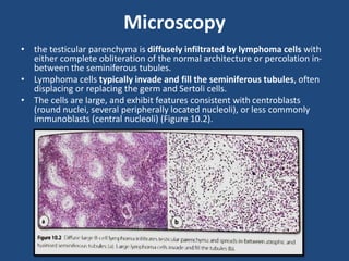
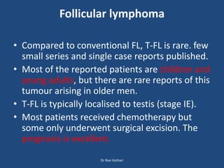
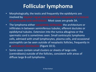
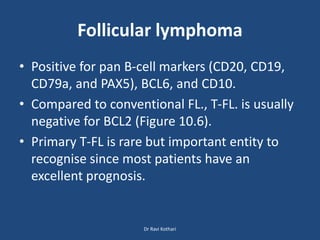
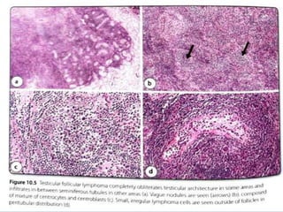
Lymphoma involving Testis
Recent advances in
Histopathology 25
Dr Ravi Kothari
Lymphoma involving Testis
• Introduction
• Testis Diffuse large B cell lymphoma(T-DLBCL)
• Other high grade B cell lyphoma
• Testis Follicular lymphoma(T-FL)
• Other low grade B cell lymphoma
• NK/T cell lymphoma
• Differential Diagnosis
• Key Points
Dr Ravi Kothari
Introduction
• Uncommon site(1-2%) , 5% of all testicular
tumors.
• More common in younger then older men.
• However in Men > 50 yrs lymphoma is most
common testicular tumors.
• Most common - Diffuse large B cell
lymphoma(DLBCL)
• DLBCL (80-90%) > Follicular lymphoma(FL)
(2%) > NHL
Dr Ravi Kothari
Testis Diffuse large B cell lymphoma
(T-DLBCL)
• Common in older, Mean age – 60 yrs.
• Can be seen in yonger, risk factor – HIV.
• White Men > Black
• The testis and central nervous system (CNS)
have been regarded as immune- privileged
sites. Testicular DLBCL shares many features
(i.e. immunophenotype, genetic and
molecular) with primary CNS large B-cell
lymphoma.
Dr Ravi Kothari
Pathogenesis
• Immune escape
• Loss of HLA gene which leads to reduced
expression of MHC 1 & 2.
• Other frequent abnormalities include gains
and amplifications of 9p24.1 locus which lead
to increased expression of PD-L1 and PD-L2
proteins and contribute to evasion of an anti-
tumour immune response.
Dr Ravi Kothari
Clinical Features
• Firm painless scrotal mass.
• 10-15% B/L.
• 50% localized to testis (Stage I)
• Can involve LN & extranodal.
• In 30% CNS involvement.
• 20-30% shows B symptoms (fever, night sweat,
wt. loss)
Dr Ravi Kothari
Diagnosis
Prophylactic Treatment
CNS targeted immuno-chemotherapy Contralateral orchidectomy
Check CNS involvement
Imaging CSF immunophenotyping
Initial diagnosis on orchidectomy specimen
Stagging by PET Check Contralateral Testis
Prognosis
•More aggressive than DLBCL NOS
•Low clinical stage – better prognosis
•CNS involvement – poor prognosis
Dr Ravi Kothari
Gross
• T-DLBCL frequently replaces most of the testicular
parenchyma (average size approximately 6 cm),
• invades tunica albuginea, usually involves epididymis, and
sometimes the spermatic cord.
• The lymphoma is usually fleshy, tan or pinkish in colour
with a uniform appearance on cut surface (Figure 10.1).
Dr Ravi Kothari
Microscopy
• the testicular parenchyma is diffusely infiltrated by lymphoma cells with
either complete obliteration of the normal architecture or percolation in-
between the seminiferous tubules.
• Lymphoma cells typically invade and fill the seminiferous tubules, often
displacing or replacing the germ and Sertoli cells.
• The cells are large, and exhibit features consistent with centroblasts
(round nuclei, several peripherally located nucleoli), or less commonly
immunoblasts (central nucleoli) (Figure 10.2).
IHC
• Positive – Pan B cell markers(CD19, CD20, CD 79a,
PAX 5)
• Negative – CD 10 , BCL6; MUM1- Positive.
• Rarely CD 10 Positive – Secondary involvement of
Testis.
• BCL2 expression seen.
• Ki 67 index high(>70%)
• OCT4 positive in 18%, DD of Seminoma
Dr Ravi Kothari
Other high grade lymphomas involving Testis
• Plasmablastic lymphoma
• Burkitt’s lymphoma
• B lymphoblastic leukemia/lymphoma(B-ALL)
Dr Ravi Kothari
Plasmablastic lymphoma
• Reported in HIV-positive with EBV infection as well as HIV-
negative patients.
• Aggressive lymphoma that frequently occurs in HIV-infected
individuals.
• Site : oral cavity (MC) > gastrointestinal tract; patients
frequently present with disseminated (stage III/IV) disease.
• Two reported cases in HIV-positive patients interestingly
documented the testis as the primary site of disease.
• a lymphoma that consists of immunoblastic/plasmablastic
cells with prominent central nucleoli.
• Plasma cell like IHC
 Positive for CD138, CD38, and MUMI.
 Usually negative or weakly positive for B-cell markers (CD20
and PAXS).
Dr Ravi Kothari
Burkitt lymphoma
• mostly as a part of a systemic disease and very
rarely as an isolated/primary site of involvement.
• Majority in Children.
• Aggressive but curable lymphoma if treated with
intensive chemotherapy regimens.
• 3 clinical subtypes endemic (seen mainly in
equatorial Africa), sporadic and
immunodeficiency associated, which is seen most
commonly in HIV-positive individuals.
Dr Ravi Kothari
Burkitt lymphoma
• Diffuse infiltrate of monotonous medium-sized
lymphoma cells with fine chromatin and small nucleoli.
• Scattered tingible-body macrophages impart the so-
called “starry- sky appearance.”
• Positivity for pan B-cell markers, CD10, BCL6, and MYC
protein.
• BCL2 is typically negative.
• Moreover, the proliferation index (by Ki67
immunostaining) approaches 100%. The signature
genetic abnormality in Burkitt lymphoma is the
translocation of the MYC gene (8q24) to the IGH
(14432)-(8:14).
Dr Ravi Kothari
B-lymphoblastic
leukaemia/lymphoma (B-ALL)
• Testicular involvement at diagnosis is mainly seen in
young boys in 1.2-2.4% of cases but is very rare in adult
men
• Morphologically. B-ALL shows diffuse testicular
involvement by sheets of blasts with displacement of
seminiferous tubules (Figure 10.4).
• Positive for B-cell markers including CD19, cCD79a, and
cCD22. Other CD10 and TdT.
• CD34 and CD20 show variable expression.
• Cytogenetic and molecular studies are routinely done
in all cases of B-ALL at the time of diagnosis for further
risk stratification and treatment planning
Dr Ravi Kothari
Follicular lymphoma
• Compared to conventional FL, T-FL is rare. few
small series and single case reports published.
• Most of the reported patients are children and
young adults, but there are rare reports of this
tumour arising in older men.
• T-FL is typically localised to testis (stage IE).
• Most patients received chemotherapy but
some only underwent surgical excision. The
prognosis is excellent.
Dr Ravi Kothari
Follicular lymphoma
• Morphologically, the testis and frequently the epididymis are
involved by atypical vague nodules/follicles composed of a mixture
of centrocytes and centroblasts. Most cases are grade 3A.
• The lymphoma either completely obliterates the architecture or
infiltrates in between seminiferous tubules, efferent ductules or
epididymal tubules. Extension into the tunica albuginea or the
spermatic cord is sometimes seen. Small centrocytic lymphoma
cells, admixed with small lymphocytes, plasma cells, and occasional
eosinophils can be seen outside of neoplastic follicles, frequently in
a peritubular distribution (Figure 10.5).
• Some cases contain small clusters or sheets of large cells
(centroblasts) outside of the follicles, consistent with areas of
diffuse large B-cell lymphoma.
Dr Ravi Kothari
Follicular lymphoma
• Positive for pan B-cell markers (CD20, CD19,
CD79a, and PAX5), BCL6, and CD10.
• Compared to conventional FL., T-FL. is usually
negative for BCL2 (Figure 10.6).
• Primary T-FL is rare but important entity to
recognise since most patients have an
excellent prognosis.
Dr Ravi Kothari
Other low-grade B-cell lymphomas
involving the testis
• Various low-grade B-cell lymphomas can
involve the testis, usually as part of a systemic
disease, and rarely as a primary or isolated
disease site. Similar to the higher grade B-cell
lymphomas, complete clinical, laboratory and
radiologic staging is needed to evaluate the
extent of disease.
Dr Ravi Kothari
Other low-grade B-cell lymphomas
involving the testis
• An FL of conventional type was reported in the testis.
In comparison to T-FL described above, this particular
case showed extensive systemic disease, was grade 1
histologically, and expressed BCL2.
• Chronic lymphocytic leukaemia/small lymphocytic
lymphoma (CLL/SLL) can involve a variety of extra-
nodal sites, including the skin, gastrointestinal the
frequency of testicular involvement is difficult to
estimate.
• An interesting case of a 72-year-old man with testicular
involvement by CLL/SLL transforming into DLBCL (so-
called "Richter transformation")
Dr Ravi Kothari
Other low-grade B-cell lymphomas
involving the testis
• We have encountered a case of mantle cell
lymphoma that involved the testis in a nodular
pattern (Figure 10.8).
• Plasma cell neoplasms
• NK/T-cell lymphomas involving the testis
• peripheral T-cell lymphoma
• anaplastic large cell lymphoma
• T-lymphoblastic lymphoma
Dr Ravi Kothari
Mantle cell lymphoma
Plasma cell neoplasms
• involve the testis, either as part of systemic disease in
plasma cell myeloma, or as solitary site of involvement
(i.e. extramedullary plasmacytoma).
• Histologically, these lesions appear as a diffuse
infiltrate of plasma cells with varying degree of atypia.
The plasma cells are positive for CD138 and show x or λ
light chain restriction.
• These neoplasms, when diagnosed in the absence of a
previous clinical history of plasma cell myeloma, are
best called "plasma cell neoplasms"
Dr Ravi Kothari
NK/T-cell lymphomas involving the
testis
• Comprise approximately 15% of non-Hodgkin lymphomas,
somewhat more common in Asian countries.
• These rare lymphomas can occur in very uncommon sites
such as the testis where they can pose a significant
diagnostic challenge even for seasoned lymphoma
pathologists.
• As emphasised with B-cell neoplasms, when NK-/T-cell
lymphomas disease. are diagnosed in the testis, clinical
staging is needed to evaluate for systemic Extranodal NK/T-
cell lymphoma (ENKTCL), nasal type is the only lymphoma
from this group that can occur as a primary testicular
lymphoma.
Dr Ravi Kothari
NK/T-cell lymphomas involving the
testis
• The lymphoma cell infiltrate is diffuse. frequently
obliterating seminiferous tubules.
• Angioinvasion, neural invasion and necrosis are
commonly seen. Lymphoma cells vary from small to
large and may be even anaplastic.
• lymphoma cells are positive for cytoplasmic CD3-
epsilon (surface CD3 is negative), CD56, CD2, and
cytotoxic molecules (TIA-1. granzyme B and perforin).
• In rare cases that show cytotoxic T-cell and not NK-cell
origin, lymphoma cells are positive for CD8, CD5 and T-
cell receptor (TCR) a and ẞ ory and 5.
• always positive for the EBV.
Dr Ravi Kothari
NK/T-cell lymphomas involving the
testis
• Clinically, ENKTCL are aggressive lymphomas
that require multimodal treatment approach
(i.e. surgery, radiation, and chemotherapy).
• Patients with testicular ENKTCL must be
completely staged with careful examination of
the upper aerodigestive tract, since occult
lymphoma can be found in some cases.
Dr Ravi Kothari
Differential diagnosis of testicular
lymphoma
• Seminoma
Rarely, the tumour can be so obscured by the
lymphocyte population, that it can be confused
for a lymphoid neoplasm.
 Seminomas are positive for SALL4, CD117, OCT-
4, D2-40 and negative for typical lymphoid
markers (Figure 10.9).
Of note, OCT-4 was reported to stain up to 18%
of T-DLBCL cases which is important to keep in
mind to avoid confusion with germ cell tumour.
Dr Ravi Kothari
Differential diagnosis of testicular
lymphoma
• Spermatocytic tumour
 usually arises in men older than 45 years of age.
Histologically, these tumours consist of sheets
of variably sized cells with a high nuclear:
cytoplasmic ratio.
spermatocytic tumour are typically negative
for lymphoid markers and are often positive
for CD117 and SALL4.
Dr Ravi Kothari
Differential diagnosis of testicular
lymphoma
• Malignant teratomas
can sometimes undergo secondary malignant
transformation to entities such as
rhabdomyosarcoma which could be confused
for a lymphoma.
Unlike lymphomas, these tumours will react
immunohistochemically with antibodies to
desmin, myogenin and MyoD1
Dr Ravi Kothari
Differential diagnosis of testicular
lymphoma
• Myeloid sarcoma
 a tumour mass consisting of myeloid blasts occurring in
extramedullary location, can occasionally involve the testis.
 Morphologically, sheets of usually monotonous blasts, frequently
with monoblastic morphology, are seen. Maturing myeloid
elements are sometimes present, including eosinophilic precursor
which are helpful morphologic 'hint’ Fig 10.12
 Myeloid blasts are usually positive for CD33, CD117, CD34, and
MPO.
 In monoblastic variant CD68 and CD163 are consistently expressed,
but MPO and CD34 are negative.
 Finally, abundant chronic inflammation seen in lymphocytic orchitis
from infection (e.g. viral orchitis) can also mimic a
haematolymphoid neoplasm.
Dr Ravi Kothari
Key points
• uncommon testicular tumours,
• most commonly seen in men > 50 years
• Testicular diffuse large B-cell lymphomas accounts for over
80%.
• Testicular follicular lymphoma is a rare tumour that usually
occurs in children and young adults. It is important to
recognise since it carries excellent prognosis even with
conservative therapy.
• Among NK-/T-cell lymphomas, extranodal NK-/T-cell
lymphoma, nasal type can occur as a primary testicular
lymphoma.
• Mimickers of testicular lymphomas include germ-cell
tumours, inflammatory conditions and myeloid sarcoma
Dr Ravi Kothari
Thank you

Testis lymphoma RA 25-1.pptx

  • 1.
    Lymphoma involving Testis Recentadvances in Histopathology 25 Dr Ravi Kothari
  • 2.
    Lymphoma involving Testis •Introduction • Testis Diffuse large B cell lymphoma(T-DLBCL) • Other high grade B cell lyphoma • Testis Follicular lymphoma(T-FL) • Other low grade B cell lymphoma • NK/T cell lymphoma • Differential Diagnosis • Key Points Dr Ravi Kothari
  • 3.
    Introduction • Uncommon site(1-2%), 5% of all testicular tumors. • More common in younger then older men. • However in Men > 50 yrs lymphoma is most common testicular tumors. • Most common - Diffuse large B cell lymphoma(DLBCL) • DLBCL (80-90%) > Follicular lymphoma(FL) (2%) > NHL Dr Ravi Kothari
  • 4.
    Testis Diffuse largeB cell lymphoma (T-DLBCL) • Common in older, Mean age – 60 yrs. • Can be seen in yonger, risk factor – HIV. • White Men > Black • The testis and central nervous system (CNS) have been regarded as immune- privileged sites. Testicular DLBCL shares many features (i.e. immunophenotype, genetic and molecular) with primary CNS large B-cell lymphoma. Dr Ravi Kothari
  • 5.
    Pathogenesis • Immune escape •Loss of HLA gene which leads to reduced expression of MHC 1 & 2. • Other frequent abnormalities include gains and amplifications of 9p24.1 locus which lead to increased expression of PD-L1 and PD-L2 proteins and contribute to evasion of an anti- tumour immune response. Dr Ravi Kothari
  • 6.
    Clinical Features • Firmpainless scrotal mass. • 10-15% B/L. • 50% localized to testis (Stage I) • Can involve LN & extranodal. • In 30% CNS involvement. • 20-30% shows B symptoms (fever, night sweat, wt. loss) Dr Ravi Kothari
  • 7.
    Diagnosis Prophylactic Treatment CNS targetedimmuno-chemotherapy Contralateral orchidectomy Check CNS involvement Imaging CSF immunophenotyping Initial diagnosis on orchidectomy specimen Stagging by PET Check Contralateral Testis Prognosis •More aggressive than DLBCL NOS •Low clinical stage – better prognosis •CNS involvement – poor prognosis Dr Ravi Kothari
  • 8.
    Gross • T-DLBCL frequentlyreplaces most of the testicular parenchyma (average size approximately 6 cm), • invades tunica albuginea, usually involves epididymis, and sometimes the spermatic cord. • The lymphoma is usually fleshy, tan or pinkish in colour with a uniform appearance on cut surface (Figure 10.1). Dr Ravi Kothari
  • 9.
    Microscopy • the testicularparenchyma is diffusely infiltrated by lymphoma cells with either complete obliteration of the normal architecture or percolation in- between the seminiferous tubules. • Lymphoma cells typically invade and fill the seminiferous tubules, often displacing or replacing the germ and Sertoli cells. • The cells are large, and exhibit features consistent with centroblasts (round nuclei, several peripherally located nucleoli), or less commonly immunoblasts (central nucleoli) (Figure 10.2).
  • 10.
    IHC • Positive –Pan B cell markers(CD19, CD20, CD 79a, PAX 5) • Negative – CD 10 , BCL6; MUM1- Positive. • Rarely CD 10 Positive – Secondary involvement of Testis. • BCL2 expression seen. • Ki 67 index high(>70%) • OCT4 positive in 18%, DD of Seminoma Dr Ravi Kothari
  • 11.
    Other high gradelymphomas involving Testis • Plasmablastic lymphoma • Burkitt’s lymphoma • B lymphoblastic leukemia/lymphoma(B-ALL) Dr Ravi Kothari
  • 12.
    Plasmablastic lymphoma • Reportedin HIV-positive with EBV infection as well as HIV- negative patients. • Aggressive lymphoma that frequently occurs in HIV-infected individuals. • Site : oral cavity (MC) > gastrointestinal tract; patients frequently present with disseminated (stage III/IV) disease. • Two reported cases in HIV-positive patients interestingly documented the testis as the primary site of disease. • a lymphoma that consists of immunoblastic/plasmablastic cells with prominent central nucleoli. • Plasma cell like IHC  Positive for CD138, CD38, and MUMI.  Usually negative or weakly positive for B-cell markers (CD20 and PAXS). Dr Ravi Kothari
  • 13.
    Burkitt lymphoma • mostlyas a part of a systemic disease and very rarely as an isolated/primary site of involvement. • Majority in Children. • Aggressive but curable lymphoma if treated with intensive chemotherapy regimens. • 3 clinical subtypes endemic (seen mainly in equatorial Africa), sporadic and immunodeficiency associated, which is seen most commonly in HIV-positive individuals. Dr Ravi Kothari
  • 14.
    Burkitt lymphoma • Diffuseinfiltrate of monotonous medium-sized lymphoma cells with fine chromatin and small nucleoli. • Scattered tingible-body macrophages impart the so- called “starry- sky appearance.” • Positivity for pan B-cell markers, CD10, BCL6, and MYC protein. • BCL2 is typically negative. • Moreover, the proliferation index (by Ki67 immunostaining) approaches 100%. The signature genetic abnormality in Burkitt lymphoma is the translocation of the MYC gene (8q24) to the IGH (14432)-(8:14). Dr Ravi Kothari
  • 15.
    B-lymphoblastic leukaemia/lymphoma (B-ALL) • Testicularinvolvement at diagnosis is mainly seen in young boys in 1.2-2.4% of cases but is very rare in adult men • Morphologically. B-ALL shows diffuse testicular involvement by sheets of blasts with displacement of seminiferous tubules (Figure 10.4). • Positive for B-cell markers including CD19, cCD79a, and cCD22. Other CD10 and TdT. • CD34 and CD20 show variable expression. • Cytogenetic and molecular studies are routinely done in all cases of B-ALL at the time of diagnosis for further risk stratification and treatment planning Dr Ravi Kothari
  • 16.
    Follicular lymphoma • Comparedto conventional FL, T-FL is rare. few small series and single case reports published. • Most of the reported patients are children and young adults, but there are rare reports of this tumour arising in older men. • T-FL is typically localised to testis (stage IE). • Most patients received chemotherapy but some only underwent surgical excision. The prognosis is excellent. Dr Ravi Kothari
  • 17.
    Follicular lymphoma • Morphologically,the testis and frequently the epididymis are involved by atypical vague nodules/follicles composed of a mixture of centrocytes and centroblasts. Most cases are grade 3A. • The lymphoma either completely obliterates the architecture or infiltrates in between seminiferous tubules, efferent ductules or epididymal tubules. Extension into the tunica albuginea or the spermatic cord is sometimes seen. Small centrocytic lymphoma cells, admixed with small lymphocytes, plasma cells, and occasional eosinophils can be seen outside of neoplastic follicles, frequently in a peritubular distribution (Figure 10.5). • Some cases contain small clusters or sheets of large cells (centroblasts) outside of the follicles, consistent with areas of diffuse large B-cell lymphoma. Dr Ravi Kothari
  • 18.
    Follicular lymphoma • Positivefor pan B-cell markers (CD20, CD19, CD79a, and PAX5), BCL6, and CD10. • Compared to conventional FL., T-FL. is usually negative for BCL2 (Figure 10.6). • Primary T-FL is rare but important entity to recognise since most patients have an excellent prognosis. Dr Ravi Kothari
  • 20.
    Other low-grade B-celllymphomas involving the testis • Various low-grade B-cell lymphomas can involve the testis, usually as part of a systemic disease, and rarely as a primary or isolated disease site. Similar to the higher grade B-cell lymphomas, complete clinical, laboratory and radiologic staging is needed to evaluate the extent of disease. Dr Ravi Kothari
  • 21.
    Other low-grade B-celllymphomas involving the testis • An FL of conventional type was reported in the testis. In comparison to T-FL described above, this particular case showed extensive systemic disease, was grade 1 histologically, and expressed BCL2. • Chronic lymphocytic leukaemia/small lymphocytic lymphoma (CLL/SLL) can involve a variety of extra- nodal sites, including the skin, gastrointestinal the frequency of testicular involvement is difficult to estimate. • An interesting case of a 72-year-old man with testicular involvement by CLL/SLL transforming into DLBCL (so- called "Richter transformation") Dr Ravi Kothari
  • 22.
    Other low-grade B-celllymphomas involving the testis • We have encountered a case of mantle cell lymphoma that involved the testis in a nodular pattern (Figure 10.8). • Plasma cell neoplasms • NK/T-cell lymphomas involving the testis • peripheral T-cell lymphoma • anaplastic large cell lymphoma • T-lymphoblastic lymphoma Dr Ravi Kothari
  • 23.
  • 24.
    Plasma cell neoplasms •involve the testis, either as part of systemic disease in plasma cell myeloma, or as solitary site of involvement (i.e. extramedullary plasmacytoma). • Histologically, these lesions appear as a diffuse infiltrate of plasma cells with varying degree of atypia. The plasma cells are positive for CD138 and show x or λ light chain restriction. • These neoplasms, when diagnosed in the absence of a previous clinical history of plasma cell myeloma, are best called "plasma cell neoplasms" Dr Ravi Kothari
  • 25.
    NK/T-cell lymphomas involvingthe testis • Comprise approximately 15% of non-Hodgkin lymphomas, somewhat more common in Asian countries. • These rare lymphomas can occur in very uncommon sites such as the testis where they can pose a significant diagnostic challenge even for seasoned lymphoma pathologists. • As emphasised with B-cell neoplasms, when NK-/T-cell lymphomas disease. are diagnosed in the testis, clinical staging is needed to evaluate for systemic Extranodal NK/T- cell lymphoma (ENKTCL), nasal type is the only lymphoma from this group that can occur as a primary testicular lymphoma. Dr Ravi Kothari
  • 26.
    NK/T-cell lymphomas involvingthe testis • The lymphoma cell infiltrate is diffuse. frequently obliterating seminiferous tubules. • Angioinvasion, neural invasion and necrosis are commonly seen. Lymphoma cells vary from small to large and may be even anaplastic. • lymphoma cells are positive for cytoplasmic CD3- epsilon (surface CD3 is negative), CD56, CD2, and cytotoxic molecules (TIA-1. granzyme B and perforin). • In rare cases that show cytotoxic T-cell and not NK-cell origin, lymphoma cells are positive for CD8, CD5 and T- cell receptor (TCR) a and ẞ ory and 5. • always positive for the EBV. Dr Ravi Kothari
  • 27.
    NK/T-cell lymphomas involvingthe testis • Clinically, ENKTCL are aggressive lymphomas that require multimodal treatment approach (i.e. surgery, radiation, and chemotherapy). • Patients with testicular ENKTCL must be completely staged with careful examination of the upper aerodigestive tract, since occult lymphoma can be found in some cases. Dr Ravi Kothari
  • 28.
    Differential diagnosis oftesticular lymphoma • Seminoma Rarely, the tumour can be so obscured by the lymphocyte population, that it can be confused for a lymphoid neoplasm.  Seminomas are positive for SALL4, CD117, OCT- 4, D2-40 and negative for typical lymphoid markers (Figure 10.9). Of note, OCT-4 was reported to stain up to 18% of T-DLBCL cases which is important to keep in mind to avoid confusion with germ cell tumour. Dr Ravi Kothari
  • 29.
    Differential diagnosis oftesticular lymphoma • Spermatocytic tumour  usually arises in men older than 45 years of age. Histologically, these tumours consist of sheets of variably sized cells with a high nuclear: cytoplasmic ratio. spermatocytic tumour are typically negative for lymphoid markers and are often positive for CD117 and SALL4. Dr Ravi Kothari
  • 30.
    Differential diagnosis oftesticular lymphoma • Malignant teratomas can sometimes undergo secondary malignant transformation to entities such as rhabdomyosarcoma which could be confused for a lymphoma. Unlike lymphomas, these tumours will react immunohistochemically with antibodies to desmin, myogenin and MyoD1 Dr Ravi Kothari
  • 31.
    Differential diagnosis oftesticular lymphoma • Myeloid sarcoma  a tumour mass consisting of myeloid blasts occurring in extramedullary location, can occasionally involve the testis.  Morphologically, sheets of usually monotonous blasts, frequently with monoblastic morphology, are seen. Maturing myeloid elements are sometimes present, including eosinophilic precursor which are helpful morphologic 'hint’ Fig 10.12  Myeloid blasts are usually positive for CD33, CD117, CD34, and MPO.  In monoblastic variant CD68 and CD163 are consistently expressed, but MPO and CD34 are negative.  Finally, abundant chronic inflammation seen in lymphocytic orchitis from infection (e.g. viral orchitis) can also mimic a haematolymphoid neoplasm. Dr Ravi Kothari
  • 32.
    Key points • uncommontesticular tumours, • most commonly seen in men > 50 years • Testicular diffuse large B-cell lymphomas accounts for over 80%. • Testicular follicular lymphoma is a rare tumour that usually occurs in children and young adults. It is important to recognise since it carries excellent prognosis even with conservative therapy. • Among NK-/T-cell lymphomas, extranodal NK-/T-cell lymphoma, nasal type can occur as a primary testicular lymphoma. • Mimickers of testicular lymphomas include germ-cell tumours, inflammatory conditions and myeloid sarcoma Dr Ravi Kothari
  • 33.