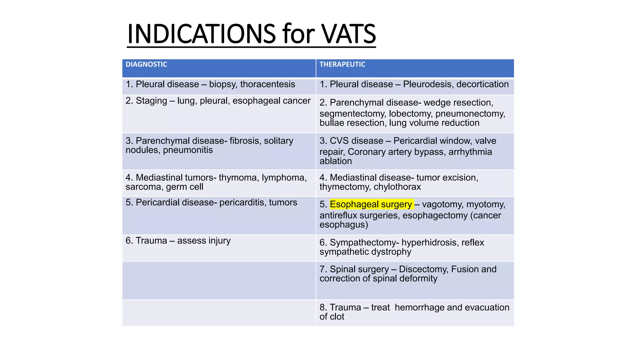
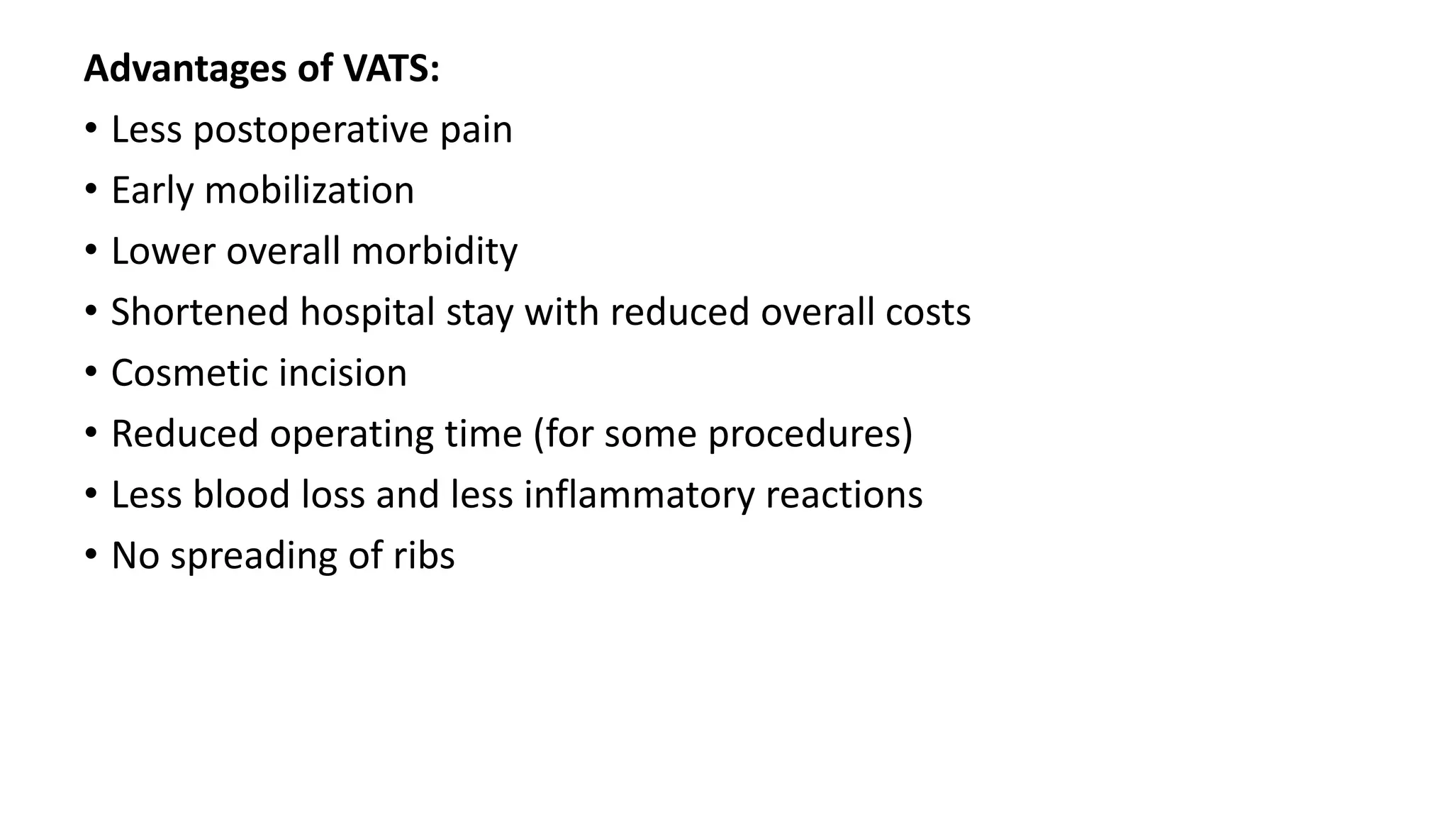
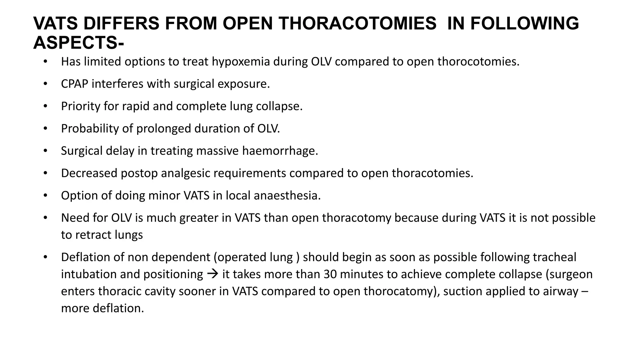
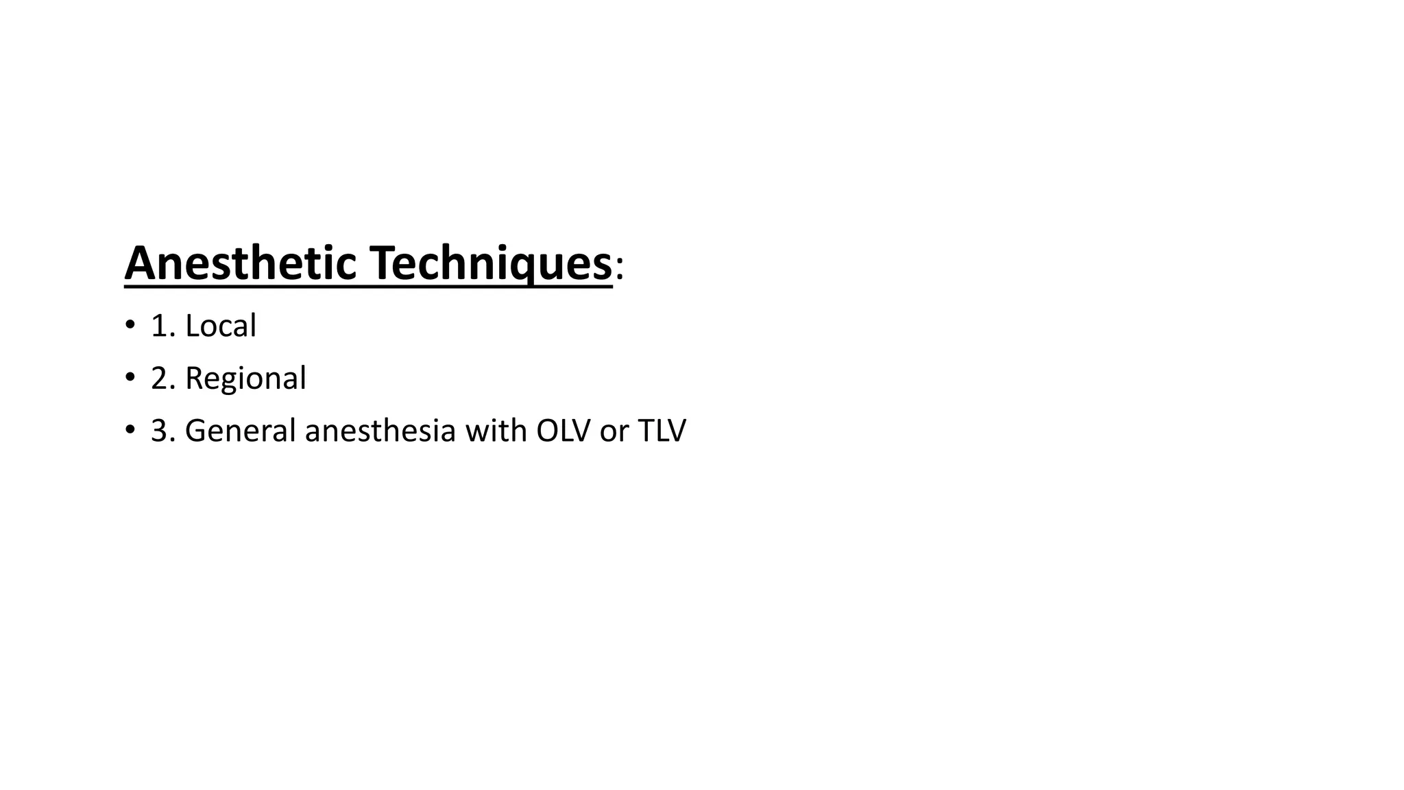
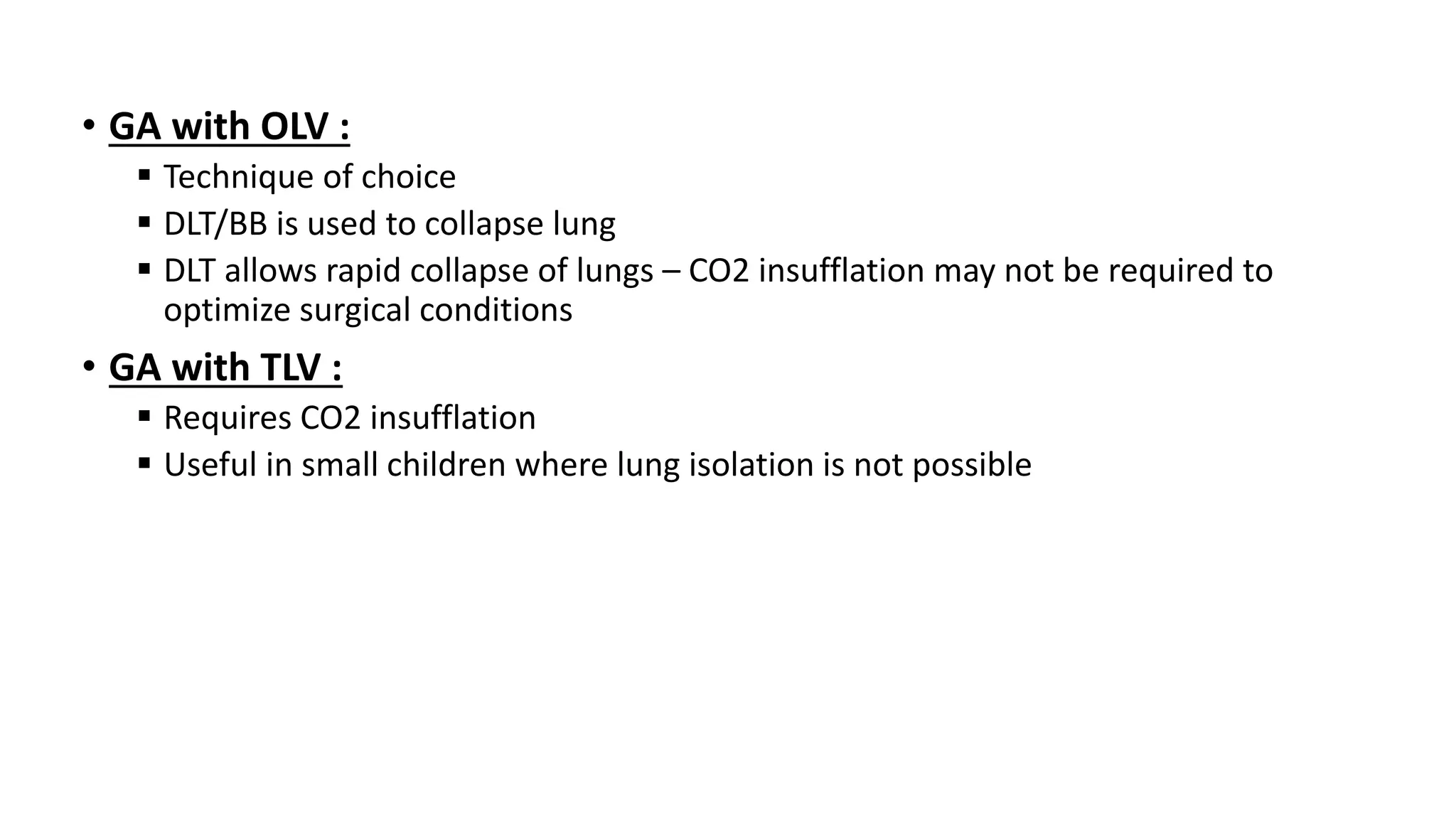
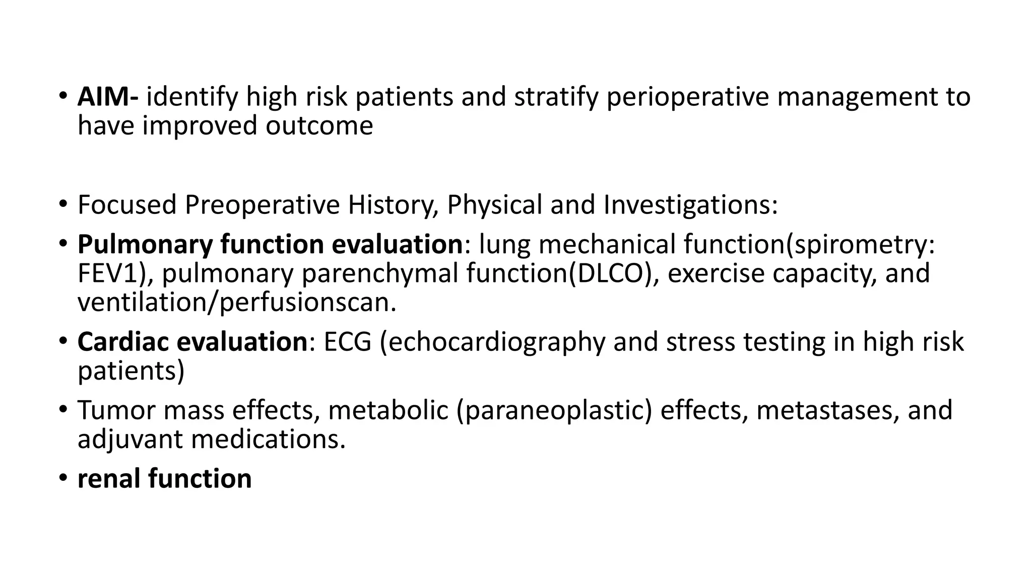
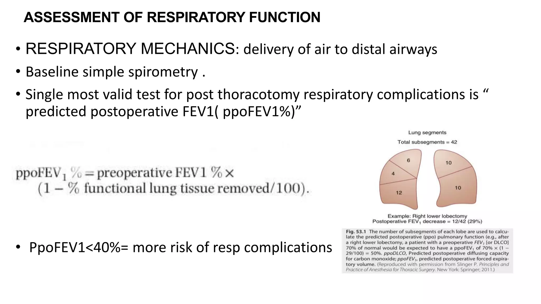
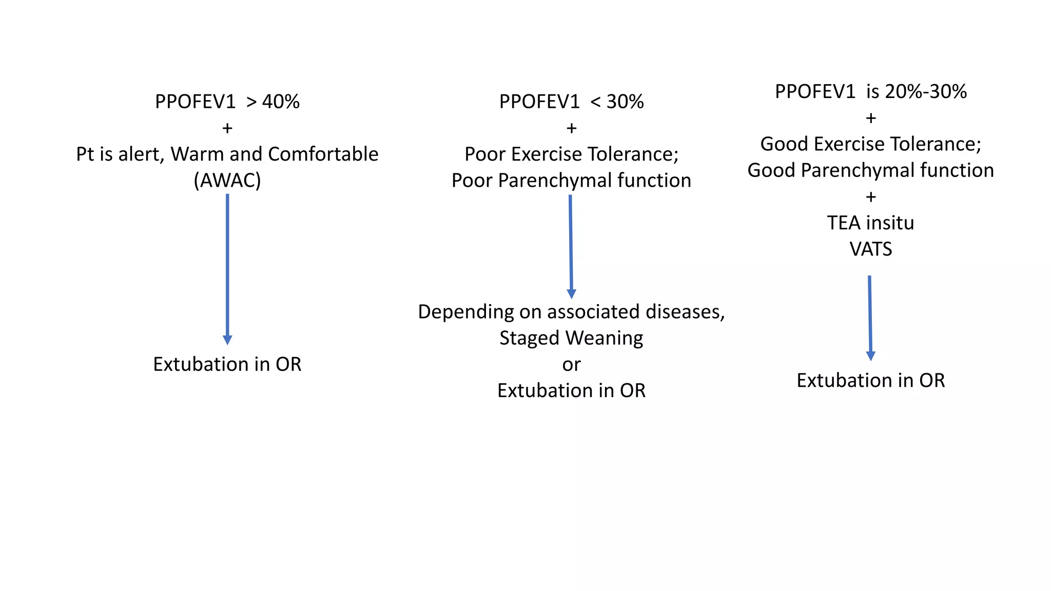
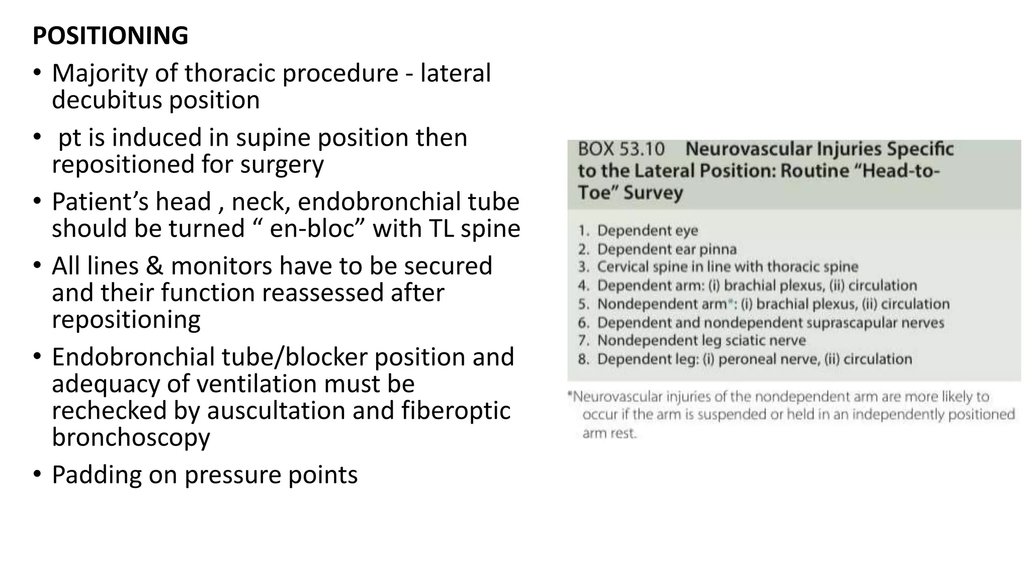
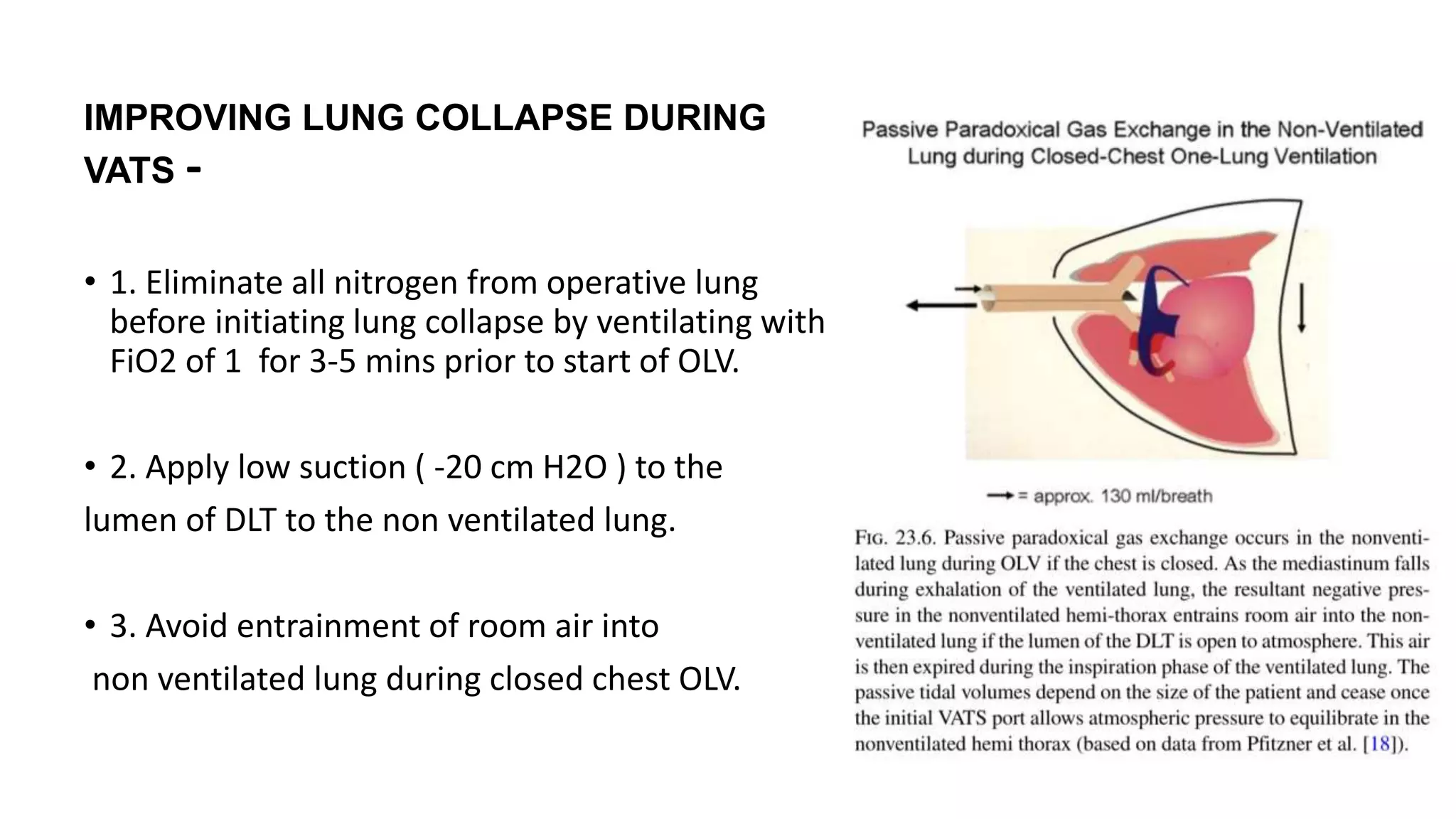
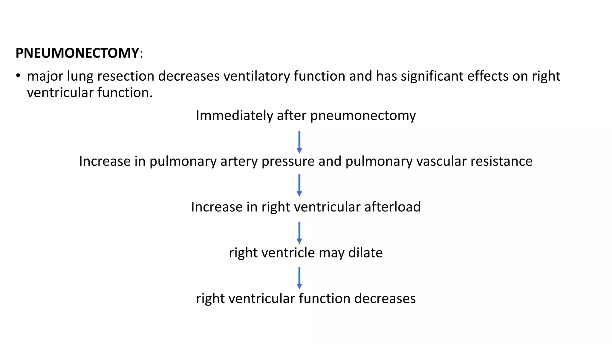
The document discusses anesthesia considerations for thoracoscopy and VATS procedures. It covers preoperative assessment and optimization, intraoperative anesthetic management including lung isolation techniques, ventilation strategies, positioning, and management of issues like hypoxemia. Protective lung ventilation principles with low tidal volumes, PEEP, and recruitment maneuvers are emphasized for lung protection during one-lung ventilation.
Intro to Hypoxic pulmonary vasoconstriction Arun Shetty
Hypoxic pulmonary vasoconstriction, a seldom heard phenomenon but very effective physiologic property which helps lungs utilise ventilation to the maximum
Intro to Hypoxic pulmonary vasoconstriction Arun Shetty
Hypoxic pulmonary vasoconstriction, a seldom heard phenomenon but very effective physiologic property which helps lungs utilise ventilation to the maximum
We understand the unique challenges pickleball players face and are committed to helping you stay healthy and active. In this presentation, we’ll explore the three most common pickleball injuries and provide strategies for prevention and treatment.
India Clinical Trials Market: Industry Size and Growth Trends [2030] Analyzed...Kumar Satyam
According to TechSci Research report, "India Clinical Trials Market- By Region, Competition, Forecast & Opportunities, 2030F," the India Clinical Trials Market was valued at USD 2.05 billion in 2024 and is projected to grow at a compound annual growth rate (CAGR) of 8.64% through 2030. The market is driven by a variety of factors, making India an attractive destination for pharmaceutical companies and researchers. India's vast and diverse patient population, cost-effective operational environment, and a large pool of skilled medical professionals contribute significantly to the market's growth. Additionally, increasing government support in streamlining regulations and the growing prevalence of lifestyle diseases further propel the clinical trials market.
Growing Prevalence of Lifestyle Diseases
The rising incidence of lifestyle diseases such as diabetes, cardiovascular diseases, and cancer is a major trend driving the clinical trials market in India. These conditions necessitate the development and testing of new treatment methods, creating a robust demand for clinical trials. The increasing burden of these diseases highlights the need for innovative therapies and underscores the importance of India as a key player in global clinical research.
CHAPTER 1 SEMESTER V - ROLE OF PEADIATRIC NURSE.pdfSachin Sharma
Pediatric nurses play a vital role in the health and well-being of children. Their responsibilities are wide-ranging, and their objectives can be categorized into several key areas:
1. Direct Patient Care:
Objective: Provide comprehensive and compassionate care to infants, children, and adolescents in various healthcare settings (hospitals, clinics, etc.).
This includes tasks like:
Monitoring vital signs and physical condition.
Administering medications and treatments.
Performing procedures as directed by doctors.
Assisting with daily living activities (bathing, feeding).
Providing emotional support and pain management.
2. Health Promotion and Education:
Objective: Promote healthy behaviors and educate children, families, and communities about preventive healthcare.
This includes tasks like:
Administering vaccinations.
Providing education on nutrition, hygiene, and development.
Offering breastfeeding and childbirth support.
Counseling families on safety and injury prevention.
3. Collaboration and Advocacy:
Objective: Collaborate effectively with doctors, social workers, therapists, and other healthcare professionals to ensure coordinated care for children.
Objective: Advocate for the rights and best interests of their patients, especially when children cannot speak for themselves.
This includes tasks like:
Communicating effectively with healthcare teams.
Identifying and addressing potential risks to child welfare.
Educating families about their child's condition and treatment options.
4. Professional Development and Research:
Objective: Stay up-to-date on the latest advancements in pediatric healthcare through continuing education and research.
Objective: Contribute to improving the quality of care for children by participating in research initiatives.
This includes tasks like:
Attending workshops and conferences on pediatric nursing.
Participating in clinical trials related to child health.
Implementing evidence-based practices into their daily routines.
By fulfilling these objectives, pediatric nurses play a crucial role in ensuring the optimal health and well-being of children throughout all stages of their development.
R3 Stem Cells and Kidney Repair A New Horizon in Nephrology.pptxR3 Stem Cell
R3 Stem Cells and Kidney Repair: A New Horizon in Nephrology" explores groundbreaking advancements in the use of R3 stem cells for kidney disease treatment. This insightful piece delves into the potential of these cells to regenerate damaged kidney tissue, offering new hope for patients and reshaping the future of nephrology.
Antibiotic Stewardship by Anushri Srivastava.pptxAnushriSrivastav
Stewardship is the act of taking good care of something.
Antimicrobial stewardship is a coordinated program that promotes the appropriate use of antimicrobials (including antibiotics), improves patient outcomes, reduces microbial resistance, and decreases the spread of infections caused by multidrug-resistant organisms.
WHO launched the Global Antimicrobial Resistance and Use Surveillance System (GLASS) in 2015 to fill knowledge gaps and inform strategies at all levels.
ACCORDING TO apic.org,
Antimicrobial stewardship is a coordinated program that promotes the appropriate use of antimicrobials (including antibiotics), improves patient outcomes, reduces microbial resistance, and decreases the spread of infections caused by multidrug-resistant organisms.
ACCORDING TO pewtrusts.org,
Antibiotic stewardship refers to efforts in doctors’ offices, hospitals, long term care facilities, and other health care settings to ensure that antibiotics are used only when necessary and appropriate
According to WHO,
Antimicrobial stewardship is a systematic approach to educate and support health care professionals to follow evidence-based guidelines for prescribing and administering antimicrobials
In 1996, John McGowan and Dale Gerding first applied the term antimicrobial stewardship, where they suggested a causal association between antimicrobial agent use and resistance. They also focused on the urgency of large-scale controlled trials of antimicrobial-use regulation employing sophisticated epidemiologic methods, molecular typing, and precise resistance mechanism analysis.
Antimicrobial Stewardship(AMS) refers to the optimal selection, dosing, and duration of antimicrobial treatment resulting in the best clinical outcome with minimal side effects to the patients and minimal impact on subsequent resistance.
According to the 2019 report, in the US, more than 2.8 million antibiotic-resistant infections occur each year, and more than 35000 people die. In addition to this, it also mentioned that 223,900 cases of Clostridoides difficile occurred in 2017, of which 12800 people died. The report did not include viruses or parasites
VISION
Being proactive
Supporting optimal animal and human health
Exploring ways to reduce overall use of antimicrobials
Using the drugs that prevent and treat disease by killing microscopic organisms in a responsible way
GOAL
to prevent the generation and spread of antimicrobial resistance (AMR). Doing so will preserve the effectiveness of these drugs in animals and humans for years to come.
being to preserve human and animal health and the effectiveness of antimicrobial medications.
to implement a multidisciplinary approach in assembling a stewardship team to include an infectious disease physician, a clinical pharmacist with infectious diseases training, infection preventionist, and a close collaboration with the staff in the clinical microbiology laboratory
to prevent antimicrobial overuse, misuse and abuse.
to minimize the developme
Navigating Challenges: Mental Health, Legislation, and the Prison System in B...Guillermo Rivera
This conference will delve into the intricate intersections between mental health, legal frameworks, and the prison system in Bolivia. It aims to provide a comprehensive overview of the current challenges faced by mental health professionals working within the legislative and correctional landscapes. Topics of discussion will include the prevalence and impact of mental health issues among the incarcerated population, the effectiveness of existing mental health policies and legislation, and potential reforms to enhance the mental health support system within prisons.
Navigating the Health Insurance Market_ Understanding Trends and Options.pdfEnterprise Wired
From navigating policy options to staying informed about industry trends, this comprehensive guide explores everything you need to know about the health insurance market.
CHAPTER 1 SEMESTER V PREVENTIVE-PEDIATRICS.pdfSachin Sharma
This content provides an overview of preventive pediatrics. It defines preventive pediatrics as preventing disease and promoting children's physical, mental, and social well-being to achieve positive health. It discusses antenatal, postnatal, and social preventive pediatrics. It also covers various child health programs like immunization, breastfeeding, ICDS, and the roles of organizations like WHO, UNICEF, and nurses in preventive pediatrics.
2. Objectives
• INTRODUCTION
• PREANESTHETIC ASSESSMENT
• PREANESTHESIA PREPARATION
• INTRAOPERATIVE ANESTHETIC MANAGEMENT
• Monitoring
• Induction and maintenance
• Airway management and surgical bronchoscopy
• Positioning
• ventilation
• Management of OLV
• Fluid & temp management
• Prevention of bronchospasm
• LUNG RESECTION SURGERIES
• VATS for ESOPHAGEAL surgeries
• ROBOTIC ASSISTED THOROCOSCOPIC SURGERY
• NON-INTUBATED ASSISTED THOROCOSCOPIC SURGEY
• POST OPERATIVE ANALGESIA
3. INTRODUCTION
• Thoracic anaestheisa includes variety of diagnostic and therapeutic
procedures involving LUNGS,AIRWAYS & other intrathoracic
structures
• The MOST COMMON INDICATIONS for thoracic surgery are
malignancy related to Lungs, Esophagus and Mediastinum
4. • Thoracoscopy involves intentionally creating a pneumothorax and then
introducing an instrument through the chest wall to visualize the intra-
thoracic structures.
• VATS ( video assissted thorocoscopic surgery) is the procedure of choice for
the diagnosis and management of -
• Diseases of the pleura
• Nondiagnosed peripheral pulmonary nodules
• Interstitial lung disease.
• It has become the first-choice technique for lung biopsies,
pleurectomies,sympathectomies, and other various pulmonary disorders
MINIMALLY INVASIVE THORACOSCOPIC SURGERIES (VATS)
6. CONTRAINDICATIONS for VATS
ABSOLUTE:
Inability to achieve adequate visualization of the hemithorax.
RELATIVE:
bronchoplastic procedures
chest wall deformities limiting visualization,
large lesions that limit visibility and would ultimately require a large incision
and rib spreading for extraction,
central/hilar lesions requiring proximal and/or intrapericardial dissection,
dense adhesions requiring decortication
neoadjuvant chemotherapy or radiation with challenging dissection
extensive chest wall involvement
7. Advantages of VATS:
• Less postoperative pain
• Early mobilization
• Lower overall morbidity
• Shortened hospital stay with reduced overall costs
• Cosmetic incision
• Reduced operating time (for some procedures)
• Less blood loss and less inflammatory reactions
• No spreading of ribs
8. VATS DIFFERS FROM OPEN THORACOTOMIES IN FOLLOWING
ASPECTS-
• Has limited options to treat hypoxemia during OLV compared to open thorocotomies.
• CPAP interferes with surgical exposure.
• Priority for rapid and complete lung collapse.
• Probability of prolonged duration of OLV.
• Surgical delay in treating massive haemorrhage.
• Decreased postop analgesic requirements compared to open thoracotomies.
• Option of doing minor VATS in local anaesthesia.
• Need for OLV is much greater in VATS than open thoracotomy because during VATS it is not possible
to retract lungs
• Deflation of non dependent (operated lung ) should begin as soon as possible following tracheal
intubation and positioning it takes more than 30 minutes to achieve complete collapse (surgeon
enters thoracic cavity sooner in VATS compared to open thorocatomy), suction applied to airway –
more deflation.
10. • GA with OLV :
Technique of choice
DLT/BB is used to collapse lung
DLT allows rapid collapse of lungs – CO2 insufflation may not be required to
optimize surgical conditions
• GA with TLV :
Requires CO2 insufflation
Useful in small children where lung isolation is not possible
12. • AIM- identify high risk patients and stratify perioperative management to
have improved outcome
• Focused Preoperative History, Physical and Investigations:
• Pulmonary function evaluation: lung mechanical function(spirometry:
FEV1), pulmonary parenchymal function(DLCO), exercise capacity, and
ventilation/perfusionscan.
• Cardiac evaluation: ECG (echocardiography and stress testing in high risk
patients)
• Tumor mass effects, metabolic (paraneoplastic) effects, metastases, and
adjuvant medications.
• renal function
13. INVESTIGATIONS
1. CBP
2. LFT,RFT
3. COAGULATION PROFILE
4. ECG - screening test for myocardial ischemia and arrhythmias and provides a baseline for comparison in the event
of perioperative cardiac complications. Most patients will receive chemoradiation, which may impact cardiac
function
5. CHEST X RAY - Preoperative CXR may reveal evidence of aspiration as well as coexisting pulmonary and cardiac
disease
6. 2D ECHO IF CARDIAC ISSUES ARE PRESENT
7. PULMONARY FUNCTION TEST and ABG
8. CT/MRI – to localise the lesion
14. • RESPIRATORY MECHANICS: delivery of air to distal airways
• Baseline simple spirometry .
• Single most valid test for post thoracotomy respiratory complications is “
predicted postoperative FEV1( ppoFEV1%)”
• PpoFEV1<40%= more risk of resp complications
ASSESSMENT OF RESPIRATORY FUNCTION
15. LUNG PARENCHYMAL FUNCTION: ability for oxygenation
• Most useful test-->diffusing capacity for CO( DLCO).
• Correlates with functioning surface area of alveolar capillary interface
• It is negatively affected by preop chemotherapy
• DLCO < 40% = more Respiratory & cardiac complications.
16. Cardiopulmonary interaction:
• Laboratory exercise testing-gold standard
• Maximal o2 consumption(VO2max)-most useful predictor of post-
thoracotomy outcome.
• Estimated VO2max =6MWT in meters/30
• Preop VO2max <15ml/kg/min-high risk for morbidity and mortality.
• Reduced saturation>4% during exercise –high risk
19. CONCOMITANT MEDICAL CONDITIONS
• ISCHEMIA-elective pulmonary resection is “ intermediate risk” for peri-op
ischemia
• Appropriate time interval for lung resection:
• 4-6weeks, in a medically stable and optimized patient after MI.
• 4-6 weeks after bare metal stents and
• 12 months after drug eluting stents.
• CONGESTIVE HEART FAILURE- patients H/O CCF poorly tolerate the obligate
20-30% shunt during OLV leading to exaggerated fall in Pao2.
CARDIAC DISEASE
20. • PULMONARY HYPERTENSION: mean PAP > 25mm hg OR systolic PAP >35 mmhg
• Patients with PAH —>increase risk of resp complications & need for prolonged
intubation
PAH
↓
↑Transmural Pressure
↓
↓Perfusion to RV by RCA +
Hypotension
↓
PAP>>Systemic Pressures
↓
↓↓Perfusion to RV
21. COPD:
• Intensive chest physiotherapy preoperatively
• Different modalities -cough, deep breathing ,incentive spirometry ,PEEP ,
CPAP
• Severe COPD—>physiotherapy+exercise+nutrition+education+smoking
cessation
• Smoking cessation— >8 wks preop and post op.
• Smoking leads to prolonged period of tissue hypoxemia and correlates
with wound healing and infection chances
22. Factors associated with an increased risk of post-thoracotomy renal
impairment:
• Pre-op renal dysfunction +intra-op HES
• Preop ACE inhibitors/ARB therapy
• DM
• CVA
• Low serum albumin level
Renal dysfunction
23.
24. PPOFEV1 > 40%
+
Pt is alert, Warm and Comfortable
(AWAC)
Extubation in OR
PPOFEV1 < 30%
+
Poor Exercise Tolerance;
Poor Parenchymal function
Depending on associated diseases,
Staged Weaning
or
Extubation in OR
PPOFEV1 is 20%-30%
+
Good Exercise Tolerance;
Good Parenchymal function
+
TEA insitu
VATS
Extubation in OR
25. • BLEOMYCIN- exacerbate o2
induced pulmonary toxicity.
Safest mx= lowest FIO2
consistent with patient
safety & close monitoring of
pulse oximetry
• CISPLATIN-pt who use this
preop develop raised sr.creat
when given NSAIDs post op
28. Preoperative Optimization and Premedication:
• H2 receptor antagonists or proton pump inhibitors reduce gastric volume and
acidity - reduce the incidence and severity of pneumonitis due to aspiration
• Anxiolysis – short acting sedatives (iv midazolam)
• Adequate blood is kept ready – massive bleeding is possible intraoperatively
• Perioperative bronchodilator and steroid therapy are continued
• Antibiotic prophylaxis
• Chest physiotherapy
• At least one large bore iv access is required as there is high risk of bleeding
• Achalasia, has been associated with spontaneous aspiration pneumonitis -
longer periods of NPO status
• To minimize the risk of tracheal aspiration in patients undergoing esophageal
surgery - preoperative suctioning of the NGT if present
29. Preoperative Preparation
1. Airway: endotracheal tubes, double lumen tubes, bronchial blockers,
videolaryngoscope, flexible fibreoptic bronchoscope
2. Warming devices to prevent hypothermia
3. Preparation for regional anesthetic technique as planned:
Thoracic epidural or paravertebral analgesia
Paravertebral block (PVB), either as a single injection or as a continuous
infusion, is ideally suited to VATS.
31. Monitors
1.ECG,
2. Pulse oximetry
3. ETCO2 monitoring
4. Radial artery of dependent arm – continuous BP monitoring , intermittent sampling of
ABGs
5. Central venous catheter – placed on side of thoracoscopy to avoid bilateral
pneumothorax
6. Temperature monitoring
7. Urine output monitoring
8. Neuromuscular monitoring
32. AIRWAY MANAGEMENT
Airway compromise - Posterior mediastinal masses (esophageal origin and the
dilated esophagus) trachea gets easily compressed posteriorly because of
the lack of cartilaginous support near complete expiratory obstruction
If there is any difficult intubation –awake intubation
For easy intubation- a rapid sequence induction is better to reduce aspiration
risk.
Intravenous induction: smooth and rapid, with propofol, thiopental or
etomidate in conjunction with a rapidly acting NMB such as succinylcholine or
rocuronium.
Intubatation: appropriate size of DLT /Bronchial blocker for lung isolation and
maintain OLV
33. Maintenance of Anesthesia:
Balanced anesthesia with 0.5 -1 MAC isoflurane or sevoflurane or
TIVA is used
TIVA may be advantageous as volatile anesthetics inhibit HPV
Halothane and N20 are avoided
Intermittent boluses of fentanyl – analgesia
Intermediate acting NMBAs – to maintain neuromuscular paralysis
34. POSITIONING
• Majority of thoracic procedure - lateral
decubitus position
• pt is induced in supine position then
repositioned for surgery
• Patient’s head , neck, endobronchial tube
should be turned “ en-bloc” with TL spine
• All lines & monitors have to be secured
and their function reassessed after
repositioning
• Endobronchial tube/blocker position and
adequacy of ventilation must be
rechecked by auscultation and fiberoptic
bronchoscopy
• Padding on pressure points
35. IMPROVING LUNG COLLAPSE DURING
VATS -
• 1. Eliminate all nitrogen from operative lung
before initiating lung collapse by ventilating with
FiO2 of 1 for 3-5 mins prior to start of OLV.
• 2. Apply low suction ( -20 cm H2O ) to the
lumen of DLT to the non ventilated lung.
• 3. Avoid entrainment of room air into
non ventilated lung during closed chest OLV.
36. • GOAL during OLV-
• maximize PVR in non ventilated lung
• Minimise PVR in ventilated lung
is achieved by understanding the correlation
between PVR & lung volumes
• Thus, maintain
• ventilated lung close to FRC
• Non ventilated close to RV ( collapse)
37. Ventilatory management:
• ventilation strategy during OLV should be adjusted to overcome two
different challenges: oxygenation and lung protection
• High FiO2 (1.0) should be avoided unless necessary.
• Protective ventilation has three intraoperative components: low TV,
RM and PEEP.
• The combined use of these three components can help prevent both
hypoxemia and ALI
• CPAP to the non-dependent lung is beneficial for both challenges
38. A: The traditional setting for OLV can lead to lung injury via its four
classical pathways: Baro-, volu-, atelecto- and biotrauma. It is to be
noted that, during expiration (dashed line) the dependent lung can
collapse entirely: during inspiration, it is over-expanded due to the
high tidal volume (TV), which is normally applied to two lungs. The
non-dependent lung is also fully collapsed.
B: Current suggestion for OLV can prevent lung injury: During
expiration (dashed line), PEEP can prevent a total collapse; during
inspiration, lower TV can achieve a more appropriate ventilation.
CPAP to non-dependent lung is beneficial for both oxygenation and
lung protection.
39.
40. ANESTHETIC CONSIDERATIONS FOR VATS -
• Hypoxemia
• Massive haemorhage and inability to control large blood vessels.
• May require conversion to an open thoracotomy in case of uncontrolled
bleeding or surgical inaccessibility.
41. • It is predictable, preventable & treatable in majority of cases
• Spo2>= 90% (pao2 >60mmhg) is commonly accepted
• Incidence of hypoxemia can be reduced by
• (1)improved lung isolation techniques such as fiberoptic bronchoscopy
to prevent lobar obstruction from DLT
• (2)usage of anesthetic drugs that cause less inhibition of HPV
• (3)understanding of pathophysiology of OLV
HYPOXEMIA
42. • Most useful prophylactic measure-
• CPAP(2-5) to non ventilated lung
And/or
• PEEP application to dependent Lung
Predictor of desaturation
during OLV
43. TREATMENT OF HYPOXEMIA DURING VATS –
• Severe or acute desaturation: resume two- lung ventilation
• Gradual desaturation:
• Assure Fio2 =1.0
• Check double lumen tube or bronchial blocker placement with foberoptic
bronchoscopy
• Optimize cardiac output
• Recruitment maneuver of the ventilated lung
• Apply PEEP 5cmH2O to ventilated lung( expect moderate – severe COPD
patients)
• Partial ventilation of the non-ventilated lung-
• segmental re-inflation (with fiberoptic bronchoscopy)
• high- frequency jet ventilation
44. Fluid management
• Iv fluids are to replace deficit and for
maintenance only .
• No volume is given for third space losses.
• d/t hydrostatic effects, excess
administration of fluids cause increased
shunting and subsequently lead to
pulmonary edema of dependent lung,
impairing gas exchange.
45. Temperature:
• Maintenance of body temp is an issue due to heat loss from open hemithorax
• particularly a problem in extremes of age.
• most of body’s physiological functions ,including HPV are inhibited during hypothermia
• Prevent intra-op hypothermia- increase ambient room temperature, fluid warmers, use of
lower and/or upper body forced-air patient warmers.
Bronchospasm:
• High incidence= coexisting reactive airway disease+ airway manipulation d/t DLT
• Avoid airway manipulation in light plane , avoid histamine releasing drugs
• Induction—> use bronchodilating anaesthetics= propofol/ketamine & inhalational agents
• Maintenance=propofol and/or volatile anesthetics( potent bronchodilator=sevoflurane)
46. Specific concerns of Lung resection surgeries
• LOBECTOMY: once the lobectomy has been performed, the bronchial
stump is usually tested with 30 cm H2O positive pressure to detect the
presence of air leak
• after the lobe and blood vessels have been dissected, a test maneuver is
performed by clamping the surgical bronchus to confirm that the specific
lobe is extirpated.
• SLEEVE RESECTION: High-frequency jet ventilation can be used for
resections done close to the tracheal carina.
• For a sleeve lobectomy involving resectioning of vessels, heparinisation is
necessary.
47. PNEUMONECTOMY:
• major lung resection decreases ventilatory function and has significant effects on right
ventricular function.
Immediately after pneumonectomy
Increase in pulmonary artery pressure and pulmonary vascular resistance
Increase in right ventricular afterload
right ventricle may dilate
right ventricular function decreases
48. Management of post pneumonectomy space:
• Important to keep the mediastinum balanced and trachea is in midline
• Suction/standard chest drain may cause mediastinal shift with
hemodynamic collapse
• Specifically desgined post pneumonectomy chest drainage system with
both high and low pressure underwater relief valves to balance the
mediastinum can be used
• Post operatively CXRAY is MANDATORY to assess the mediastinal shift
49. • significant loss of blood caused by chest wall vessel involvement.
• During tumor dissection, venous return to the heart may be
compromised due to blood loss, compression effect by the tumor in
superior vena cava, or surgical causes.
• Because of extensive tumor resection and the pericardial resection in
right-sided surgery, cardiac herniation or hemodynamic instability can
occur postoperatively after the patient is turned from the lateral
decubitus to the supine position.
Management -
• a CVP catheter be used to guide intravascular fluid administration
• blood loss must be replaced to maintain an acceptable hematocrit level
and the coagulation profile kept within normal limits.
51. Preoperative Assessment
Thorough history and physical examination
Comorbid conditions - evaluated and optimized prior to surgery
Particular attention -signs and symptoms of esophageal obstruction, GERD, and silent aspiration.
• Obstruction - dysphagia and odynophagia, may lead to reduced oral intake and malnutrition which can
lead to increased morbidity and mortality
Thorough Preoperative evaluation of the cardiopulmonary system
The chemotherapeutic agents used to treat esophageal cancer bone marrow suppression
anemia and thrombocytopenia.
52. Ventilatory Management
Use of protective lung strategies during OLV decreases
proinflammatory systemic response
can be achieved by delivering 5 ml/kg of tidal volume, plateau
pressures below 35 cmH2O, PEEP of 5 cm H20 to dependent
lung.
53. Extubation
• Early extubation is preferred
• Functional ICD with under water seal is inserted prior to extubation for safe
extubation
• Bilateral air entry checked at the time of extubation
• NGT and oral suction prior to extubation – to prevent aspiration
• Extubate when patient is fully awake and reversed
• Avoid coughing and straining at extubation
• Placing the patient in a 30° head-up position may improve pulmonary ventilation
and decrease aspiration risk
55. ENCHANCED RECOVERY AFTER
SURGERY(ERAS)
• Combines multispeciality approach to periop management
• Concept is to mitigate stress response to surgery & have faster recovery
• ERAS pathways in lung cancer surgery are associated with reduced
complications, shorter stay and cost savings
56.
57.
58. • RESPIRATORY FAILURE: leading cause of postop morbidity & mortality
• Definition-acute onset of hypoxemia(pao2<60 mm hg ) or
hypercapnia(paco2 >45mmhg) or use of postop mechaical ventilation
for> 24hrs
• Preop predictors: decreased respiratory function preop
Elderly
CAD
More extent of lung resection
• Intraop predictors: crossover contamination d/t failed lung isolation
• Postop predictors: mechanical ventilation is associated with risk of
nosocomial pneumonia & bronchopleural fistula
EARLY MAJOR COMPLICATIONS
59. Management:
• (1) early extubation in cases of uncomplicated lung resection.
• (2)chest physiotherapy ,incentive spirometry, early ambulation
• (3)Thoracic Epidural Analgesia
Treatment:
• Supportive therapy
• Better oxygenation
• Treat infection
• Provide vital organ support
60. CARDIAC HERNIATION: Complication of pneumonectomy.
Occurs after chest closure because of the pressure difference between the two
hemi thoraces.
Heart being extruded through a pericardial defect.
↓
Cardiac herniation
After left Pneumonectomy
↓
Less Cardiac rotation but edge
of Pericardium compresses
myocardium
↓
⁻ MI
⁻ Arrythmias
After right Pneumonectomy
↓
Torsion of heart
↓
⁻ Impairment of VR
⁻ Increase CVP
⁻ Tachycardia
⁻ Hypotension
⁻ Shock
61. Management:
• Dire emergent surgery
• Relocation of heart to its anatomical position with repair of pericardial
defect is key to patient survival
• All precautions should be taken for a redo exploration=IV & arterial
access
• Careful lateral positioning( to avoid hypotension)
• As time is crucial, SLT is used
• vasopressors/inotropes - support circulation
• TEE- to ensure there is no excessive compression of hear during repair
62. Complications specific to thoracoscopy:
hemorrhage
visceral damage : RLN, aorta or SVC, trachea, diaphram
port site recurrence of pulmonary or esophageal tumors
Chronic pain due to compression of intercoastal nerves with thoracoscope
63. Complications specific to surgery :
Esophageal Anastomotic Leaks- frequent and serious
complication of esophagectomy,
o Preoperative factors: diabetes, pulmonary disease, and cardiovascular
disease
o Intraoperative : surgical and technical factors
o postoperative factors: gastric distension, prolonged ventilatory support,
and hypoxia.
respiratory : pneumonia, aspiration pneumonitis, acute lung
injury (ALI), ARDS, bronchopleural fistula, atelectasis, pulmonary
embolism, pulmonary edema
64. ARRTHYMIA:
• AF M/C complication.
• 30-50% incidence in first week post op.
CAUSE
(1) Inc flow resistance through pulm vascular bed --> strain on rt heart
(2) Inc sympathetic stimuli and o2 requirement as the patient becomes mobile on day 2 post op.
Penumonectomy
Intrapericardial dissection
Extrapleural Pneumonectomy are high riskfor Post-op Arrythmia
Blood loss
Aged
• Diltiazem- most useful drug for arrhthymia prophylaxis.
65. Complications due to CO2 insufflation:
Venous gas embolism
Tension capnothorax – especially in robotic surgeries
Compression of the mediastinal structures like the atrium and vena cava decreased
venous return and cardiac output
Higher incidence of arrythmias
rise in PaCO2, has been shown to decrease pulmonary compliance –
permissive hypercapnia is tolerated in order to maintain a protective lung ventilation
strategy during OLV As long as the patient does not have severe comorbidities
PULMONARY COMPLICATIONS AND ANASTOMOTIC LEAK IS RELATIVELY LOW IN VATS
COMPARED TO OPEN THORACOTOMY
67. • Improved postop care ,specifically pain
management reduce the resp.complications
Sensory afferents that transmit nociceptive
stimuli:
• Incision -- Intercostal nerves -- T4-T6
• Chest drains -- Intercostal nerves -- T7-T8
• Mediastinal pleura -- Vagus nerve, CN X
• Central diaphragmatic pleura -- Phrenic nerve,
C3-C5
• Ipsilateral shoulder -- Brachial plexus
• So, analgesia should be multimodal
68. Post thoracotomy analgesia :
(1)Opioids: Effective in controlling background pain
(2)Non-steroidal Anti-inflammatory agents:
• Reduce opioid consumption by 30%
• Useful in treating the ipsilateral shoulder pain
(3)Local anesthetics:
• Intercostal Nerve Blocks
• Epidural Analgesia
• Paravertebral Block
69. Intercostal Nerve Blocks
• Blocks the intercostal nerves supplying the dermatomes of the surgical
incision
• Can be done percutaneously or under direct vision when the chest is open
• should be placed at post axillary line to block lateral cut branch of
intercostal nerve
• Useful for the pain associated with the multiple small-port incisions and
chest drains after VATS
• Dose: 1 mg/kg of bupivacaine
Disadvantages :
• Accidental injection into intercostal vessels
• Requirements of repeated doses
70. Epidural Analgesia
• TEA for thoracic surgery with infusion of LA & opioids is GOLD STANDARD
• Epidural techniques reduce the incidence of respiratory complications
⁻Preservation of FRC
⁻Efficient Mucociliary clearance
⁻Reduce the inhibitory reflexes on diaphragm
Atelectasis and
secondary infections
71. • Paravertebral Block
• Paravertebral space -- potential
space deep to the endothoracic
fascia that the intercostal nerve
traverses
Can be approached by
• Percutaneous method
• Direct vision with open chest
• Ultrasound guidance
• Provides reliable, multi level,and
unilateral intercostal blockade
73. • SERRATUS ANTERIOR PLANE BLOCK:
• At the level of 5th rib in midsaxillary plane
• Needle insertion can be inplane or out of plane f/b injection of LA
above/below the muscle
• Showed to improve analgesia by patient controlled morphine
• ERECTOR SPINAE BLOCK:
• Variant of paravertebral block
• both acute &chronic post thoracotomy pain
• 20ml of LA is injected into plane deep to ES muscle at level of T5
transverse process & catheter is passes 5cm distal to tip of needle &
infusion is begun
• Spread of injectate=C7-T8
Ultrasound guided blocks
74. Ketamine:
• reduces acute post thoracotomy pain
• Can be started intraop as low dose bolus or infusions and continues
postop
• Post op infusion- 0.1-0.15mg/kg/hr
• Psychomimetic effects is rarely seen with analgesic, subanesthetic doses
Dexmedetomidine:
• Selective adrenergic α2- agonist
• Used in combination with epidural local anesthetics
• Decrease the requirement for opioids
• Maintenance infusion dose= 0.3-0.4ug/kg/hr
75. Non-Intubated Thoracic Anesthesia
LOCAL/REGIONAL :
• Local infiltration ,
• intercoastal nerve blocks ( given at level of incision and two spaces above
and below) ,
• thoracic epidural,
• paravertebral blocks,
• intrapleural blocks
• Sedation – short acting drugs (remifentanil, propofal)
76. • Partial collapse of lung on the side of surgery occurs when air enters the
pleural cavity
• Do not insufflate gases into hemithorax in awake patients
• High Fio2 should be delivered via facemask to overcome shunt because of
loss in lung volume caused by unavoidable pneumothorax
Concerns:
• Manipulation of the lung in awake patients Stimulation of Cough reflex
hindrance during surgery due to moment of surgical field
• Techniques to suppress cough reflex
• Inhalation of aerosolized lidocaine or application of LA spray on surface of the lung
• Stellate ganglion block
• Vagus nerve block
• Should be aware of aspiration
77. Lung recruitment during awake
Open Pneumothorax
↓
Shifting of mediastinum
↓
Dependent lung compression
↓
Atelectasis and
making spontaneous breathing difficult
↓
Worsening hypoxemia
– Apply PEEP by using NIV
NIV
– Mitigate the effects of Pneumothorax
– Decrease in LV after load Increased CO
78. Advantages of NI-VATS:
• Reduces the chances of post-op nosocomial infections
• Decreased ICU and hospital stay
• Reduction in overall morbidity and mortality
Postoperatively, continue NIV to prevent atelectasis and Pneumonia.
79. Pediatric Thoracic Anesthesia:
• Preoperative evaluation: neonatal history as this may indicate comorbid
pulmonary and cardiac disease and linked syndromes which must be
investigated.
• Lung isolation is not always necessary in pediatric thoracic surgery.
• Physiologic manifestation of one-lung ventilation may be more
pronounced in children than in adults.
• The compliant rib cage, compressible lung parenchyma, reduced FRC
under anesthesia, and higher oxygen consumption in the child
contribute to aggravate hypoxemia during lung isolation.
• Alveolar collapse occurs more readily in infants as FRC approaches
residual volume.
80. • Slow insufflation of co2 should be done with flowrate 1 l/min and
limitation of inflation pressure to 4–6 mmHg should be ensured.
• Younger children absorb proportionately moreCO2 than older
children, leading to hypercarbia and acidosis.
• Management of hypoxemia, hypercapnia and aletectasis is
mandatory.
• Children are prone to hypothermia during thoracoscopy with creation
of capnothorax due to cold CO2 gas insufflation, so temperature
monitoring is important and corrective measures should be taken.
• Detection and treatment of arrhythmias is important.·
• Aspiration prior to injection and slow creation of artificial
pneumothorax will limit in advertent CO2 embolism.
81. Robotic Thoracic Surgery:
Although the first surgical robot system was constructed in 1983, it was not
until 1992 that the first robotic operation, a prostatectomy, was performed
Robot-assisted thoracoscopic esophagectomy was first described in 2004 .
A robotic system is superior to traditional video-assisted thoracoscopic
surgery (VATS) because
• (I) it provides a three-dimensional view of the surgical field with magnification
and
• (II) the articulating limbs of the robot allow micromanipulation and facilitate
navigation in difficult to access spaces.
For small lesions, mediastinal tumor resection using robotic-assisted VATS
has been shown to be safe, technically easier to perform, and avoids a
median sternotomy
The basic pre-anesthesia evaluation is same as VATS.
A patient who is high risk for open thoracic surgery is also high risk for
robotic surgery.
82. • Robotic thoracic Surgery:
• Room layout:
The operating surgeon is seated at a console several feet away from the patient.
The surgical assistant stands to one side of the patient to assist with placement of trocars,
changing the robotic instruments, and manipulating additional endoscopic instruments as
needed.
The scrub technician and instrument trays are located on the opposite side of the assistant
surgeon.
The anesthesiologist and anesthesia machine remain at the head of the patient
83. Positioning and Access:
Lateral decubitus : most common position for robotic lung resection,
diaphragmatic repair and the thoracic portion of esophagectomy
Careful attention must be paid to patient positioning in order to enable
optimal surgical access and also prevent patient injury.
Some procedures, such as esophagectomy : may require multiple
position changes, increasing the risk of patient injury
Any portion of the patient body that may come in contact with robotic
arms needs to be protected with gel pads or sponge pads and constantly
monitored to prevent injury from the large forces used to move the
robot arms
84.
85. CONCLUSION
• VATS has replaced many diagnostic and therapeutic
procedures previously performed by traditional
thoracotomy
• The anesthetic management of VATS involves the
ability to separate the lungs to provide safe and
effective OLV.
• Minimising unnecessary fluid administration, adequate
pain management, hypotension, and protective lung
ventilation – can improve outcome after surgery