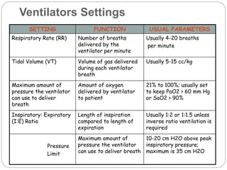
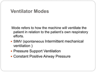
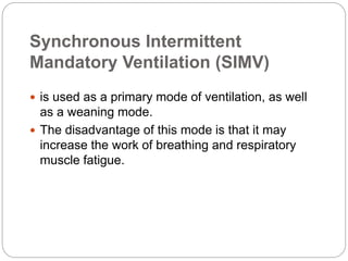
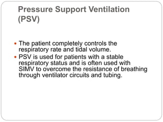
Mechanical
Ventilation
Management
Airway Management & Mechanical
Ventilation Basics
 The goal of airway management is to ensure that
the patient has a patent airway through which
effective ventilation can take place.
 An obstructed airway causes the body to be
deprived of oxygen and, if ventilation isn’t
reestablished, causes brain death within minutes.
Indications
1. The patient experiencing respiratory failure
or arrest .
2. Any patient experiencing respiratory
distress with impincreased aired gas
exchange or work of breathing before they
reach the point of respiratory arrest.
3. Hypercabnia & hypoxemia.
Role of the Nurse
 Monitoring the patient’s respiratory status.
 Keep an eye on any equipment required by
the patient, includingventilators and
monitoring equipment, and to respond to
monitor alarms.
 Notifying the respiratory therapist when
mechanical problems occur with the ventilator,
and when there are new physician orders that
call for changes in the settings or the alarm
parameters
 The nurse is responsible for documenting
frequent respiratory assessments.
Role of the Nurse
Ventilators
There are two general kinds of ventilators:
Pressure-cycle
Volume-cycle
Operation and Maintenance
 Many ventilators are now computerized and
have a user-friendly control panel. To activate
various modes, settings, and alarms, the
appropriate key need only be pressed.
 Ventilators are electrical equipment and must
be plugged in.
Operation and Maintenance
 There should be a manual resuscitation bag
at the bedside of every patient receiving
mechanical ventilation, so they can be
manually ventilated if needed.
 When mechanical ventilation is initiated, the
ventilator goes through a self-test to ensure
that it’s working properly.
Operation and Maintenance
 The ventilator tubing should be changed every
24 hours and another self-test run afterwards.
 The bacteria filters should be checked for
occlusions or tears and the water traps
Ventilator Settings
 Ventilator settings are ordered by the
physician and are individualized for each
patient.
 Ventilators are designed to monitor many
components of the patient’s respiratory status.
Various alarms and parameters can be set to
warn healthcare providers that the patient is
having difficulty with the settings.
Respiratory Rate (RR)
 The respiratory rate is the number of breaths
that the ventilator delivers to the patient each
minute.
 The rate chosen depends on the tidal volume,
the type of pulmonary pathology, and the
patient’s target PaCO2.
Tidal Volume (VT)
 The tidal volume is the volume of gas the
ventilator delivers to the patient with each
breath.
 The usual setting is 5-15 cc/kg, based on
compliance, resistance, and type of pathology.
Fractional Inspired Oxygen
(FIO2)
 The fractional inspired oxygen is the amount
of oxygen delivered to the patient.
 It can range from 21% (room air) to 100%.
 It’s recommended that the FIO2 be set at 1.0
(100%) upon the initiation of mechanical
ventilation.
Fractional Inspired Oxygen (FIO2)
 Most ventilators have a temporary 100%
oxygen setting that delivers 100% oxygen for
only a few breaths. This should always be
used prior to and after suctioning; during
bronchoscopy, chest physio-therapy, or other
stressful procedures; and during patient
transport.
Inspiratory: Expiratory
(I:E) Ratio
 The I:E ratio is usually set at 1:2 or 1:1.5 to
approximate the normal physiology of
inspiration and expiration.
Pressure Limit
 The pressure limit regulates the amount of
pressure the volume-cycled ventilator can
generate to deliver the preset tidal volume.
 Because high pressures can cause lung
injury, it’s recommended that the plateau
pressure not exceed 35 cm H20.
Pressure Limit
 High pressure can be that the patient’s airway
is obstructed with mucus.
 It can also be caused by the patient coughing,
biting on the ETT, breathing against the
ventilator, or by a kink in the ventilator tubing.
 The high pressure is usually resolved with
suctioning
Ventilators Settings
SETTING FUNCTION USUAL PARAMETERS
Respiratory Rate (RR) Number of breaths
delivered by the
ventilator per minute
Usually 4-20 breaths
per minute
Tidal Volume (VT) Volume of gas delivered
during each ventilator
breath
Usually 5-15 cc/kg
Maximum amount of
pressure the ventilator
can use to deliver
breath
Amount of oxygen
delivered by ventilator
to patient
21% to 100%; usually set
to keep PaO2 > 60 mm Hg
or SaO2 > 90%
Inspiratory: Expiratory
(I:E) Ratio
Length of inspiration
compared to length of
expiration
Usually 1:2 or 1:1.5 unless
inverse ratio ventilation is
required
Pressure
Limit
Maximum amount of
pressure the ventilator
can use to deliver breath
10-20 cm H2O above peak
inspiratory pressure;
maximum is 35 cm H2O
Ventilator Modes
Mode refers to how the machine will ventilate the
patient in relation to the patient’s own respiratory
efforts.
 SIMV (spontaneous Intermittent mechanical
ventilation )
 Pressure Support Ventilation
 Constant Positive Airway Pressure
Synchronous Intermittent
Mandatory Ventilation (SIMV)
 is used as a primary mode of ventilation, as well
as a weaning mode.
 The disadvantage of this mode is that it may
increase the work of breathing and respiratory
muscle fatigue.
Pressure Support Ventilation
(PSV)
 The patient completely controls the
respiratory rate and tidal volume.
 PSV is used for patients with a stable
respiratory status and is often used with
SIMV to overcome the resistance of breathing
through ventilator circuits and tubing.
Positive End Expiratory Pressure
(PEEP)
 PEEP is positive pressure that is applied by
the ventilator at the end of expiration.
 Complications from the increased pressure
can include decreased cardiac output,
pneumothorax, and increased intracranial
pressure.
Constant Positive Airway Pressure
(CPAP)
 CPAP is similar to PEEP except that it works
only for patients who are breathing
spontaneously.
 CPAP can also be administered using a mask
and CPAP machine for patients who
do not require mechanical ventilation, but
who need respiratory support
Alarms and Common Causes
High Pressure
Limit
Low Pressure High Respiratory
Rate
Low Exhaled
Volume
• Secretions
in ETT/airway or
condensation in
tubing
• Kink in vent
tubing
• Patient biting on
ETT
• Patient
coughing, gagging,
or trying to talk
• Increased
airway pressure
from
bronchospasm or
pneumothorax
• Ventilator
tubing not
connected
• Displaced ETT
or tracheostomy
tube
• Patient anxiety
or pain
• Secretions in
ETT/airway
• Hypoxia
• Hypercabnia
• Ventilator
tubing not
connected
• Leak in cuff or
inadequate cuff
seal
• Occurrence of
another alarm
preventing full
delivery of breath
Weaning & Extubation
 It is usually a gradual process.
 The modes of mechanical ventilation are
gradually changed to allow the patient to
initiate more breaths while the ventilator
provides less.
 Weaning should not be attempted until the
patient’s respiratory status is stable and they
are arousals and able to follow
commands.
Weaning & Extubation
 Weaning is accomplished by decreasing the
number of breaths supplied by the ventilator, as
well as by changing the way in which those
breaths are delivered to the patient.
Extubation
 the nurse should obtain the ABG prior to the
weaning.
 Once the physician’s order to extubate is
received, the nurse and RT coordinate a time
when they can both be in the patient’s room.
 The RT is usually responsible for assembling the
oxygen delivery system to be used after
extubation.
Extubation
 The nurse should explain the procedure to the
patient and prepare suction. The patient should
be sitting up at least 45 degrees.
 Prior to extubating, the patient should be
suctioned both via the ETT and orally.
 All fasteners holding the ETT should be
loosened.
Extubation
 A sterile suction catheter should be inserted into
the ETT and withdrawn as the tube is removed.
 The ETT should be removed in a steady, quick
motion as the patient will likely cough and gag.
Extubation
 The patient should be asked to cough and speak.
Quite often, the patient’s first request is for water
because of a dry, sore throat. Generally, you can
immediately swab the patient’s mouth with an oral
swab dipped in water.
Post-Extubation Care
 Humidified oxygen
 Respiratory exercises
 Assessment and monitoring
 Prepare for intubation

Mechanical Ventilation (1).ppt

  • 1.
  • 2.
    Airway Management &Mechanical Ventilation Basics  The goal of airway management is to ensure that the patient has a patent airway through which effective ventilation can take place.  An obstructed airway causes the body to be deprived of oxygen and, if ventilation isn’t reestablished, causes brain death within minutes.
  • 3.
    Indications 1. The patientexperiencing respiratory failure or arrest . 2. Any patient experiencing respiratory distress with impincreased aired gas exchange or work of breathing before they reach the point of respiratory arrest. 3. Hypercabnia & hypoxemia.
  • 4.
    Role of theNurse  Monitoring the patient’s respiratory status.  Keep an eye on any equipment required by the patient, includingventilators and monitoring equipment, and to respond to monitor alarms.
  • 5.
     Notifying therespiratory therapist when mechanical problems occur with the ventilator, and when there are new physician orders that call for changes in the settings or the alarm parameters  The nurse is responsible for documenting frequent respiratory assessments. Role of the Nurse
  • 6.
    Ventilators There are twogeneral kinds of ventilators: Pressure-cycle Volume-cycle
  • 7.
    Operation and Maintenance Many ventilators are now computerized and have a user-friendly control panel. To activate various modes, settings, and alarms, the appropriate key need only be pressed.  Ventilators are electrical equipment and must be plugged in.
  • 8.
    Operation and Maintenance There should be a manual resuscitation bag at the bedside of every patient receiving mechanical ventilation, so they can be manually ventilated if needed.  When mechanical ventilation is initiated, the ventilator goes through a self-test to ensure that it’s working properly.
  • 9.
    Operation and Maintenance The ventilator tubing should be changed every 24 hours and another self-test run afterwards.  The bacteria filters should be checked for occlusions or tears and the water traps
  • 10.
    Ventilator Settings  Ventilatorsettings are ordered by the physician and are individualized for each patient.  Ventilators are designed to monitor many components of the patient’s respiratory status. Various alarms and parameters can be set to warn healthcare providers that the patient is having difficulty with the settings.
  • 11.
    Respiratory Rate (RR) The respiratory rate is the number of breaths that the ventilator delivers to the patient each minute.  The rate chosen depends on the tidal volume, the type of pulmonary pathology, and the patient’s target PaCO2.
  • 12.
    Tidal Volume (VT) The tidal volume is the volume of gas the ventilator delivers to the patient with each breath.  The usual setting is 5-15 cc/kg, based on compliance, resistance, and type of pathology.
  • 13.
    Fractional Inspired Oxygen (FIO2) The fractional inspired oxygen is the amount of oxygen delivered to the patient.  It can range from 21% (room air) to 100%.  It’s recommended that the FIO2 be set at 1.0 (100%) upon the initiation of mechanical ventilation.
  • 14.
    Fractional Inspired Oxygen(FIO2)  Most ventilators have a temporary 100% oxygen setting that delivers 100% oxygen for only a few breaths. This should always be used prior to and after suctioning; during bronchoscopy, chest physio-therapy, or other stressful procedures; and during patient transport.
  • 15.
    Inspiratory: Expiratory (I:E) Ratio The I:E ratio is usually set at 1:2 or 1:1.5 to approximate the normal physiology of inspiration and expiration.
  • 16.
    Pressure Limit  Thepressure limit regulates the amount of pressure the volume-cycled ventilator can generate to deliver the preset tidal volume.  Because high pressures can cause lung injury, it’s recommended that the plateau pressure not exceed 35 cm H20.
  • 17.
    Pressure Limit  Highpressure can be that the patient’s airway is obstructed with mucus.  It can also be caused by the patient coughing, biting on the ETT, breathing against the ventilator, or by a kink in the ventilator tubing.  The high pressure is usually resolved with suctioning
  • 18.
    Ventilators Settings SETTING FUNCTIONUSUAL PARAMETERS Respiratory Rate (RR) Number of breaths delivered by the ventilator per minute Usually 4-20 breaths per minute Tidal Volume (VT) Volume of gas delivered during each ventilator breath Usually 5-15 cc/kg Maximum amount of pressure the ventilator can use to deliver breath Amount of oxygen delivered by ventilator to patient 21% to 100%; usually set to keep PaO2 > 60 mm Hg or SaO2 > 90% Inspiratory: Expiratory (I:E) Ratio Length of inspiration compared to length of expiration Usually 1:2 or 1:1.5 unless inverse ratio ventilation is required Pressure Limit Maximum amount of pressure the ventilator can use to deliver breath 10-20 cm H2O above peak inspiratory pressure; maximum is 35 cm H2O
  • 19.
    Ventilator Modes Mode refersto how the machine will ventilate the patient in relation to the patient’s own respiratory efforts.  SIMV (spontaneous Intermittent mechanical ventilation )  Pressure Support Ventilation  Constant Positive Airway Pressure
  • 20.
    Synchronous Intermittent Mandatory Ventilation(SIMV)  is used as a primary mode of ventilation, as well as a weaning mode.  The disadvantage of this mode is that it may increase the work of breathing and respiratory muscle fatigue.
  • 21.
    Pressure Support Ventilation (PSV) The patient completely controls the respiratory rate and tidal volume.  PSV is used for patients with a stable respiratory status and is often used with SIMV to overcome the resistance of breathing through ventilator circuits and tubing.
  • 22.
    Positive End ExpiratoryPressure (PEEP)  PEEP is positive pressure that is applied by the ventilator at the end of expiration.  Complications from the increased pressure can include decreased cardiac output, pneumothorax, and increased intracranial pressure.
  • 23.
    Constant Positive AirwayPressure (CPAP)  CPAP is similar to PEEP except that it works only for patients who are breathing spontaneously.  CPAP can also be administered using a mask and CPAP machine for patients who do not require mechanical ventilation, but who need respiratory support
  • 24.
    Alarms and CommonCauses High Pressure Limit Low Pressure High Respiratory Rate Low Exhaled Volume • Secretions in ETT/airway or condensation in tubing • Kink in vent tubing • Patient biting on ETT • Patient coughing, gagging, or trying to talk • Increased airway pressure from bronchospasm or pneumothorax • Ventilator tubing not connected • Displaced ETT or tracheostomy tube • Patient anxiety or pain • Secretions in ETT/airway • Hypoxia • Hypercabnia • Ventilator tubing not connected • Leak in cuff or inadequate cuff seal • Occurrence of another alarm preventing full delivery of breath
  • 25.
    Weaning & Extubation It is usually a gradual process.  The modes of mechanical ventilation are gradually changed to allow the patient to initiate more breaths while the ventilator provides less.  Weaning should not be attempted until the patient’s respiratory status is stable and they are arousals and able to follow commands.
  • 26.
    Weaning & Extubation Weaning is accomplished by decreasing the number of breaths supplied by the ventilator, as well as by changing the way in which those breaths are delivered to the patient.
  • 27.
    Extubation  the nurseshould obtain the ABG prior to the weaning.  Once the physician’s order to extubate is received, the nurse and RT coordinate a time when they can both be in the patient’s room.  The RT is usually responsible for assembling the oxygen delivery system to be used after extubation.
  • 28.
    Extubation  The nurseshould explain the procedure to the patient and prepare suction. The patient should be sitting up at least 45 degrees.  Prior to extubating, the patient should be suctioned both via the ETT and orally.  All fasteners holding the ETT should be loosened.
  • 29.
    Extubation  A sterilesuction catheter should be inserted into the ETT and withdrawn as the tube is removed.  The ETT should be removed in a steady, quick motion as the patient will likely cough and gag.
  • 30.
    Extubation  The patientshould be asked to cough and speak. Quite often, the patient’s first request is for water because of a dry, sore throat. Generally, you can immediately swab the patient’s mouth with an oral swab dipped in water.
  • 31.
    Post-Extubation Care  Humidifiedoxygen  Respiratory exercises  Assessment and monitoring  Prepare for intubation