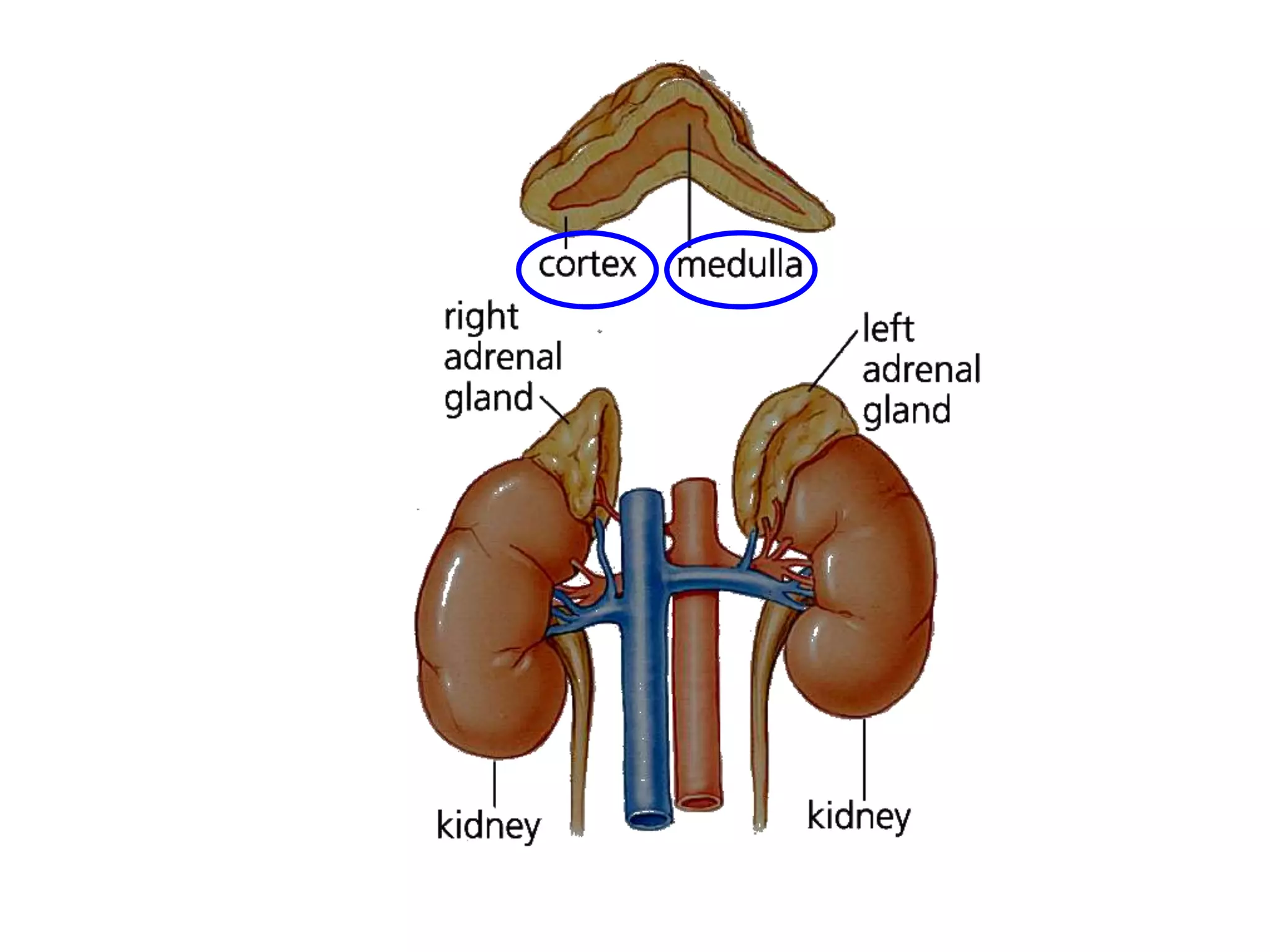
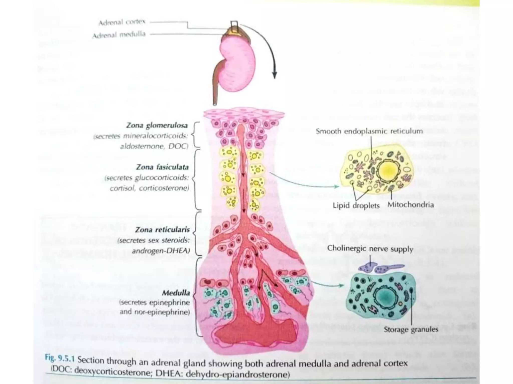
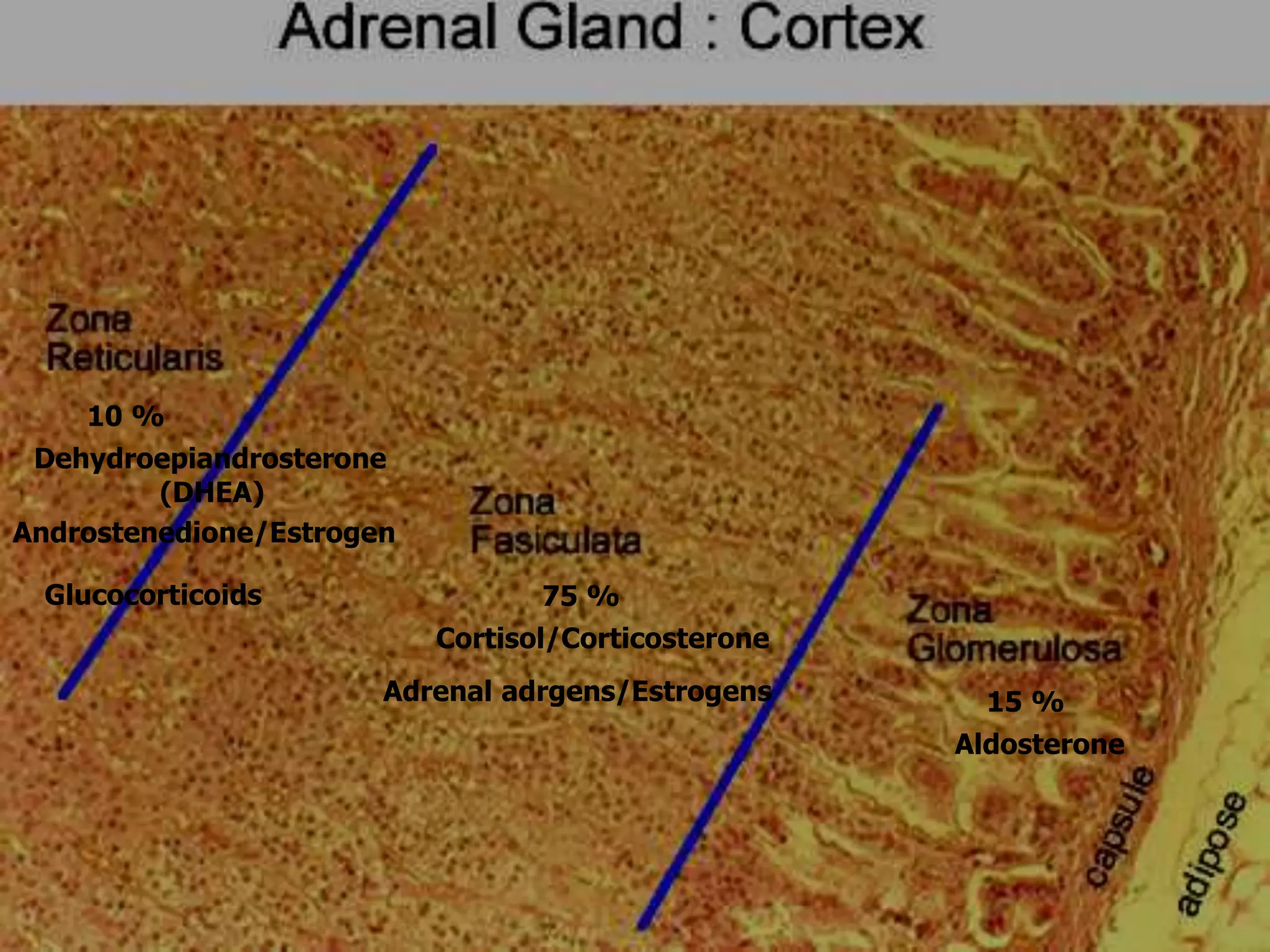
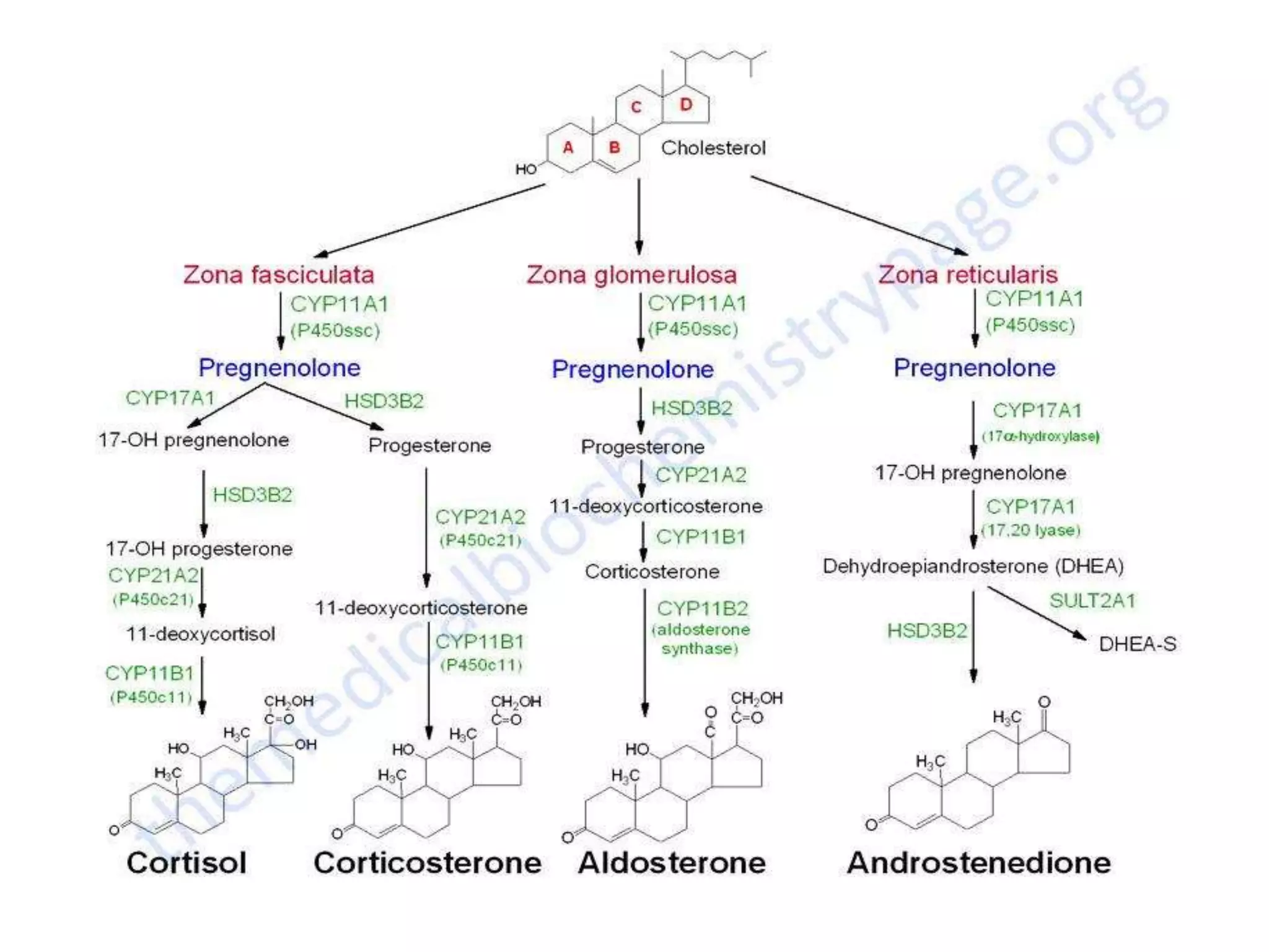
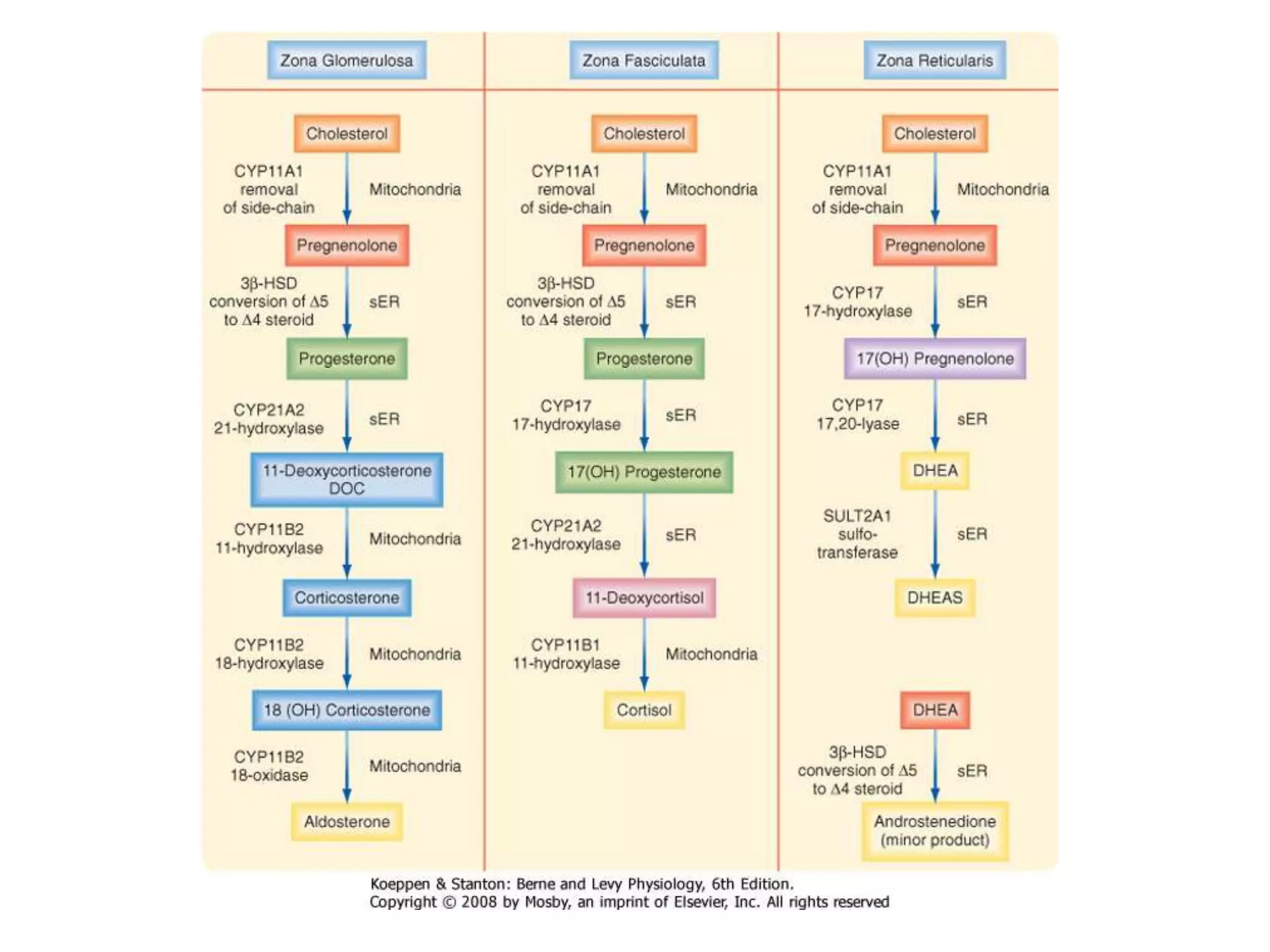
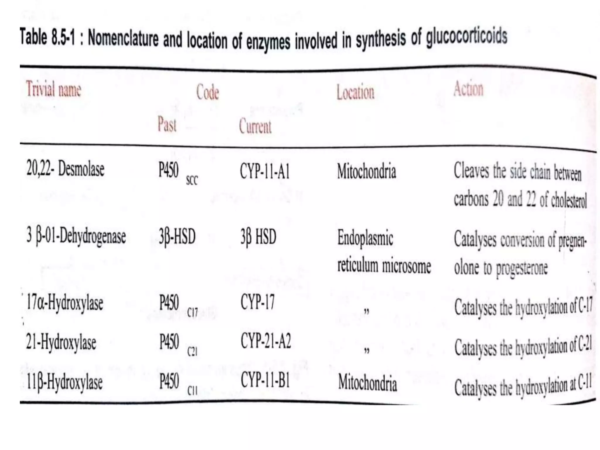
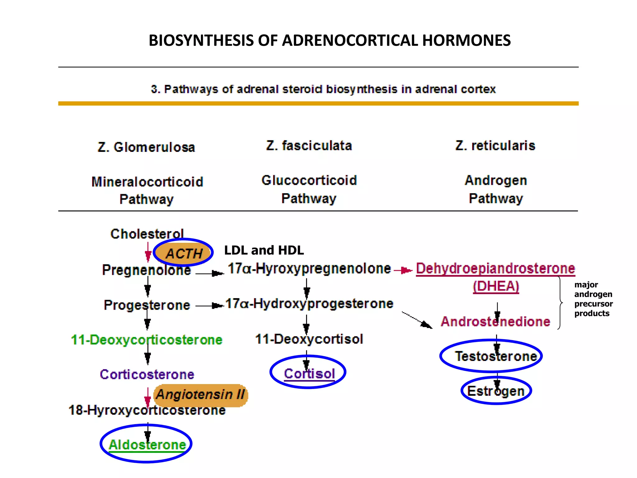
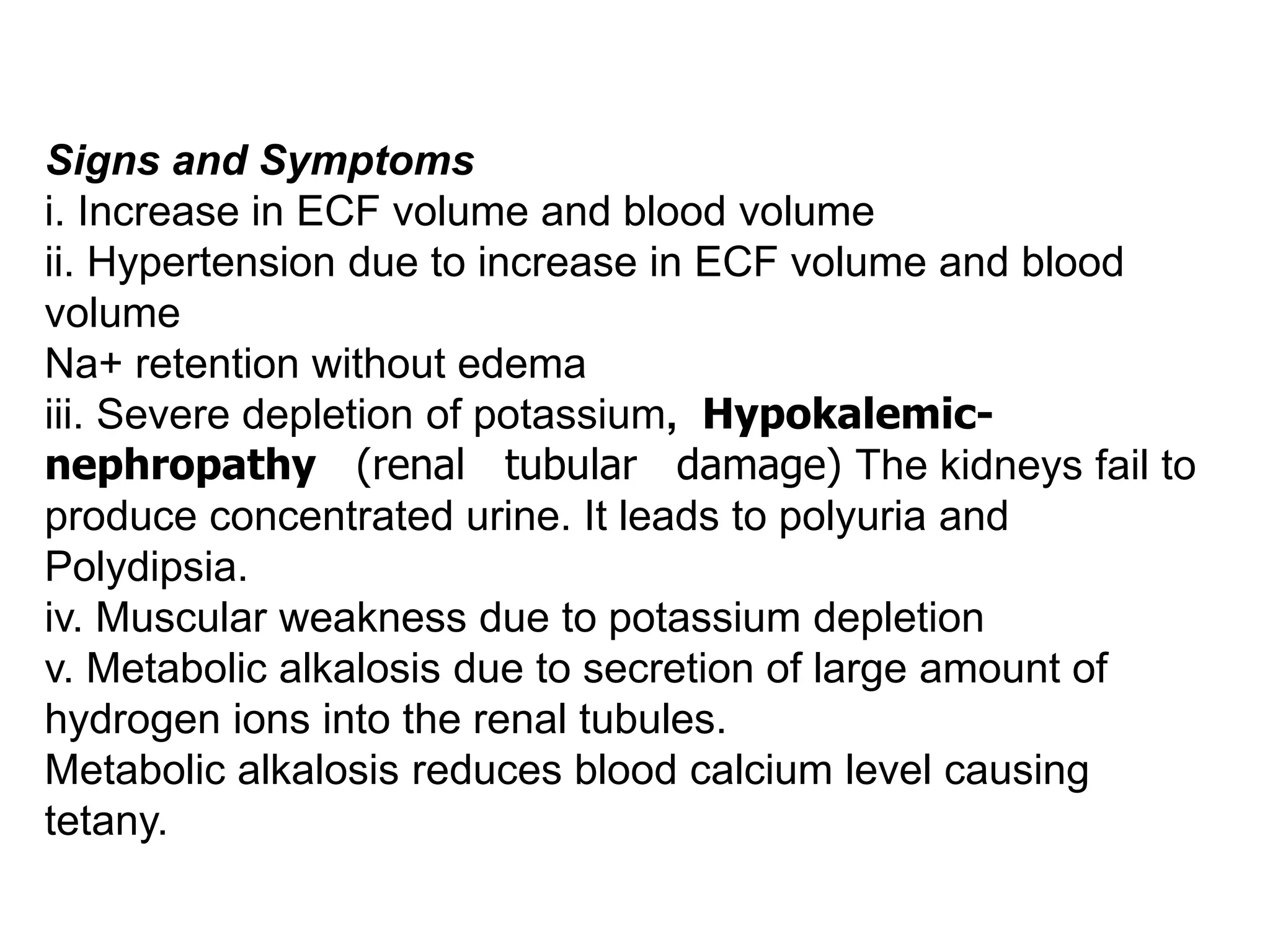
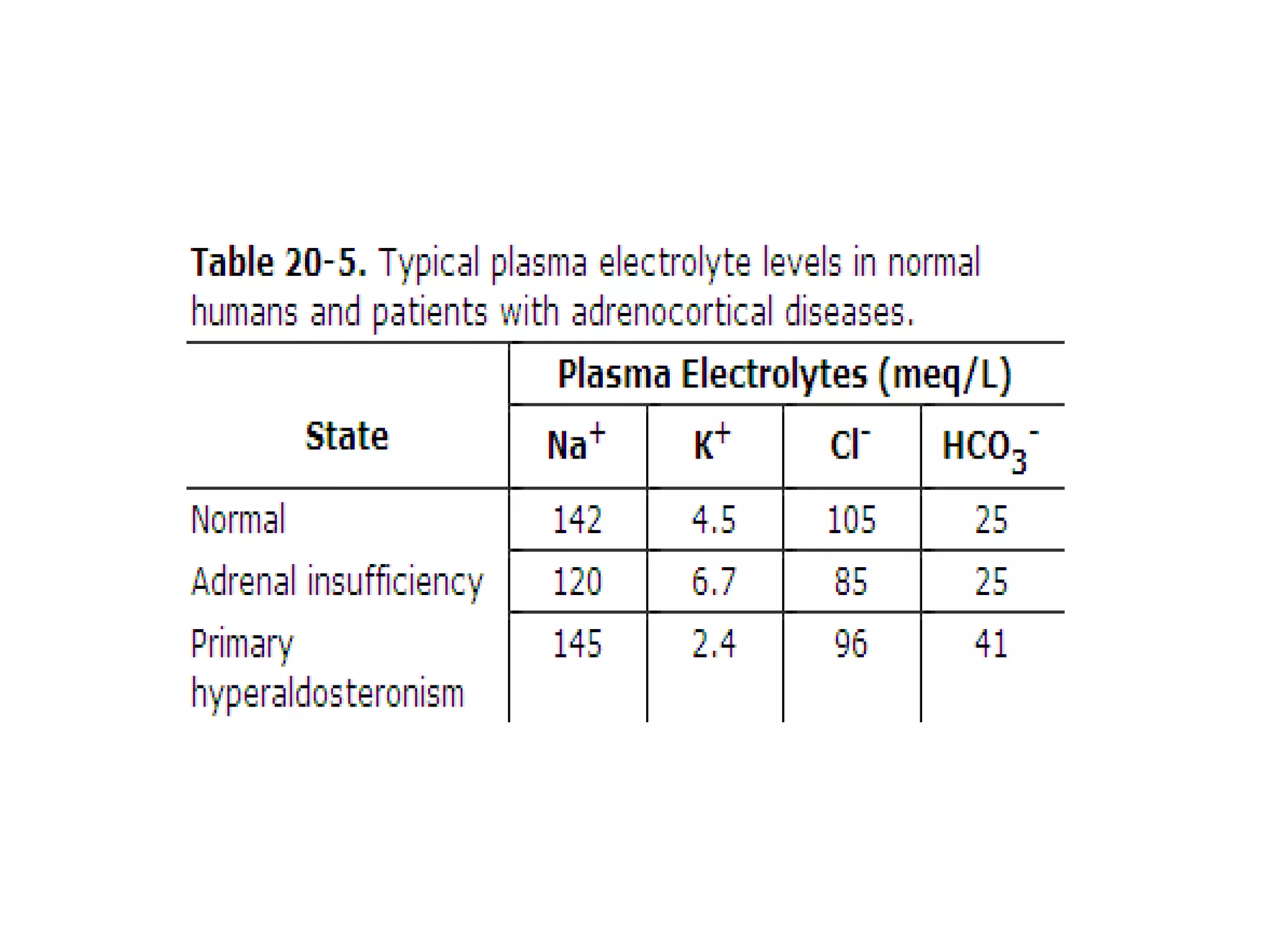
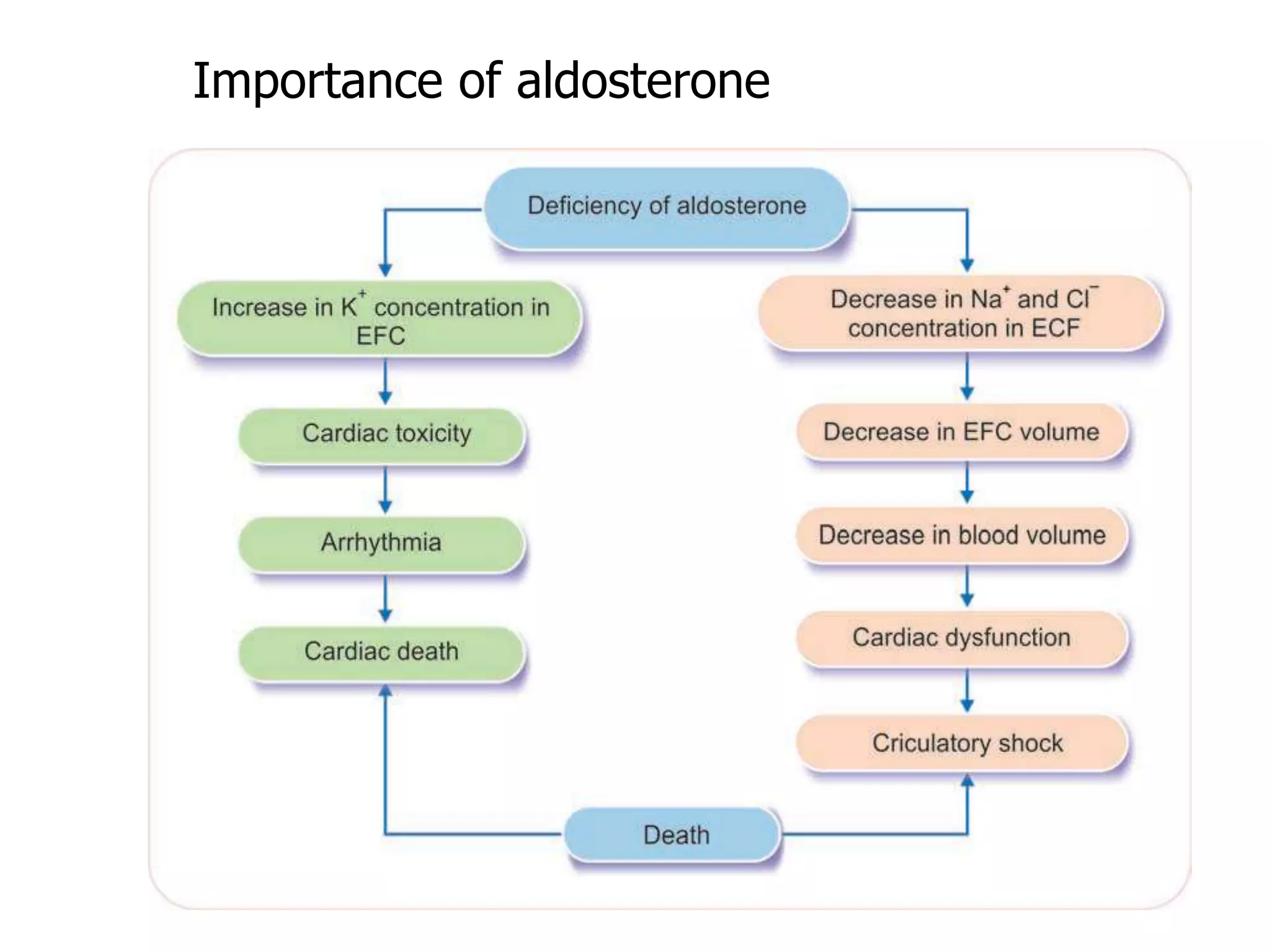
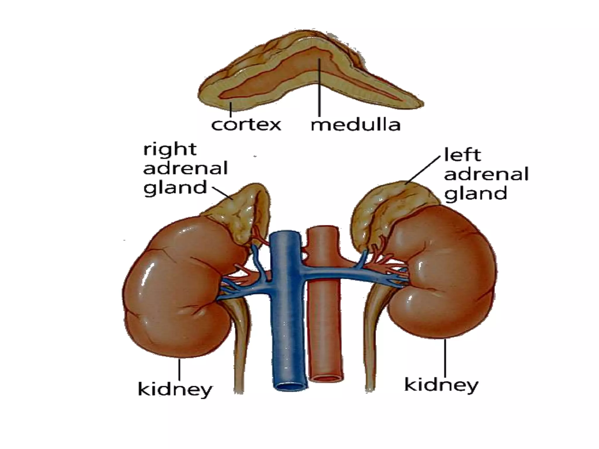
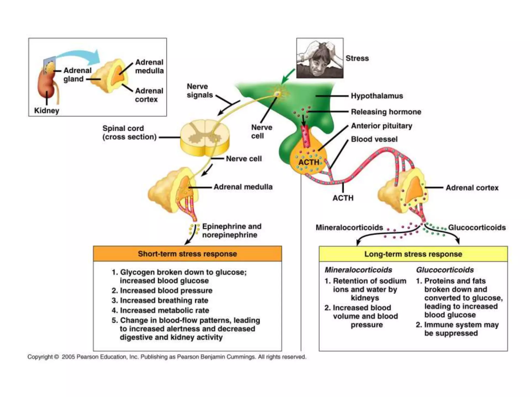
Dr. RAJNEE
• Adrenal glands are called the ‘life-saving glands’ or
‘essential endocrine glands’ or gland of emergency. It
is because the absence of adrenocortical hormones
causes death within 3 to 15 days.
10- 20 %
(Neuroectodermal tissue)
80 - 90 %
(mesodermal tissue)
Epinephrine
Norepinephrine
CATECHOLAMINES
CORTICOSTEROIDS
Mineralocorticoid
Glucocorticoid
Androgen
The Adrenal Gland
Anatomy was first described in 1563.
Is located above (or attached to) the upper
pole of the kidney.
Is pyramidal in structure and weights about four
grams.
Consists of the adrenal cortex and adrenal
medulla
Activities are regulation of fluid volume and
stress response
Adrenal Gland
• Adrenal cortex
– Secretes lipid-based steroid hormones, called
“corticosteroids” – “cortico” as in “cortex”
• MINERALOCORTICOIDS
– Aldosterone is the main one
• GLUCOCORTICOIDS
– Cortisol (hydrocortisone) is the main one
• Adrenal medulla
– Secretes epinephrine and norepinephrine
6
Aorta, Renal arteries and phrenic arteries
Adrenal Cortex
• Is divided into 3 zones in the adult gland: Zona
Glomerulosa, Zona Fasciculata, Zona Rericularis.
• Is divided onto 4 zones in the fetal gland.
• The three zones of the permanent cortex
constitutes only 20% of the fetal gland’s size. The
remaining zone (fetal cortex) comprises up to 80%
of gland’s size during fetal life.
15 %
Aldosterone
75 %
Cortisol/Corticosterone
Adrenal adrgens/Estrogens
10 %
Dehydroepiandrosterone
(DHEA)
Androstenedione/Estrogen
Glucocorticoids
PHYSIOLOGICAL ANATOMY
ADRENAL CORTEX
1. Adrenal (or Suprarenal Glands ): 2 parts
• Outer: Adrenal cortex (80% of total gland): Essential to Life .
• Inner: Adrenal medulla.
2. Adrenal cortex: 3 zones (from outer to inner)
– Zona glomerulosa - mineralocorticoids
– Zona fasciculata - glucocorticoids. together called
cortiosteroides)
– Zona reticulata - sex steroids.
4. characteristic features - adrenal cortex.
I. In foetus (during 3-4 months of intrauterine Iife);
adrenals are larger than the kidney.
- secrete DHEA.
I. Three differentiated zones are not formed until the
3rd year of life.
II. Adrenal cortical cells contain high amounts of lipids
(specially cholesterol) and vitamin C
III. Zona glomerulosa can form the other two layers.
IV. All its hormones being steroids and lipid soluble
can diffuse across the cell membranes into target
organs.
SYNTHESIS
• All adrenocortical hormones are steroid in nature
and are synthesized mainly from cholesterol (CPPP
ring)
cyclo pentano perhydro phenanthrene nucleus that
is absorbed directly from the circulating blood. Small
quantity of cholesterol is also synthesized within the
cortical cells from acetylcoenzyme A (acetyl-CoA).
BIOSYNTHESIS OF ADRENOCORTICAL HORMONES
LDL and HDL
major
androgen
precursor
products
REGULATION
• CORTISOL SECRETION
– Hypothalamic-Pituitary Axis (CRH-ACTH axis)
• ADROGEN SECRETION
– Hypothalamic-Pituitary Axis (ACTH)
– Cortical Adrogen-Stimulating Hormone
• ALDOSTERONE SECRETION
– ECF Potassium Ion Concentration ( K+)
– Renin-Angiotensin-Aldosterone System (AII)
– ECF Sodium Ion Concentration ( Na+)
– Hypothalamic-Pituitary Axis (ACTH)
(has little effect on controlling the rate of secretion)
MINERALOCORTICOIDS
• Aldosterone - principal mineralocorticoid
deoxycorticosterone
• secreted by zona glomerulosa ofadrenal cortex.
• Life-saving Hormone
• accounts for about 90% mineralocorticoid activity
– 60% (protein-bound mainly globulins. ), 40% (free form)
– Half life – 20 minutes
– destroyed mainly in the liver (bile/feces, 25%), mainly
excreted in the urine 75%
• mainly affect electrolytes (minerals K+/Na+) of the ECF.
Aldosterone is very essential for life and it maintains the
osmolarity and volume of ECF.
• BP regulation
Synthesis of aldosterone
Aldosterone, the main mineralocorticoid
• Secreted by adrenal cortex in response to a
decline in either blood volume or blood
pressure (e.g. severe hemorrhage)
– Is terminal hormone in renin-angiotensin
mechanism
• Prompts distal and collecting tubules in kidney
to reabsorb more sodium
– Water passively follows
– Blood volume thus increases
25
Biologic Effects
•  renal tubular reabsorption of sodium (DCT and
CD)
•  renal tubular secretion of potassium (DCT and
CD)
•  renal tubular secretion of hydrogen (DCT and
CD)
• maintenance of ECF volume (principal function)
• same effect (sweat glands, salivary glands,
intestinal epithelial cells (colon)
1. On Sodium Ions
Aldosterone acts on the DCT & CT and
increases the reabsorption of sodium.
Hypersecretion- the loss of sodium through
urine is only few mg/day.
Hyposecretion- the loss of sodium through
urine increases (hypernatriuria) up to about
20 g/day.
It proves the importance of aldosterone in
regulation of sodium ion concentration and
osmolality in the body.
2. On Extracellular Fluid Volume
When sodium ions are reabsorbed from the renal
tubules, simultaneously water is also reabsorbed.
Water reabsorption is almost equal to sodium
reabsorption; so the net result is the increase in ECF
volume.
Even though aldosterone increases the sodium
reabsorption from renal tubules, the concentration
of sodium in the body does not increase very much
because water is also reabsorbed simultaneously.
But still, there is a possibility for mild increase in
concentration of sodium in blood (mild hypernatremia).
It induces thirst, leading to intake of water which again
increases the ECF volume and blood volume.
3. On Blood Pressure
Increase in ECF volume and the blood volume finally
leads to increase in blood pressure.
Aldosterone escape or escape phenomenon
Aldosterone escape refers to escape of the kidney
from salt-retaining effects of excess
administration or secretion of aldosterone, as in the
case of primary hyperaldosteronism.
Significance of aldosterone escape
Because of aldosterone escape, edema does not
occur.
Aldosterone escape
4. On Potassium Ions
Aldosterone increases the potassium excretion
through the renal tubules.
Hyposecretion the potassium ion
concentration in ECF increases leading to
hyperkalemia.
Hyperkalemia results in serious cardiac
toxicity, with weak contractions of heart and
development of arrhythmia. In very severe
conditions, it may cause cardiac death.
Hypersecretion - leads to hypokalemia and
muscular weakness.
5. On Hydrogen Ion Concentration
While increasing the sodium reabsorption from
renal tubules, aldosterone causes tubular secretion
of hydrogen ions. To some extent, secretion of
hydrogen ions is in exchange for sodium ions. It
obviously reduces the hydrogen ion concentration
in the ECF. In normal conditions, aldosterone is
essential to maintain acid base balance in the body.
In hypersecretion, it causes alkalosis and in
hyposecretion, it causes acidosis.
6. On Sweat Glands and Salivary Glands
Sodium is reabsorbed from sweat glands under the
influence of aldosterone, thus the loss of sodium
from the body is prevented. Same effect is shown
on saliva also. Thus, aldosterone helps in the
conservation of sodium in the body.
7. On Intestine
Aldosterone increases sodium absorption from the
intestine, especially from colon and prevents loss of
sodium through feces. Aldosterone deficiency leads
todiarrhea, with loss of sodium and water.
Mode Of Action
Aldosterone acts through the
messengerRNA(mRNA)mechanism.
Sequence of Events
1. Since aldosterone is lipid soluble, it diffuses readily into the
cytoplasm of the tubular epithelial cells through the lipid layer of
the cell membrane
2. In the cytoplasm, aldosterone binds with the specific receptor
protein
3. Aldosterone-receptor complex diffuses into the nucleus where
it binds to deoxyribonucleic acid (DNA) and causes formation of
mRNA
4. The mRNA diffuses back into the cytoplasm and causes
protein synthesis along with ribosomes.
Most of the synthesized proteins are in the form of enzymes.
One of such enzymes is sodium-potassium ATPase, which helps
in the transport of sodium and potassium.
Binding of Cortisol to the
Mineralocorticoid Receptor
• In the laboratory, cortisol(GC) can bind very effectively to
the mineralocorticoid receptor, resulting in activation.
• However, in most tissues due to presence of 11-beta
hydroxysteroid dehydrogenase enzyme, most of the cortisol
(GC)is locally converted to cortisone(11-oxy derivative)
• Cortisone does not bind to the mineralocorticoid receptor
very well.
• Deficiency in 11-beta hydroxysteroid dehydrogenase leads
to high blood pressure, due to increased mineralocorticoid
activity of cortisol (genetic defect).
Role of 11-beta Hydroxysteroid
Dehydrogenase in Cortisol Metabolism
cortisol mineralocorticoid receptor
increased retention of sodium
and water from kidneycortisone
11-beta HSD
REGULATION OF SECRETION
Aldosterone secretion is regulated by four important factors
which are given below in the order of their potency:
1. Increase in potassium ion (K+) concentration in ECF
2.Decrease in sodium ion (Na+) concentration in ECF
3. Decrease in ECF volume
4. Renin angiotensin system
5. Adrenocorticotropic hormone (ACTH).
Increase in the concentration of potassium ions is the most
effective stimulant for aldosterone secretion. It acts directly on
the zona glomerulosa and increases the secretion of
aldosterone.
Regulation of Aldosterone Production
• The main stimulator of aldosterone production is
angiotensin II, not ACTH.
• ACTH does have a trophic (stimulatory) effect on
the zona glomerulosa, preventing atrophy.
The Renin-Angiotensin System
• The juxtaglomerular cells of the kidney
release renin in response to decreased
blood pressure, decreased sodium levels,
or sympathetic stimulation.
Regulation of aldosterone secretion
↓ ABP
HYPERALDOSTERONISM
Increased secretion of aldosterone is called hyperaldosteronism.
Causes and Types
Depending upon the causes, hyperaldosteronism is classified into
two types:
i. Primary hyperaldosteronism
ii. Secondary hyperaldosteronism.
Primary Hyperaldosteronism
(Conn’s syndrome)
Primary hyperaldosteronism develops due to tumor in zona
glomerulosa of adrenal cortex. In primary hyperaldosteronism,
edema does not occur because of escape phenomenon. Renin
secretion decreases.
Secondary Hyperaldosteronism
occurs due to extra adrenal causes such as:
i. Congestive cardiac failure
ii. Nephrosis
iii. Toxemia of pregnancy
iv. Cirrhosis of liver.
Features
1.  level of angiotensin II and renin.
2.  BP with oedema (due to Na+ and water
retention).
Signs and Symptoms
i. Increase in ECF volume and blood volume
ii. Hypertension due to increase in ECF volume and blood
volume
Na+ retention without edema
iii. Severe depletion of potassium, Hypokalemic-
nephropathy (renal tubular damage) The kidneys fail to
produce concentrated urine. It leads to polyuria and
Polydipsia.
iv. Muscular weakness due to potassium depletion
v. Metabolic alkalosis due to secretion of large amount of
hydrogen ions into the renal tubules.
Metabolic alkalosis reduces blood calcium level causing
tetany.
HYPERALDOSTERONISM
 ABP
 ECFV
 RBF
 Sympathetic discharge
KIDNEY
RENIN
ANGOITENSINOGEN ANGOITENSIN I
ACE (LUNGS)
ANGIOTENSIN II
potent vasoconstrictor
total peripheral
resistance
 ABP
(+)
ADRENAL CORTEX
ALDOSTERONE
 sodium reabsorption
 potassium secretion
 hydrogen secretion
hypernatremia
hypokalemia
mild alkalosis
hypertension
 ECFV
–  aldosterone level  renin level
 Secondary Aldosteronism
-  aldosterone level  renin level
negative feedback
tumor (kidney)
Primary Hyperaldosteronism
HYPOACTIVITY OF ADRENAL CORTEX
Hyposecretion of adrenocortical hormones leads to the
following conditions:
1. Addison disease or chronic adrenal insufficiency
2. Congenital adrenal hyperplasia.
Addison Disease Or Chronic Adrenal Insufficiency
Addison disease is the failure of adrenal cortex to secrete
corticosteroids.
Types of Addison Disease
i. Primary Addison disease due to adrenal cause
ii. Secondary Addison disease due to failure of anterior pituitary
to secrete ACTH
iii. Tertiary Addison disease due failure of hypothalamus to
secrete corticotropin-releasing factor (CRF).
• Causes for Primary Addison Disease
• i. Atrophy of adrenal cortex due to autoimmune diseases
• ii. Destruction of the gland because of tuberculosis
• iii. Destruction of hormone-secreting cells in adrenal
cortex by malignant tissues
• iv. Congenital failure to secrete cortisol
• v. Adrenalectomy and failure to take hormonetherapy.
Signs and Symptoms
Signs and symptoms develop in Addison disease because of
deficiency of both cortisol and aldosterone.
Common signs and symptom are:
i. Pigmentation of skin and mucous membrane due to excess
ACTH secretion, induced by cortisol deficiency. ACTH causes
pigmentation by its melanocyte-stimulating action
ii. Muscular weakness
iii. Dehydration with loss of sodium
iv. Hypotension
v. Decreased cardiac output and decreased workload of the
heart, leading to decrease in size of the heart
vi. Hypoglycemia
vii. Nausea, vomiting and diarrhea. Prolonged vomiting and
diarrhea cause dehydration and loss of body weight
viii. Susceptibility to any type of infection
ix. Inability to withstand any stress, resulting in Addisonian
crisis
Tests for Addison Disease
i.Measurement of blood level of cortisol and aldosterone
ii.ii. Measurement of amount of steroids excreted in urine.
Addisonian Crisis or Adrenal Crisis
or Acute Adrenal Insufficiency
Adrenal crisis is a common symptom of Addison disease,
characterized by sudden collapse associated with an
increase in need for large quantities of glucocorticoids.
The condition becomes fatal if not treated in time.
Causes
i. Exposure to even mild stress
ii. Hypoglycemia due to fasting
iii. Trauma
iv. Surgical operation
v. Sudden withdrawal of glucocorticoid treatment.
Importance of aldosterone
GLUCOCORTICOIDS
Aorta, Renal arteries and phrenic arteries
GLUCOCORTICOIDS
• Glucocorticoids are secreted mainly by
zona fasciculata of adrenal cortex. A small
quantity of glucocorticoids is also secreted
by zona reticularis.
• Glucocorticoids act mainly on glucose
metabolism.
• Glucocorticoids are steroids having 21
carbon atoms.
1.Cortisol (hydrocortisone) – principal GC
accounts for about 95% of all glucocorticoid activity
90 - 95% (protein bound), mainly to globulin (cortisol
binding globulin or transcortin) and 6-10% (free
form)
Half life – 60 to 90 minutes
destroyed mainly in the liver (bile/feces, 25%), mainly
excreted in the urine
2.Corticosterone-4%
Half life -- 50 minutes.
3. Cortisone -1%
Half-life of cortisone is not known.
Daily output and plasma level of glucocorticoids are
Cortisol -10.0 (μg) 13.9 (μg/dl)
Corticosterone - 3.0 (μg) 0.4 (μg/dl)
• average amount of derivatives excreted in urine
for 24 hours;
• free cortisol-0.03 mg
• glucocorticoids conjugate-14mg
• keto-steroids - 1.0mg
• Unidentified metabolites-7mg
Cortisol, the most important glucocorticoid
• It is essential for life
• has slight mineralocorticoid activity
Helps the body deal with stressful situations within
minutes
– Physical: trauma, surgery, exercise
– Psychological: anxiety, depression, crowding
– Physiological: fasting, hypoglycemia, fever,
infection
• Regulates or supports a variety of important
cardiovascular, metabolic, immunologic, and
homeostatic functions including water balance.
People with adrenal insufficiency: these stresses can
cause hypotension, shock and death:
t/t give glucocorticoids, eg for surgery , infection etc
• Cortisol is not stored in the adrenocortical
cells. Hence, an acute need for increased amounts
of circulating cortisol requires rapid activation by
ACTH.
• In women, the adrenal glands ultimately supply 50
– 60% of the androgenic hormone requirements.
Mechanism of Action of Glucocorticoids;
being a steroid, acts by stimulating DNA
dependent mRNA Synthesis in the nuclei of their
target cells.
Hormone Effects on Gene Activity
Cortisol
Regulation of Glucocorticoids
secretion
Two mechanisms
1. by ACTH
2. by Glucocorticoids feedback mechanism.
HPA axis (hypothalamic/pituitary/adrenal axis)
Role of Anterior Pituitary – ACTH
Anterior pituitary controls the activities of adrenal
cortex by secreting ACTH. ACTH is mainly concerned
with the regulation of cortisol secretion and it plays only
a minor role in the regulation of mineralocorticoid
secretion.
Source of secretion
ACTH is secreted by the basophilic chromophilic cells
of anterior pituitary.
Chemistry, plasma level and half-life
ACTH is a single straight chain polypeptide with 39 AA
. MW-4600.The sequence of 4-11 AA of ACTH is
identical with β-MCH
daily output is 10 ng
concentration in plasma is 3 ng/dL.
Half-life is 10 minutes.
ACTH
• Acts on adrenal cortex
– stimulates growth of cortex (trophic action)
– Stimulates steroid hormone synthesis
• Lack of negative feedback from cortisol results in
aberrantly high ACTH, elevated levels of other adrenal
corticosteroids– adrenal androgens
• Adrenogenital syndrome: masculization of female fetus
Synthesis
ACTH is synthesized from a protein called
preproopiomelanocortin (POMC). Along with
ACTH, the POMC gives rise to some more
byproducts called β-lipotropin, γ-lipotropin and β-
endorphin. Two more by products,namely α-
melanocyte-stimulating hormone (α-MSH)and
β-melanocyte-stimulating hormone (β-MSH) are
also secreted.
Mode of action of ACTH
ACTH acts by the formation of cyclic AMP.
Actions
1. Maintenance of structural integrity and vascularization
of zona fasciculata and zona reticularis of adrenal
cortex. In hypophysectomy, these two layers in the
adrenal cortex are atrophied.
2. Conversion of cholesterol into pregnenolone, which is
the precursor of glucocorticoids. Thus, ACTH is
responsible for the synthesis of glucocorticoids
3. Release of glucocorticoids
4. Prolongation of glucocorticoid action on various cells.
Other (Non-adrenal) actions of ACTH
1. Mobilization of fats from tissues
2. Melanocyte-stimulating effect-because of structural
similarity with MSH, ACTH causes darkening of skin by
acting on melanophores, which are the cutaneous
pigment cells containing melanin.
Feedback Control
Cortisol regulates its own secretion through
negative feedback control by inhibiting the
release of CRF from hypothalamus and ACTH
from anterior pituitary. CRF- 41AA poly peptide
Circadian rhythm of ACTH
ACTH secretion follows circadian rhythm i.e. it
varies in different periods of the day. The rate of
secretion of both ACTH and CRF is high in the
morning and low in the evening. Hypothalamus
plays an important role in the circadian
fluctuations of ACTH secretion.
Control of Cortisol Secretion: Feedback Loops
• External stimuli
• Hypothalamic
• Anterior Pituitary
• Adrenal cortex
• Tissues
Figure 23-3: The control pathway for cortisol
Actions of Glucocorticoids
1. On Protein Metabolism: Catabolic ( breakdown )
leading to:
i. Decrease in cellular proteins
ii. Increase in plasma level of amino acids By releasing amino
acids from body cells (except liver cells), into the blood.
iii. Increase in protein content in liver. . By increasing the uptake
of amino acids by hepatic cells from blood.
By promoting gluconeogenesis (glucose formation) in liver from
amino acids.
Hypersecretion of glucocorticoids, there is excess catabolism of
proteins, resulting in muscular wasting and negative nitrogen
balance.
2. On Carbohydrate Metabolism - Hyperglycemia
by:
(i) increases hepatic output of glucose
(ii) decreases peripheral utilization of glucose anti
insulin action
(iii) increases glycogen synthesis and decreases
glycolysis in the liver.
• Hypersecretion of glucocorticoids increases the
blood glucose level, resulting in hyperglycemia,
glucosuria and adrenal diabetes.
• Hyposecretion of these hormones causes
hypoglycemia and fasting during adrenal
insufficiency will be fatal. It decreases blood glucose
level to a great extent, resulting in death.
3. On Fat Metabolism: Lipolytic action.
(i) increases lipase activity by potentiating action of
GH, catecholamines, glucagon and thyroid hormones.
(ii) Favours mobilization of fatty acids from adipose
tissue to liver.
(iii) Increasing the concentration of fatty acids in blood
(iv) increases gluconeogenesis.
(v) Increasing the utilization of fat for energy.
• energy is liberated. It leads to the formation of a large
amount of ketone bodies. It is called ketogenic effect
of glucocorticoids.
• Hypersecretion of glucocorticoids causes an
abnormal type of obesity by increasing the
deposition of fat in certain areas such as abdomen,
chest, face and buttocks.
4. On Electrolyte and Water Metabolism: Paradoxical
effects
(i) Due to its mild mineralocorticoid activity increases Na+
retention and increases K+ excretion by the kidney.
(ii) Antagonises the action of ADH on renal tubules diuresis.
In healthy person (i) and (ii) balance each other.
Adrenal cortex insufficiency
• Decreases diuretic effect - water intoxication - water
enters the brain cells - convulsions, unconsciousness
and death.
• Hypersecretion of GC causes edema ,hypertension, hypokalemia
and muscular weakness.
• GC decrease the blood calcium by inhibiting its absorption from
intestine and increasing the excretion through urine.
5. On Bone-GC stimulate the bone resorption
(osteoclastic activity) and inhibit bone formation and
mineralization (osteoblastic activity). hypersecretion
of GC leads osteoporosis & tetany.
6. On Muscles-GC increase the catabolism of proteins in
muscle.
hypersecretion causes muscular weakness due to loss
of protein.
7. Restore vascular reactivity - maintain normal B.P.
Mechanism: GC -
(a) inhibit COMT which - breakdown of catecholamines
(b) increases catecholamine synthesis.
(A and b) - sensitizes arterioles to the constrictor
action of catecholamines.
8. Permissive Action of Glucocorticoids
Permissive action of glucocorticoids refers to
execution of actions of some hormones only in the
presence of glucocorticoids.
Examples:
i. Calorigenic effect of glucagon
ii. Lipolytic effect of catecholamines
iii. Vascular effects of catecholamines
iv. Bronchodilator effect of catecholamines. GC must
be present for catecholamines to produce pressor
response and broncodilatation.
9. On Resistance to Stress , stress is a normal
Physiological response to maintain against
adverse conditions.
• Exposure to any type of stress, either physical or
mental, increases the secretion of ACTH, which in
turn increases glucocorticoid secretion. (called
general adaptation syndrome). The increase in
glucocorticoid level is very essential for survival
during stress conditions, as it offers high resistance
to the body against stress.
Glucocorticoids enhance the resistance by the
following ways:
i. Immediate release and transport of amino acids from
tissues to liver cells for the synthesis of new proteins
and other substances, which are essential to withstand
the stress.
ii. Release of fatty acids from cells for the production of
more energy during stress.
iii. Enhancement of vascular response to
catecholamines and fatty acid-mobilizing action of
catecholamines, which are necessary to withstand the
stress.
iv. Prevention of severity of other changes in the body
caused by stress.
10. Anti-Inflammatory and Anti-Allergic action.
These response only seen with high out put of GC (50 -75 mg/day;
normal : 25 mg/day).
• In high dose GC - prevent the inflammatory reactions.
(i) decreases local reaction by decreasing hyperaemia , exudation,
migration and infiltration of WBCs at the site of injury.
(ii) Prevents tissue damage by stabilizing lysosomal membrane.
(iii) decreases fibroblastic activity.
(iv) decreases release of endogenous pyrogens from granulocytes-
- decreases fever
(v) decreases antibody formation by its destructive effect on fixed
lymphoid tissues.
(vi) Causing vasoconstriction through the permissive action on
catecholamines.
12. Immunosuppressive Effects
GC suppress the immune system of the body by
decreasing the number of circulating T lymphocytes. It
is done by suppressing proliferation of T cells and the
lymphoid tissues (lymph nodes and thymus) GC also
prevent the release of interleukin-2 by T cells.
• hypersecretion or excess use of GC decreases the immune
reactions against all foreign bodies entering the body,
increases the susceptibility to bacterial, fungal and
viral infection leads to severe infection causing death.
• Immunological reactions, which are common during organ
transplantation, may cause rejection of the transplanted
tissues. GC are used to suppress the immunological
reactions because of their immunosuppressive action.
13. Effects on the fetus
– facilitates maturation of the fetus (maturation of the
CNS, retina, skin, GIT and lungs)
– ↑ synthesis of surfactant, flattening of alveolar cells,
thinning of the lung septa and ↑ the rate of
development of the alveoli (last weeks of gestation)
14.Effects on CNS- GC excess decrease threshold of
electrical excitation of the brain so increase brain
excitability leads to restlessness, psychosis & convulsions.
15.Effects on GIT- increase gastric acid & pepsin secretion
and decrease gastric mucosal cell proliferation leads to
peptic ulcer formation in GC excess .
16.Effects on blood- GC excess produce
eosinopenia , basopenia ,lymphopenia,
neutrophilia, polycythaemia, increase in platelets
count. decrease in CT.
• Changes in eosinophil levels have been used
as an index of change in ACTH secretion.
Functions Of Glucocorticoids
It helps to withstand the stress and trauma in life.
Glucocorticoids have metabolic effects on
carbohydrates, proteins, fats and water.
These hormones also show mild mineralocorticoid
effect.
Removal of adrenal glands in human beings and
animals causes disturbances of metabolism.
Exposure to even mild harmful stress after
adrenalectomy, leads to collapse and death.
The General Adaptation Syndrome
CUSHING SYNDROME
Cushing syndrome is a disorder characterized by
obesity.
Causes
• Cushing syndrome is due to the hypersecretion
of glucocorticoids, particularly cortisol.
• It may be either due to pituitary origin or adrenal
origin.
• If it is due to pituitary origin, it is known as
Cushing disease.
• If it is due to adrenal origin it is called
Cushing syndrome.
Generally, these two terms are used
interchangeably.
i. Pituitary Origin
Increased secretion of ACTH causes hyperplasia of
adrenal cortex, leading to hyper-secretion of
glucocorticoid.
ACTH-dependent Cushing syndrome which is
due to hyper-secretion of ACTH
i. Tumor in pituitary cells, particularly in basophilic cells
which secrete ACTH.
ii. Malignant tumor of non-endocrine origin like cancer of
lungs or abdominal viscera.
iii. Hypothalamic disorder causing hyper secretion of
CRH.
iv. Iatrogenic- Prolonged t/t with ACTH or its synthetic
analogue which stimulates adrenal cortex to secrete
excess glucocorticoids.
Cushing syndrome is classified into two
types:
ii. Adrenal Origin - ACTH-independent Cushing
syndrome in which the secretion of ACTH is normal
or low.
• The syndrome develops due to abnormal
membrane receptors for some peptides like
interleukin-1, gonadotropin-releasing hormone
(GnRH )and gastric inhibitory polypeptide (GIP) in
the cells of zona fasciculata. The binding of these
peptides to the abnormal receptors increases
secretion of glucocorticoids, resulting in Cushing
syndrome.
• Tumor in zona fasciculata of adrenal cortex
• Carcinoma of adrenal cortex
• Iatrogenic--Prolonged treatment of chronic
inflammatory diseases like rheumatoid arthritis, with
high dose of exogenous glucocorticoids.
Signs and Symptoms
Characteristic feature of this disease is the
disproportionate centripetal distribution of body fat,
resulting in some abnormal features:
a. Moon face: The edematous facial appearance due
to fat accumulation and retention of water and salt
b. Torso: Fat accumulation in the chest and abdomen.
Arms and legs are very slim in proportion to torso (
trunk of the body)
c. Buffalo hump: Due to fat deposit on the back of
neck and shoulder
d. Pot belly: Due to fat accumulation in abdomen.
e. Atherosclerosis
ii. Purple striae: Reddish purple stripes on abdomen due to
3 reasons:
– Stretching of abdominal wall by excess subcutaneous
fat
– Rupture of sub dermal tissues due to stretching
– Deficiency of collagen fibers due to protein depletion.
iii. Thinning of extremities
iv. Thinning of skin and subcutaneous tissues due to protein
depletion caused by increased catabolism of proteins –ve
nitrogen balance leads to retardation of growth.
v. Aconthosis: Skin disease characterized by darkened skin patches
in certain areas such as axilla, neck and groin
vi. Pigmentation of skin, especially in ACTH dependent type
due to hypersecretion of ACTH which has got melanocyte-
stimulating effect
vii. Facial plethora: Facial redness, acne
viii. Hirsutism: Heavy growth of body and facial hair
ix. Weakening of muscles because of protein depletion
steroid myopathy.
x. Bone resorption and osteoporosis due to protein
depletion. Bone becomes susceptible to easy fracture
xi. Hyperglycemia due to gluconeogenesis (from
proteins) and inhibition of peripheral utilization of
glucose. Hyperglycemia leads to glucosuria and steroid
(adrenal ) diabetes.
xii. Oedema & hypertension by the mineralocorticoid
effects of glucocorticoids – retention of sodium and
water results in increase in ECF volume and blood
volume, leading to hypertension.
xiii. Immuno-suppression resulting in susceptibility for
infection.
xiv. Poor wound healing - easy bruisability
xv. Hair become thin and rough.
xvi. Blood- eosinopenia , basopenia
lymphopenia,neutrophilia, polycythamia
xvii. CNS- increase brain excitability leads to
restlessness, psychosis & convulsions.
xviii. GIT-peptic ulcer formation.
xix. Impotency & hypogonadism in males,
amenorrhoea in females.
Cushing’s Syndrome
– manifested by;
• Buffalo hump hypertension
• Moon face hypernatremia
• Truncal obesity hypokalemia
• Purplish striae mild alkalosis
• Weakness vertebral fractures
• Osteoporosis easy bruisability
• Diabetes psychiatric
disorders
Tests for Cushing Syndrome
i. Observation of external features
ii. Determination of blood sugar and cortisol levels
iii. Analysis of urine for 17-hydroxysteroids.
Treatment for Cushing Syndrome
Treatment depends upon the cause of the disease.
T/t include cortisol-inhibiting drugs, surgical removal of pituitary
or adrenal tumor, radiation or chemotherapy.
Nelson syndrome Nelson syndrome is a disorder that
develops after surgical removal of both adrenal glands. It is
because of the growth of pituitary tumor that secretes excess
ACTH.
The features include headache and visual problems.
Nelson syndrome can be treated with radiation or surgical
removal of the pituitary gland.
SYNTHETIC STEROIDS
Synthetic steroids that are commonly used are:
1. Cortisone and hydrocortisone, which are used
for
replacement therapy have both glucocorticoid and
mineralocorticoid effects
2. Prednisolone has more glucocorticoid activity
than
mineralocorticoid activity
3. Fludrocortisone (9-fluorocortisol) has more
mineralocorticoid activity than glucocorticoid activity.
It has most potent mineralocorticoid effect.
4. Dexamethasone has only glucocorticoid
effect.
Corticosteroid therapy and its uses
1) in adrenal insufficiency
2) treatment of rheumatoid arthritis and collagen
disorders
3) in organ transplant recipients for reducing graft
rejection
4) used in bronchial asthma and skin disease
5) used in leukaemis
6) to treat cerebral edema and in treatment of
circulatory shock
(Q) Prolonged t/t with GC decreases ACTH synthesis
and produces adrenocortical atrophy. Sudden
stoppage of such therapy is lethal & person may
die of adrenal crisis. This complication can be
avoided by slowed decreasing the GC dose over a
long period.
(Q) GC when used in patient with bacterial infections
must be used along with antibiotics otherwise
signs and symptoms get masked by GC t/t thus
there may be serious & even fetal delays in
diagnosis & t/t of infection.
Adrenal Hormones- Androgens
Androgens
• Androgens are secreted mainly by zona
reticularis. Zona fasciculata also secretes small
quantities of androgens.
• In foetus, during 3rd - 4th month of IUL adrenals
are larger than the kidney. A major function of
foetal adrenal is secretion of
Dehydroepiandrosterone (DHEA) sulphate
conjugate of androgen which is converted in the
placenta to androgen and oestrogen and enters
the maternal circulation. After birth adrenal cortex
regresses & secretes less sex hormones. At
puberty in both sexes adrenal androgen secretion
again increases.
ANDROGENS
• Dehydroepiandrosterone (DHEA) and
androstenedione
– principal cortical androgens
• responsible for the early development of male
sex organs.
• converted to testosterone (potent)
• “musculinizing effect”
• also secrete estrogen (estradiol) and
progesterone in female
• Adrenal sex hormones are transported by another
special plasma protein known as sex hormone-
binding globulin.
• DHEA is the most active adrenal androgen.
Androgens, in general, are responsible for
masculine features of the body . But in
normal conditions, the adrenal androgens
have insignificant physiological effects,
because of the low amount of secretion
both in males and females.
• Androgen (DHEA) Its activity is < 20% of
testosterone activity.
• Normal plasma levels of DHEA is 150-200
µg/dl.
Mechanism of Action of androgen ;
being a steroid, acts by stimulating DNA
dependent mRNA Synthesis in the nuclei of their
target cells.
Synthesis of androgen in zona reticularis
Regulation of adrenal androgen
• 1) by ACTH
• 2) by AASH (adrenal androgen
stimulating hormone) & not by GnRH
ADRENOGENITAL SYNDROME
Under normal conditions, adrenal cortex secretes
small quantities of androgens which do not have any
significant effect on sex organs or sexual function.
However, secretion of abnormal quantities of adrenal
androgens develops adrenogenital syndrome.
Testosterone is responsible for the androgenic
activity in adrenogenital syndrome.
Causes
Adrenogenital syndrome is due to the tumor of zona
reticularis in adrenal cortex.
Symptoms
Adrenogenital syndrome is characterized by the
tendency for the development of secondary sexual
character of opposite sex.
Symptoms in females
Increased secretion of androgens causes
development of male secondary sexual characters.
The condition is called adrenal virilism.
Symptoms are:
i. Masculinisation due to increased muscular growth
ii. Deepening of voice
iii. Primary amenorrhea -Loss of regular of menses
iv. Male type of hair growth (diamond shaped pubic
hairs), Growth of beard, Baldness
v. Regression of breast tissue
Symptoms in males
Sometimes, the tumor of estrogen secreting
cells produces more than normal quantity of
estrogens in males. It produces some
symptoms such as:
i. Feminization
ii. Gynecomastia (enlargement of breast)
iii. Atrophy of testis
CONGENITAL ADRENAL HYPERPLASIA
• Congenital adrenal hyperplasia is a congenital
disorder, characterized by increase in size of
adrenal cortex. Size increases due to abnormal
increase in the number of steroid-secreting cortical
cells.
• Excess quantity of androgens is secreted. In
males, it does not produce any special effect
because, large quantity of androgens are
produced by testes also. But in females, the
androgens produce masculine features. Some of
the androgens are converted into testosterone.
Testosterone is responsible for the androgenic
activity in congenital adrenal hyperplasia
Causes
Even though the size of the gland increases,
aldosterone &cortisol secretion decreases. It is
because of the congenital deficiency of the enzymes
necessary for the synthesis of cortisol, particularly, 21β-
hydroxylase.&11β-hydroxylase necessary for the
synthesis of aldosterone .Lack of this enzyme reduces
the synthesis of cortisol and aldosterone resulting in
ACTH secretion from pituitary by feedback mechanism.
ACTH stimulates the adrenal cortex causing
hyperplasia, with accumulation of lipid droplets. Hence,
it is also called congenital lipid adrenal hyperplasia.
Therefore, due to the constant simulation of adrenal
cortex by ACTH, the secretion of androgens increases.
It results in sexual abnormalities such as virilism.
Symptoms
In boys
Adrenal hyperplasia produces a condition known as
Precocious Pseudo Puberty.
Precocious Pseudo Puberty-Early development of
secondary sexual characters without gametogenesis
. It is due to abnormal exposure of immature males
to androgens .
.
In girls
Increased secretion of androgens causes
development of male secondary sexual
characters. The condition is called adrenal
virilism.
• Increased secretion of androgens before
12 week IUL female foetus , causes the
female child born with external genitalia
of male type. This condition is called
pseudohermaphroditism.
• In majority of cases associated
aldosterone deficiency causes loss of
Na+ result in hypovolemia .
• Medulla is the inner part of adrenal gland
and it forms 20% of the mass of adrenal
gland. It is made up of interlacing cords of
cells known as chromaffin cells or
pheochrome cells or chromophil cells.
ADRENAL MEDULLA
• sympathetic ganglion in which the
postganglionic neurons have lost their axons
and become secretory cells.
• Adrenal medulla is formed by two types of chromaffin
cells.
– Epinephrine-secreting type (80-90%)
– Norepinephrine-secreting type (10-20%)
Catecholamines secreted by adrenal medulla
1. Adrenaline or epinephrine
2. Noradrenaline or norepinephrine
3. Dopamine.
Adrenal Medulla: Modified Sympathetic Ganglion
• Sympathetic stimulation
– Catecholamine release to blood
• Epinephrine
• Norepinephrine
– Travel to:
• Multiple targets
• Distant targets
The Adrenal Medulla
Review of Efferent Pathways: Motor and Autonomic
• PLASMA LEVEL OF CATECHOLAMINES
• 1. Adrenaline : 3 μg/dL
• 2. Noradrenaline : 30 μg/dL
• 3. Dopamine : 3.5 μg/dL
• „HALF-LIFE OF CATECHOLAMINES
• Half-life of catecholamines is about 2 minutes.
Synthesis of catecholamines. DOPA = Dihydroxyphenylalanine,
PNMT = Phenylethanolamine-N-methyl transferase.
COMT =Catechol-O-methyltransferase,
MAO = Monoamine oxidase
Metabolism of catecholamines.
DEGRADATION
OF
CATECHOLAMINES
Removal of Catecholamines
Catecholamines are removed from body through
urine
in three forms:
i. 15% as free adrenaline and free noradrenaline
ii. 50% as free or conjugated meta-adrenaline
(metanephrine)and meta-noradrenaline
(normetanephrine)
iii. 35% as vanillylmandelic acid (VMA).
„
Sympathetic receptors
Adrenergic
receptors
Alpha Beta
Alpha 1 Alpha 2 Beta 1
Beta 3
Beta 2
Mechanism of Action
• receptor mediated – adrenergic receptors
• peripheral effects are dependent upon the
type and ratio of receptors in target tissues
Receptor  
Norepinephrine +++++ ++
Epinephrine ++++ ++++
Relative effects of epinephrine and norepinephrine on  and  adrenergic receptors.
Effect of nor epinephrine – is prolonged and it
defuse in blood from adrenergic nerve ending and
epinephrine comes from adrenal medulla .
• Adrenaline acts through both alpha and beta
receptors equally.
• Noradrenaline acts mainly through alpha receptors
and occasionally through beta receptors. (β1)
A. Situation with which individual is familiar
increase NE secretion.
– Eg.-anger, challenging situation
B. Situation with which individual is not
familiar increase E secretion.
– Eg.- anxiety, tension & threatening situation.
Receptor Mode of action Response
Alpha-1 receptor Activates IP3 through
phospholipase C
Mediates more of
noradrenaline actions than
adrenaline actions
Alpha-2 receptor Inhibits adenyl cyclase and
cAMP
Mediates more of
noradrenaline actions than
adrenaline actions
Beta-1 receptor Activates adenyl cyclase
and cAMP
Mediates actions of
adrenaline and
noradrenaline equally
Beta-2 receptor Activates adenyl cyclase
and cAMP
Mediates more of
adrenaline actions than
noradrenaline actions
Adrenergic receptors
IP3 = Inositol triphosphate
ACTIONS
• Circulating adrenaline and noradrenaline have
similar effect of sympathetic stimulation. But, the
effect of adrenal hormones is prolonged 10 times
more than that of sympathetic stimulation. It is
because of the slow inactivation, slow degradation
and slow removal of these hormone.
• Adrenaline and noradrenaline stimulate the
nervous system. Adrenaline has significant effects
on metabolic functions and both adrenaline and
noradrenaline have significant effects on
cardiovascular system.
1. On Metabolism (via Alpha and Beta Receptors)
Adrenaline influences the metabolic functions more than
noradrenaline.
i. General metabolism: Adrenaline increases O2
consumption and carbon CO2 removal. It increases
BMR. So, it is said to be a calorigenic hormone.
ii. Carbohydrate metabolism: Adrenaline increases the
blood glucose level by increasing the glycogenolysis in
liver and muscle. So, a large quantity of glucose enters
the circulation.
iii. Fat metabolism: Adrenaline causes mobilization of
free fatty acids from adipose tissues.
Catecholamines need the presence of glucocorticoids for
this action.
2. On Blood (via Beta Receptors)
• Adrenaline decreases blood coagulation time.
• It increases RBC count in blood by contracting
smooth muscles of splenic capsule and releasing
RBCs from spleen into circulation.
3. On Heart (via Beta Receptors)
Adrenaline has stronger effects on heart than
noradrenaline.
• It increases overall activity of the heart, i.e.
i. Heart rate (chronotropic effect)
ii. Force of contraction (inotropic effect)
iii. Excitability of heart muscle (bathmotropic effect)
iv. Conductivity in heart muscle (dromotropic effect).
4. On Blood Vessels (via Alpha and Beta-2 Receptors)
 Noradrenaline has strong effects on blood vessels.
It causes constriction of blood vessels throughout
the body via alpha receptors. So it is called ‘general
vasoconstrictor’. Vasoconstrictor effect of
noradrenaline increases total peripheral resistance.
 Adrenaline also causes constriction of blood vessels.
However, it causes dilatation of blood vessels in skeletal
muscle, liver and heart through beta-2 receptors. So, the
total peripheral resistance is decreased by adrenaline.
 Catecholamines need the presence of glucocorticoids, for
these vascular effects.
5. On Blood Pressure (via Alpha and Beta Receptors)
• Adrenaline increases systolic blood pressure by
increasing the force of contraction of the heart and
cardiac output. But, it decreases diastolic blood
pressure by reducing the total peripheral
resistance.
• Noradrenaline increases diastolic pressure due to
general vasoconstrictor effect by increasing the total
peripheral resistance. It also increases the systolic
blood pressure to a slight extent by its actions on
heart. Thus, hypersecretion of catecholamines leads to
hypertension.
• The action of catecholamines on blood pressure needs
the presence of glucocorticoids.
6. On Respiration (via Beta-2 Receptors)
Adrenaline increases rate and force of respiration.
Adrenaline injection produces apnea, which is known as
adrenaline apnea. It also causes bronchodilation.
7. On Skin (via Alpha and Beta-2 Receptors)
Adrenaline causes contraction of pilo erector. It also
increases the secretion of sweat.
8. On Skeletal Muscle (via Alpha and Beta-2
Receptors)
Adrenaline causes severe contraction and quick fatigue
of skeletal muscle. It increases glycogenolysis and
release of glucose from muscle into blood. It also
causes vasodilatation in skeletal muscles.
9. On Smooth Muscle (via Alpha & Beta Receptors)
A. Catecholamines cause contraction of smooth
muscles
in the following organs:
i. Splenic capsule
ii. Sphincters of gastrointestinal (GI) tract
iii. pilo erector of skin
iv. Gallbladder
v. Uterus
B. Catecholamines cause relaxation of smooth
muscles in the following organs:
i. Non-sphincteric part of GI tract (esophagus, stomach and
intestine)
ii. Bronchioles
iii. Urinary bladder. - β2 receptor cause relaxation of
detrusor muscle , α1 receptor cause contraction of
sphincter.
10. On Central Nervous System
(via Beta Receptors)
Adrenaline increases the activity of brain. Adrenaline
secretion increases during ‘fight or flight reactions’
after exposure to stress. It enhances the cortical
arousal and other facilitatory functions of CNS.
11. Other Effects of Catecholamines
i. On salivary glands (via alpha and beta-2 receptors):
Cause vasoconstriction in salivary gland, leading to mild
increase in salivary Secretion.
ii. On sweat glands (via beta-2 receptors): Increase
the secretion of apocrine sweat glands
iii. On lacrimal glands (via alpha receptors): Increase the
secretion of tears.
iv. On ACTH secretion (via alpha receptors):
Adrenaline increases ACTH secretion
v. On nerve fibers (via alpha receptors): Adrenaline
decreases the latency of action potential in the nerve fibers,
i.e. electrical activity is accelerated.
vi. On renin secretion (via beta receptors): Increase
the rennin secretion from JG apparatus of the kidney.
Differences between Epinephrine and
Norepinephrine
• Epinephrine >> norepinephrine – in terms of cardiac
stimulation leading to greater cardiac output (
stimulation).
• Epinephrine < norepinephrine – in terms of
constriction of blood vessels – leading to increased
peripheral resistance – increased arterial pressure.
• Epinephrine >> norepinephrine –in terms of
increasing metabolism.
Catechalomines: Activity
• Stimulates the “fight or fight” reaction
• Increased plasma glucose levels
• Increased cardiovascular function
• Increased metabolic function
• O2 consumption increases
• Decreased gastrointestinal and genitourinary
function
• Heightens your senses, tenses your muscles, openings
breathing passages, etc.
• In response to stress take less than 30 seconds to kick
in and last several minutes
Activity of Epinephrine
Regulation
1) Nervous control
• Splanchnic nerve
• Ach, AT-II, histamine, bradykinin
2) Indirectly by GC secretion
3) selective secretion
• In emergency or stress
4) By adrenergic receptors
• Up regulation & down regulation
• Denervation hypersensitivity
DOPAMINE
 Dopamine is secreted by adrenal medulla. Type of
cells secreting this hormone is not known.
 Dopamine is also secreted by dopaminergic
neurons in some areas of brain, particularly basal
ganglia. In brain, this hormone acts as a
neurotransmitter.
Injected dopamine produces the following effects:
1. Vasoconstriction by releasing norepinephrine
2. Vasodilatation in mesentery
3. Increase in heart rate via beta receptors
4. Increase in systolic blood pressure. Dopamine does
not affect diastolic blood pressure.
 Deficiency of dopamine in basal ganglia produces
nervous disorder called parkinsonism
APPLIED PHYSIOLOGY –
PHEOCHROMOCYTOMA
Pheochromocytoma is a condition characterized by
hypersecretion of catecholamines. (norepinephrine)
 Cause
Pheochromocytoma is caused by tumor of chromophil
cells in adrenal medulla. It is also caused rarely by tumor
of sympathetic ganglia (extra-adrenal
pheochromocytoma).
 Signs and Symptoms
Characteristic feature of pheochromocytoma is
hypertension. This type of hypertension is known as
endocrine or secondary hypertension.
Other features:
1. Anxiety
2. Chest pain
3. Fever
4. Headache
5. Hyperglycemia
6. Metabolic disorders
7. Nausea and vomiting
8. Palpitation
9. Polyuria and glucosuria
10. Sweating and flushing
11. Tachycardia
12. Weight loss.
Tests for Pheochromocytoma
Pheochromocytoma is detected by measuring
metanephrines and VMA in urine and catecholamines in
plasma.

Adrenal gland

  • 1.
  • 2.
    • Adrenal glandsare called the ‘life-saving glands’ or ‘essential endocrine glands’ or gland of emergency. It is because the absence of adrenocortical hormones causes death within 3 to 15 days.
  • 4.
    10- 20 % (Neuroectodermaltissue) 80 - 90 % (mesodermal tissue) Epinephrine Norepinephrine CATECHOLAMINES CORTICOSTEROIDS Mineralocorticoid Glucocorticoid Androgen
  • 5.
    The Adrenal Gland Anatomywas first described in 1563. Is located above (or attached to) the upper pole of the kidney. Is pyramidal in structure and weights about four grams. Consists of the adrenal cortex and adrenal medulla Activities are regulation of fluid volume and stress response
  • 6.
    Adrenal Gland • Adrenalcortex – Secretes lipid-based steroid hormones, called “corticosteroids” – “cortico” as in “cortex” • MINERALOCORTICOIDS – Aldosterone is the main one • GLUCOCORTICOIDS – Cortisol (hydrocortisone) is the main one • Adrenal medulla – Secretes epinephrine and norepinephrine 6
  • 7.
    Aorta, Renal arteriesand phrenic arteries
  • 12.
    Adrenal Cortex • Isdivided into 3 zones in the adult gland: Zona Glomerulosa, Zona Fasciculata, Zona Rericularis. • Is divided onto 4 zones in the fetal gland. • The three zones of the permanent cortex constitutes only 20% of the fetal gland’s size. The remaining zone (fetal cortex) comprises up to 80% of gland’s size during fetal life.
  • 13.
    15 % Aldosterone 75 % Cortisol/Corticosterone Adrenaladrgens/Estrogens 10 % Dehydroepiandrosterone (DHEA) Androstenedione/Estrogen Glucocorticoids
  • 14.
    PHYSIOLOGICAL ANATOMY ADRENAL CORTEX 1.Adrenal (or Suprarenal Glands ): 2 parts • Outer: Adrenal cortex (80% of total gland): Essential to Life . • Inner: Adrenal medulla. 2. Adrenal cortex: 3 zones (from outer to inner) – Zona glomerulosa - mineralocorticoids – Zona fasciculata - glucocorticoids. together called cortiosteroides) – Zona reticulata - sex steroids.
  • 15.
    4. characteristic features- adrenal cortex. I. In foetus (during 3-4 months of intrauterine Iife); adrenals are larger than the kidney. - secrete DHEA. I. Three differentiated zones are not formed until the 3rd year of life. II. Adrenal cortical cells contain high amounts of lipids (specially cholesterol) and vitamin C III. Zona glomerulosa can form the other two layers. IV. All its hormones being steroids and lipid soluble can diffuse across the cell membranes into target organs.
  • 16.
    SYNTHESIS • All adrenocorticalhormones are steroid in nature and are synthesized mainly from cholesterol (CPPP ring) cyclo pentano perhydro phenanthrene nucleus that is absorbed directly from the circulating blood. Small quantity of cholesterol is also synthesized within the cortical cells from acetylcoenzyme A (acetyl-CoA).
  • 20.
    BIOSYNTHESIS OF ADRENOCORTICALHORMONES LDL and HDL major androgen precursor products
  • 21.
    REGULATION • CORTISOL SECRETION –Hypothalamic-Pituitary Axis (CRH-ACTH axis) • ADROGEN SECRETION – Hypothalamic-Pituitary Axis (ACTH) – Cortical Adrogen-Stimulating Hormone • ALDOSTERONE SECRETION – ECF Potassium Ion Concentration ( K+) – Renin-Angiotensin-Aldosterone System (AII) – ECF Sodium Ion Concentration ( Na+) – Hypothalamic-Pituitary Axis (ACTH) (has little effect on controlling the rate of secretion)
  • 22.
    MINERALOCORTICOIDS • Aldosterone -principal mineralocorticoid deoxycorticosterone • secreted by zona glomerulosa ofadrenal cortex. • Life-saving Hormone • accounts for about 90% mineralocorticoid activity – 60% (protein-bound mainly globulins. ), 40% (free form) – Half life – 20 minutes – destroyed mainly in the liver (bile/feces, 25%), mainly excreted in the urine 75% • mainly affect electrolytes (minerals K+/Na+) of the ECF. Aldosterone is very essential for life and it maintains the osmolarity and volume of ECF. • BP regulation
  • 23.
  • 25.
    Aldosterone, the mainmineralocorticoid • Secreted by adrenal cortex in response to a decline in either blood volume or blood pressure (e.g. severe hemorrhage) – Is terminal hormone in renin-angiotensin mechanism • Prompts distal and collecting tubules in kidney to reabsorb more sodium – Water passively follows – Blood volume thus increases 25
  • 26.
    Biologic Effects • renal tubular reabsorption of sodium (DCT and CD) •  renal tubular secretion of potassium (DCT and CD) •  renal tubular secretion of hydrogen (DCT and CD) • maintenance of ECF volume (principal function) • same effect (sweat glands, salivary glands, intestinal epithelial cells (colon)
  • 27.
    1. On SodiumIons Aldosterone acts on the DCT & CT and increases the reabsorption of sodium. Hypersecretion- the loss of sodium through urine is only few mg/day. Hyposecretion- the loss of sodium through urine increases (hypernatriuria) up to about 20 g/day. It proves the importance of aldosterone in regulation of sodium ion concentration and osmolality in the body.
  • 29.
    2. On ExtracellularFluid Volume When sodium ions are reabsorbed from the renal tubules, simultaneously water is also reabsorbed. Water reabsorption is almost equal to sodium reabsorption; so the net result is the increase in ECF volume. Even though aldosterone increases the sodium reabsorption from renal tubules, the concentration of sodium in the body does not increase very much because water is also reabsorbed simultaneously. But still, there is a possibility for mild increase in concentration of sodium in blood (mild hypernatremia). It induces thirst, leading to intake of water which again increases the ECF volume and blood volume.
  • 30.
    3. On BloodPressure Increase in ECF volume and the blood volume finally leads to increase in blood pressure. Aldosterone escape or escape phenomenon Aldosterone escape refers to escape of the kidney from salt-retaining effects of excess administration or secretion of aldosterone, as in the case of primary hyperaldosteronism. Significance of aldosterone escape Because of aldosterone escape, edema does not occur.
  • 31.
  • 32.
    4. On PotassiumIons Aldosterone increases the potassium excretion through the renal tubules. Hyposecretion the potassium ion concentration in ECF increases leading to hyperkalemia. Hyperkalemia results in serious cardiac toxicity, with weak contractions of heart and development of arrhythmia. In very severe conditions, it may cause cardiac death. Hypersecretion - leads to hypokalemia and muscular weakness.
  • 33.
    5. On HydrogenIon Concentration While increasing the sodium reabsorption from renal tubules, aldosterone causes tubular secretion of hydrogen ions. To some extent, secretion of hydrogen ions is in exchange for sodium ions. It obviously reduces the hydrogen ion concentration in the ECF. In normal conditions, aldosterone is essential to maintain acid base balance in the body. In hypersecretion, it causes alkalosis and in hyposecretion, it causes acidosis.
  • 34.
    6. On SweatGlands and Salivary Glands Sodium is reabsorbed from sweat glands under the influence of aldosterone, thus the loss of sodium from the body is prevented. Same effect is shown on saliva also. Thus, aldosterone helps in the conservation of sodium in the body. 7. On Intestine Aldosterone increases sodium absorption from the intestine, especially from colon and prevents loss of sodium through feces. Aldosterone deficiency leads todiarrhea, with loss of sodium and water.
  • 35.
    Mode Of Action Aldosteroneacts through the messengerRNA(mRNA)mechanism. Sequence of Events 1. Since aldosterone is lipid soluble, it diffuses readily into the cytoplasm of the tubular epithelial cells through the lipid layer of the cell membrane 2. In the cytoplasm, aldosterone binds with the specific receptor protein 3. Aldosterone-receptor complex diffuses into the nucleus where it binds to deoxyribonucleic acid (DNA) and causes formation of mRNA 4. The mRNA diffuses back into the cytoplasm and causes protein synthesis along with ribosomes. Most of the synthesized proteins are in the form of enzymes. One of such enzymes is sodium-potassium ATPase, which helps in the transport of sodium and potassium.
  • 37.
    Binding of Cortisolto the Mineralocorticoid Receptor • In the laboratory, cortisol(GC) can bind very effectively to the mineralocorticoid receptor, resulting in activation. • However, in most tissues due to presence of 11-beta hydroxysteroid dehydrogenase enzyme, most of the cortisol (GC)is locally converted to cortisone(11-oxy derivative) • Cortisone does not bind to the mineralocorticoid receptor very well. • Deficiency in 11-beta hydroxysteroid dehydrogenase leads to high blood pressure, due to increased mineralocorticoid activity of cortisol (genetic defect).
  • 38.
    Role of 11-betaHydroxysteroid Dehydrogenase in Cortisol Metabolism cortisol mineralocorticoid receptor increased retention of sodium and water from kidneycortisone 11-beta HSD
  • 39.
    REGULATION OF SECRETION Aldosteronesecretion is regulated by four important factors which are given below in the order of their potency: 1. Increase in potassium ion (K+) concentration in ECF 2.Decrease in sodium ion (Na+) concentration in ECF 3. Decrease in ECF volume 4. Renin angiotensin system 5. Adrenocorticotropic hormone (ACTH). Increase in the concentration of potassium ions is the most effective stimulant for aldosterone secretion. It acts directly on the zona glomerulosa and increases the secretion of aldosterone.
  • 40.
    Regulation of AldosteroneProduction • The main stimulator of aldosterone production is angiotensin II, not ACTH. • ACTH does have a trophic (stimulatory) effect on the zona glomerulosa, preventing atrophy.
  • 41.
    The Renin-Angiotensin System •The juxtaglomerular cells of the kidney release renin in response to decreased blood pressure, decreased sodium levels, or sympathetic stimulation.
  • 42.
  • 43.
  • 44.
    HYPERALDOSTERONISM Increased secretion ofaldosterone is called hyperaldosteronism. Causes and Types Depending upon the causes, hyperaldosteronism is classified into two types: i. Primary hyperaldosteronism ii. Secondary hyperaldosteronism. Primary Hyperaldosteronism (Conn’s syndrome) Primary hyperaldosteronism develops due to tumor in zona glomerulosa of adrenal cortex. In primary hyperaldosteronism, edema does not occur because of escape phenomenon. Renin secretion decreases.
  • 45.
    Secondary Hyperaldosteronism occurs dueto extra adrenal causes such as: i. Congestive cardiac failure ii. Nephrosis iii. Toxemia of pregnancy iv. Cirrhosis of liver. Features 1.  level of angiotensin II and renin. 2.  BP with oedema (due to Na+ and water retention).
  • 46.
    Signs and Symptoms i.Increase in ECF volume and blood volume ii. Hypertension due to increase in ECF volume and blood volume Na+ retention without edema iii. Severe depletion of potassium, Hypokalemic- nephropathy (renal tubular damage) The kidneys fail to produce concentrated urine. It leads to polyuria and Polydipsia. iv. Muscular weakness due to potassium depletion v. Metabolic alkalosis due to secretion of large amount of hydrogen ions into the renal tubules. Metabolic alkalosis reduces blood calcium level causing tetany.
  • 48.
    HYPERALDOSTERONISM  ABP  ECFV RBF  Sympathetic discharge KIDNEY RENIN ANGOITENSINOGEN ANGOITENSIN I ACE (LUNGS) ANGIOTENSIN II potent vasoconstrictor total peripheral resistance  ABP (+) ADRENAL CORTEX ALDOSTERONE  sodium reabsorption  potassium secretion  hydrogen secretion hypernatremia hypokalemia mild alkalosis hypertension  ECFV
  • 49.
    –  aldosteronelevel  renin level  Secondary Aldosteronism -  aldosterone level  renin level negative feedback tumor (kidney) Primary Hyperaldosteronism
  • 50.
    HYPOACTIVITY OF ADRENALCORTEX Hyposecretion of adrenocortical hormones leads to the following conditions: 1. Addison disease or chronic adrenal insufficiency 2. Congenital adrenal hyperplasia. Addison Disease Or Chronic Adrenal Insufficiency Addison disease is the failure of adrenal cortex to secrete corticosteroids. Types of Addison Disease i. Primary Addison disease due to adrenal cause ii. Secondary Addison disease due to failure of anterior pituitary to secrete ACTH iii. Tertiary Addison disease due failure of hypothalamus to secrete corticotropin-releasing factor (CRF).
  • 51.
    • Causes forPrimary Addison Disease • i. Atrophy of adrenal cortex due to autoimmune diseases • ii. Destruction of the gland because of tuberculosis • iii. Destruction of hormone-secreting cells in adrenal cortex by malignant tissues • iv. Congenital failure to secrete cortisol • v. Adrenalectomy and failure to take hormonetherapy.
  • 52.
    Signs and Symptoms Signsand symptoms develop in Addison disease because of deficiency of both cortisol and aldosterone. Common signs and symptom are: i. Pigmentation of skin and mucous membrane due to excess ACTH secretion, induced by cortisol deficiency. ACTH causes pigmentation by its melanocyte-stimulating action ii. Muscular weakness iii. Dehydration with loss of sodium iv. Hypotension v. Decreased cardiac output and decreased workload of the heart, leading to decrease in size of the heart vi. Hypoglycemia vii. Nausea, vomiting and diarrhea. Prolonged vomiting and diarrhea cause dehydration and loss of body weight viii. Susceptibility to any type of infection ix. Inability to withstand any stress, resulting in Addisonian crisis
  • 53.
    Tests for AddisonDisease i.Measurement of blood level of cortisol and aldosterone ii.ii. Measurement of amount of steroids excreted in urine. Addisonian Crisis or Adrenal Crisis or Acute Adrenal Insufficiency Adrenal crisis is a common symptom of Addison disease, characterized by sudden collapse associated with an increase in need for large quantities of glucocorticoids. The condition becomes fatal if not treated in time. Causes i. Exposure to even mild stress ii. Hypoglycemia due to fasting iii. Trauma iv. Surgical operation v. Sudden withdrawal of glucocorticoid treatment.
  • 54.
  • 55.
  • 57.
    Aorta, Renal arteriesand phrenic arteries
  • 58.
    GLUCOCORTICOIDS • Glucocorticoids aresecreted mainly by zona fasciculata of adrenal cortex. A small quantity of glucocorticoids is also secreted by zona reticularis. • Glucocorticoids act mainly on glucose metabolism. • Glucocorticoids are steroids having 21 carbon atoms.
  • 61.
    1.Cortisol (hydrocortisone) –principal GC accounts for about 95% of all glucocorticoid activity 90 - 95% (protein bound), mainly to globulin (cortisol binding globulin or transcortin) and 6-10% (free form) Half life – 60 to 90 minutes destroyed mainly in the liver (bile/feces, 25%), mainly excreted in the urine 2.Corticosterone-4% Half life -- 50 minutes. 3. Cortisone -1% Half-life of cortisone is not known. Daily output and plasma level of glucocorticoids are Cortisol -10.0 (μg) 13.9 (μg/dl) Corticosterone - 3.0 (μg) 0.4 (μg/dl)
  • 63.
    • average amountof derivatives excreted in urine for 24 hours; • free cortisol-0.03 mg • glucocorticoids conjugate-14mg • keto-steroids - 1.0mg • Unidentified metabolites-7mg
  • 64.
    Cortisol, the mostimportant glucocorticoid • It is essential for life • has slight mineralocorticoid activity Helps the body deal with stressful situations within minutes – Physical: trauma, surgery, exercise – Psychological: anxiety, depression, crowding – Physiological: fasting, hypoglycemia, fever, infection • Regulates or supports a variety of important cardiovascular, metabolic, immunologic, and homeostatic functions including water balance. People with adrenal insufficiency: these stresses can cause hypotension, shock and death: t/t give glucocorticoids, eg for surgery , infection etc
  • 65.
    • Cortisol isnot stored in the adrenocortical cells. Hence, an acute need for increased amounts of circulating cortisol requires rapid activation by ACTH. • In women, the adrenal glands ultimately supply 50 – 60% of the androgenic hormone requirements.
  • 66.
    Mechanism of Actionof Glucocorticoids; being a steroid, acts by stimulating DNA dependent mRNA Synthesis in the nuclei of their target cells.
  • 67.
    Hormone Effects onGene Activity Cortisol
  • 68.
    Regulation of Glucocorticoids secretion Twomechanisms 1. by ACTH 2. by Glucocorticoids feedback mechanism. HPA axis (hypothalamic/pituitary/adrenal axis)
  • 69.
    Role of AnteriorPituitary – ACTH Anterior pituitary controls the activities of adrenal cortex by secreting ACTH. ACTH is mainly concerned with the regulation of cortisol secretion and it plays only a minor role in the regulation of mineralocorticoid secretion. Source of secretion ACTH is secreted by the basophilic chromophilic cells of anterior pituitary. Chemistry, plasma level and half-life ACTH is a single straight chain polypeptide with 39 AA . MW-4600.The sequence of 4-11 AA of ACTH is identical with β-MCH daily output is 10 ng concentration in plasma is 3 ng/dL. Half-life is 10 minutes.
  • 70.
    ACTH • Acts onadrenal cortex – stimulates growth of cortex (trophic action) – Stimulates steroid hormone synthesis • Lack of negative feedback from cortisol results in aberrantly high ACTH, elevated levels of other adrenal corticosteroids– adrenal androgens • Adrenogenital syndrome: masculization of female fetus
  • 71.
    Synthesis ACTH is synthesizedfrom a protein called preproopiomelanocortin (POMC). Along with ACTH, the POMC gives rise to some more byproducts called β-lipotropin, γ-lipotropin and β- endorphin. Two more by products,namely α- melanocyte-stimulating hormone (α-MSH)and β-melanocyte-stimulating hormone (β-MSH) are also secreted.
  • 73.
    Mode of actionof ACTH ACTH acts by the formation of cyclic AMP.
  • 74.
    Actions 1. Maintenance ofstructural integrity and vascularization of zona fasciculata and zona reticularis of adrenal cortex. In hypophysectomy, these two layers in the adrenal cortex are atrophied. 2. Conversion of cholesterol into pregnenolone, which is the precursor of glucocorticoids. Thus, ACTH is responsible for the synthesis of glucocorticoids 3. Release of glucocorticoids 4. Prolongation of glucocorticoid action on various cells. Other (Non-adrenal) actions of ACTH 1. Mobilization of fats from tissues 2. Melanocyte-stimulating effect-because of structural similarity with MSH, ACTH causes darkening of skin by acting on melanophores, which are the cutaneous pigment cells containing melanin.
  • 75.
    Feedback Control Cortisol regulatesits own secretion through negative feedback control by inhibiting the release of CRF from hypothalamus and ACTH from anterior pituitary. CRF- 41AA poly peptide Circadian rhythm of ACTH ACTH secretion follows circadian rhythm i.e. it varies in different periods of the day. The rate of secretion of both ACTH and CRF is high in the morning and low in the evening. Hypothalamus plays an important role in the circadian fluctuations of ACTH secretion.
  • 76.
    Control of CortisolSecretion: Feedback Loops • External stimuli • Hypothalamic • Anterior Pituitary • Adrenal cortex • Tissues Figure 23-3: The control pathway for cortisol
  • 79.
    Actions of Glucocorticoids 1.On Protein Metabolism: Catabolic ( breakdown ) leading to: i. Decrease in cellular proteins ii. Increase in plasma level of amino acids By releasing amino acids from body cells (except liver cells), into the blood. iii. Increase in protein content in liver. . By increasing the uptake of amino acids by hepatic cells from blood. By promoting gluconeogenesis (glucose formation) in liver from amino acids. Hypersecretion of glucocorticoids, there is excess catabolism of proteins, resulting in muscular wasting and negative nitrogen balance.
  • 80.
    2. On CarbohydrateMetabolism - Hyperglycemia by: (i) increases hepatic output of glucose (ii) decreases peripheral utilization of glucose anti insulin action (iii) increases glycogen synthesis and decreases glycolysis in the liver. • Hypersecretion of glucocorticoids increases the blood glucose level, resulting in hyperglycemia, glucosuria and adrenal diabetes. • Hyposecretion of these hormones causes hypoglycemia and fasting during adrenal insufficiency will be fatal. It decreases blood glucose level to a great extent, resulting in death.
  • 81.
    3. On FatMetabolism: Lipolytic action. (i) increases lipase activity by potentiating action of GH, catecholamines, glucagon and thyroid hormones. (ii) Favours mobilization of fatty acids from adipose tissue to liver. (iii) Increasing the concentration of fatty acids in blood (iv) increases gluconeogenesis. (v) Increasing the utilization of fat for energy. • energy is liberated. It leads to the formation of a large amount of ketone bodies. It is called ketogenic effect of glucocorticoids. • Hypersecretion of glucocorticoids causes an abnormal type of obesity by increasing the deposition of fat in certain areas such as abdomen, chest, face and buttocks.
  • 82.
    4. On Electrolyteand Water Metabolism: Paradoxical effects (i) Due to its mild mineralocorticoid activity increases Na+ retention and increases K+ excretion by the kidney. (ii) Antagonises the action of ADH on renal tubules diuresis. In healthy person (i) and (ii) balance each other. Adrenal cortex insufficiency • Decreases diuretic effect - water intoxication - water enters the brain cells - convulsions, unconsciousness and death. • Hypersecretion of GC causes edema ,hypertension, hypokalemia and muscular weakness. • GC decrease the blood calcium by inhibiting its absorption from intestine and increasing the excretion through urine.
  • 83.
    5. On Bone-GCstimulate the bone resorption (osteoclastic activity) and inhibit bone formation and mineralization (osteoblastic activity). hypersecretion of GC leads osteoporosis & tetany. 6. On Muscles-GC increase the catabolism of proteins in muscle. hypersecretion causes muscular weakness due to loss of protein. 7. Restore vascular reactivity - maintain normal B.P. Mechanism: GC - (a) inhibit COMT which - breakdown of catecholamines (b) increases catecholamine synthesis. (A and b) - sensitizes arterioles to the constrictor action of catecholamines.
  • 84.
    8. Permissive Actionof Glucocorticoids Permissive action of glucocorticoids refers to execution of actions of some hormones only in the presence of glucocorticoids. Examples: i. Calorigenic effect of glucagon ii. Lipolytic effect of catecholamines iii. Vascular effects of catecholamines iv. Bronchodilator effect of catecholamines. GC must be present for catecholamines to produce pressor response and broncodilatation.
  • 85.
    9. On Resistanceto Stress , stress is a normal Physiological response to maintain against adverse conditions. • Exposure to any type of stress, either physical or mental, increases the secretion of ACTH, which in turn increases glucocorticoid secretion. (called general adaptation syndrome). The increase in glucocorticoid level is very essential for survival during stress conditions, as it offers high resistance to the body against stress.
  • 86.
    Glucocorticoids enhance theresistance by the following ways: i. Immediate release and transport of amino acids from tissues to liver cells for the synthesis of new proteins and other substances, which are essential to withstand the stress. ii. Release of fatty acids from cells for the production of more energy during stress. iii. Enhancement of vascular response to catecholamines and fatty acid-mobilizing action of catecholamines, which are necessary to withstand the stress. iv. Prevention of severity of other changes in the body caused by stress.
  • 87.
    10. Anti-Inflammatory andAnti-Allergic action. These response only seen with high out put of GC (50 -75 mg/day; normal : 25 mg/day). • In high dose GC - prevent the inflammatory reactions. (i) decreases local reaction by decreasing hyperaemia , exudation, migration and infiltration of WBCs at the site of injury. (ii) Prevents tissue damage by stabilizing lysosomal membrane. (iii) decreases fibroblastic activity. (iv) decreases release of endogenous pyrogens from granulocytes- - decreases fever (v) decreases antibody formation by its destructive effect on fixed lymphoid tissues. (vi) Causing vasoconstriction through the permissive action on catecholamines.
  • 90.
    12. Immunosuppressive Effects GCsuppress the immune system of the body by decreasing the number of circulating T lymphocytes. It is done by suppressing proliferation of T cells and the lymphoid tissues (lymph nodes and thymus) GC also prevent the release of interleukin-2 by T cells. • hypersecretion or excess use of GC decreases the immune reactions against all foreign bodies entering the body, increases the susceptibility to bacterial, fungal and viral infection leads to severe infection causing death. • Immunological reactions, which are common during organ transplantation, may cause rejection of the transplanted tissues. GC are used to suppress the immunological reactions because of their immunosuppressive action.
  • 91.
    13. Effects onthe fetus – facilitates maturation of the fetus (maturation of the CNS, retina, skin, GIT and lungs) – ↑ synthesis of surfactant, flattening of alveolar cells, thinning of the lung septa and ↑ the rate of development of the alveoli (last weeks of gestation) 14.Effects on CNS- GC excess decrease threshold of electrical excitation of the brain so increase brain excitability leads to restlessness, psychosis & convulsions. 15.Effects on GIT- increase gastric acid & pepsin secretion and decrease gastric mucosal cell proliferation leads to peptic ulcer formation in GC excess .
  • 92.
    16.Effects on blood-GC excess produce eosinopenia , basopenia ,lymphopenia, neutrophilia, polycythaemia, increase in platelets count. decrease in CT. • Changes in eosinophil levels have been used as an index of change in ACTH secretion.
  • 93.
    Functions Of Glucocorticoids Ithelps to withstand the stress and trauma in life. Glucocorticoids have metabolic effects on carbohydrates, proteins, fats and water. These hormones also show mild mineralocorticoid effect. Removal of adrenal glands in human beings and animals causes disturbances of metabolism. Exposure to even mild harmful stress after adrenalectomy, leads to collapse and death.
  • 95.
  • 96.
  • 97.
  • 98.
    CUSHING SYNDROME Cushing syndromeis a disorder characterized by obesity. Causes • Cushing syndrome is due to the hypersecretion of glucocorticoids, particularly cortisol. • It may be either due to pituitary origin or adrenal origin. • If it is due to pituitary origin, it is known as Cushing disease. • If it is due to adrenal origin it is called Cushing syndrome. Generally, these two terms are used interchangeably.
  • 99.
    i. Pituitary Origin Increasedsecretion of ACTH causes hyperplasia of adrenal cortex, leading to hyper-secretion of glucocorticoid. ACTH-dependent Cushing syndrome which is due to hyper-secretion of ACTH i. Tumor in pituitary cells, particularly in basophilic cells which secrete ACTH. ii. Malignant tumor of non-endocrine origin like cancer of lungs or abdominal viscera. iii. Hypothalamic disorder causing hyper secretion of CRH. iv. Iatrogenic- Prolonged t/t with ACTH or its synthetic analogue which stimulates adrenal cortex to secrete excess glucocorticoids. Cushing syndrome is classified into two types:
  • 100.
    ii. Adrenal Origin- ACTH-independent Cushing syndrome in which the secretion of ACTH is normal or low. • The syndrome develops due to abnormal membrane receptors for some peptides like interleukin-1, gonadotropin-releasing hormone (GnRH )and gastric inhibitory polypeptide (GIP) in the cells of zona fasciculata. The binding of these peptides to the abnormal receptors increases secretion of glucocorticoids, resulting in Cushing syndrome. • Tumor in zona fasciculata of adrenal cortex • Carcinoma of adrenal cortex • Iatrogenic--Prolonged treatment of chronic inflammatory diseases like rheumatoid arthritis, with high dose of exogenous glucocorticoids.
  • 101.
    Signs and Symptoms Characteristicfeature of this disease is the disproportionate centripetal distribution of body fat, resulting in some abnormal features: a. Moon face: The edematous facial appearance due to fat accumulation and retention of water and salt b. Torso: Fat accumulation in the chest and abdomen. Arms and legs are very slim in proportion to torso ( trunk of the body) c. Buffalo hump: Due to fat deposit on the back of neck and shoulder d. Pot belly: Due to fat accumulation in abdomen. e. Atherosclerosis
  • 102.
    ii. Purple striae:Reddish purple stripes on abdomen due to 3 reasons: – Stretching of abdominal wall by excess subcutaneous fat – Rupture of sub dermal tissues due to stretching – Deficiency of collagen fibers due to protein depletion. iii. Thinning of extremities iv. Thinning of skin and subcutaneous tissues due to protein depletion caused by increased catabolism of proteins –ve nitrogen balance leads to retardation of growth. v. Aconthosis: Skin disease characterized by darkened skin patches in certain areas such as axilla, neck and groin
  • 104.
    vi. Pigmentation ofskin, especially in ACTH dependent type due to hypersecretion of ACTH which has got melanocyte- stimulating effect vii. Facial plethora: Facial redness, acne viii. Hirsutism: Heavy growth of body and facial hair ix. Weakening of muscles because of protein depletion steroid myopathy. x. Bone resorption and osteoporosis due to protein depletion. Bone becomes susceptible to easy fracture
  • 105.
    xi. Hyperglycemia dueto gluconeogenesis (from proteins) and inhibition of peripheral utilization of glucose. Hyperglycemia leads to glucosuria and steroid (adrenal ) diabetes. xii. Oedema & hypertension by the mineralocorticoid effects of glucocorticoids – retention of sodium and water results in increase in ECF volume and blood volume, leading to hypertension. xiii. Immuno-suppression resulting in susceptibility for infection. xiv. Poor wound healing - easy bruisability xv. Hair become thin and rough.
  • 106.
    xvi. Blood- eosinopenia, basopenia lymphopenia,neutrophilia, polycythamia xvii. CNS- increase brain excitability leads to restlessness, psychosis & convulsions. xviii. GIT-peptic ulcer formation. xix. Impotency & hypogonadism in males, amenorrhoea in females.
  • 108.
    Cushing’s Syndrome – manifestedby; • Buffalo hump hypertension • Moon face hypernatremia • Truncal obesity hypokalemia • Purplish striae mild alkalosis • Weakness vertebral fractures • Osteoporosis easy bruisability • Diabetes psychiatric disorders
  • 110.
    Tests for CushingSyndrome i. Observation of external features ii. Determination of blood sugar and cortisol levels iii. Analysis of urine for 17-hydroxysteroids. Treatment for Cushing Syndrome Treatment depends upon the cause of the disease. T/t include cortisol-inhibiting drugs, surgical removal of pituitary or adrenal tumor, radiation or chemotherapy. Nelson syndrome Nelson syndrome is a disorder that develops after surgical removal of both adrenal glands. It is because of the growth of pituitary tumor that secretes excess ACTH. The features include headache and visual problems. Nelson syndrome can be treated with radiation or surgical removal of the pituitary gland.
  • 112.
    SYNTHETIC STEROIDS Synthetic steroidsthat are commonly used are: 1. Cortisone and hydrocortisone, which are used for replacement therapy have both glucocorticoid and mineralocorticoid effects 2. Prednisolone has more glucocorticoid activity than mineralocorticoid activity 3. Fludrocortisone (9-fluorocortisol) has more mineralocorticoid activity than glucocorticoid activity. It has most potent mineralocorticoid effect. 4. Dexamethasone has only glucocorticoid effect.
  • 113.
    Corticosteroid therapy andits uses 1) in adrenal insufficiency 2) treatment of rheumatoid arthritis and collagen disorders 3) in organ transplant recipients for reducing graft rejection 4) used in bronchial asthma and skin disease 5) used in leukaemis 6) to treat cerebral edema and in treatment of circulatory shock
  • 114.
    (Q) Prolonged t/twith GC decreases ACTH synthesis and produces adrenocortical atrophy. Sudden stoppage of such therapy is lethal & person may die of adrenal crisis. This complication can be avoided by slowed decreasing the GC dose over a long period. (Q) GC when used in patient with bacterial infections must be used along with antibiotics otherwise signs and symptoms get masked by GC t/t thus there may be serious & even fetal delays in diagnosis & t/t of infection.
  • 115.
  • 116.
    Androgens • Androgens aresecreted mainly by zona reticularis. Zona fasciculata also secretes small quantities of androgens. • In foetus, during 3rd - 4th month of IUL adrenals are larger than the kidney. A major function of foetal adrenal is secretion of Dehydroepiandrosterone (DHEA) sulphate conjugate of androgen which is converted in the placenta to androgen and oestrogen and enters the maternal circulation. After birth adrenal cortex regresses & secretes less sex hormones. At puberty in both sexes adrenal androgen secretion again increases.
  • 117.
    ANDROGENS • Dehydroepiandrosterone (DHEA)and androstenedione – principal cortical androgens • responsible for the early development of male sex organs. • converted to testosterone (potent) • “musculinizing effect” • also secrete estrogen (estradiol) and progesterone in female • Adrenal sex hormones are transported by another special plasma protein known as sex hormone- binding globulin.
  • 118.
    • DHEA isthe most active adrenal androgen. Androgens, in general, are responsible for masculine features of the body . But in normal conditions, the adrenal androgens have insignificant physiological effects, because of the low amount of secretion both in males and females. • Androgen (DHEA) Its activity is < 20% of testosterone activity. • Normal plasma levels of DHEA is 150-200 µg/dl.
  • 119.
    Mechanism of Actionof androgen ; being a steroid, acts by stimulating DNA dependent mRNA Synthesis in the nuclei of their target cells.
  • 120.
    Synthesis of androgenin zona reticularis
  • 121.
    Regulation of adrenalandrogen • 1) by ACTH • 2) by AASH (adrenal androgen stimulating hormone) & not by GnRH
  • 122.
    ADRENOGENITAL SYNDROME Under normalconditions, adrenal cortex secretes small quantities of androgens which do not have any significant effect on sex organs or sexual function. However, secretion of abnormal quantities of adrenal androgens develops adrenogenital syndrome. Testosterone is responsible for the androgenic activity in adrenogenital syndrome. Causes Adrenogenital syndrome is due to the tumor of zona reticularis in adrenal cortex. Symptoms Adrenogenital syndrome is characterized by the tendency for the development of secondary sexual character of opposite sex.
  • 123.
    Symptoms in females Increasedsecretion of androgens causes development of male secondary sexual characters. The condition is called adrenal virilism. Symptoms are: i. Masculinisation due to increased muscular growth ii. Deepening of voice iii. Primary amenorrhea -Loss of regular of menses iv. Male type of hair growth (diamond shaped pubic hairs), Growth of beard, Baldness v. Regression of breast tissue
  • 124.
    Symptoms in males Sometimes,the tumor of estrogen secreting cells produces more than normal quantity of estrogens in males. It produces some symptoms such as: i. Feminization ii. Gynecomastia (enlargement of breast) iii. Atrophy of testis
  • 125.
    CONGENITAL ADRENAL HYPERPLASIA •Congenital adrenal hyperplasia is a congenital disorder, characterized by increase in size of adrenal cortex. Size increases due to abnormal increase in the number of steroid-secreting cortical cells. • Excess quantity of androgens is secreted. In males, it does not produce any special effect because, large quantity of androgens are produced by testes also. But in females, the androgens produce masculine features. Some of the androgens are converted into testosterone. Testosterone is responsible for the androgenic activity in congenital adrenal hyperplasia
  • 126.
    Causes Even though thesize of the gland increases, aldosterone &cortisol secretion decreases. It is because of the congenital deficiency of the enzymes necessary for the synthesis of cortisol, particularly, 21β- hydroxylase.&11β-hydroxylase necessary for the synthesis of aldosterone .Lack of this enzyme reduces the synthesis of cortisol and aldosterone resulting in ACTH secretion from pituitary by feedback mechanism. ACTH stimulates the adrenal cortex causing hyperplasia, with accumulation of lipid droplets. Hence, it is also called congenital lipid adrenal hyperplasia. Therefore, due to the constant simulation of adrenal cortex by ACTH, the secretion of androgens increases. It results in sexual abnormalities such as virilism.
  • 127.
    Symptoms In boys Adrenal hyperplasiaproduces a condition known as Precocious Pseudo Puberty. Precocious Pseudo Puberty-Early development of secondary sexual characters without gametogenesis . It is due to abnormal exposure of immature males to androgens . .
  • 128.
    In girls Increased secretionof androgens causes development of male secondary sexual characters. The condition is called adrenal virilism. • Increased secretion of androgens before 12 week IUL female foetus , causes the female child born with external genitalia of male type. This condition is called pseudohermaphroditism.
  • 129.
    • In majorityof cases associated aldosterone deficiency causes loss of Na+ result in hypovolemia .
  • 131.
    • Medulla isthe inner part of adrenal gland and it forms 20% of the mass of adrenal gland. It is made up of interlacing cords of cells known as chromaffin cells or pheochrome cells or chromophil cells.
  • 132.
    ADRENAL MEDULLA • sympatheticganglion in which the postganglionic neurons have lost their axons and become secretory cells. • Adrenal medulla is formed by two types of chromaffin cells. – Epinephrine-secreting type (80-90%) – Norepinephrine-secreting type (10-20%) Catecholamines secreted by adrenal medulla 1. Adrenaline or epinephrine 2. Noradrenaline or norepinephrine 3. Dopamine.
  • 133.
    Adrenal Medulla: ModifiedSympathetic Ganglion • Sympathetic stimulation – Catecholamine release to blood • Epinephrine • Norepinephrine – Travel to: • Multiple targets • Distant targets
  • 134.
  • 135.
    Review of EfferentPathways: Motor and Autonomic
  • 137.
    • PLASMA LEVELOF CATECHOLAMINES • 1. Adrenaline : 3 μg/dL • 2. Noradrenaline : 30 μg/dL • 3. Dopamine : 3.5 μg/dL • „HALF-LIFE OF CATECHOLAMINES • Half-life of catecholamines is about 2 minutes.
  • 138.
    Synthesis of catecholamines.DOPA = Dihydroxyphenylalanine, PNMT = Phenylethanolamine-N-methyl transferase.
  • 139.
    COMT =Catechol-O-methyltransferase, MAO =Monoamine oxidase Metabolism of catecholamines.
  • 140.
  • 141.
    Removal of Catecholamines Catecholaminesare removed from body through urine in three forms: i. 15% as free adrenaline and free noradrenaline ii. 50% as free or conjugated meta-adrenaline (metanephrine)and meta-noradrenaline (normetanephrine) iii. 35% as vanillylmandelic acid (VMA). „
  • 142.
  • 143.
    Mechanism of Action •receptor mediated – adrenergic receptors • peripheral effects are dependent upon the type and ratio of receptors in target tissues Receptor   Norepinephrine +++++ ++ Epinephrine ++++ ++++ Relative effects of epinephrine and norepinephrine on  and  adrenergic receptors.
  • 147.
    Effect of norepinephrine – is prolonged and it defuse in blood from adrenergic nerve ending and epinephrine comes from adrenal medulla . • Adrenaline acts through both alpha and beta receptors equally. • Noradrenaline acts mainly through alpha receptors and occasionally through beta receptors. (β1)
  • 148.
    A. Situation withwhich individual is familiar increase NE secretion. – Eg.-anger, challenging situation B. Situation with which individual is not familiar increase E secretion. – Eg.- anxiety, tension & threatening situation.
  • 149.
    Receptor Mode ofaction Response Alpha-1 receptor Activates IP3 through phospholipase C Mediates more of noradrenaline actions than adrenaline actions Alpha-2 receptor Inhibits adenyl cyclase and cAMP Mediates more of noradrenaline actions than adrenaline actions Beta-1 receptor Activates adenyl cyclase and cAMP Mediates actions of adrenaline and noradrenaline equally Beta-2 receptor Activates adenyl cyclase and cAMP Mediates more of adrenaline actions than noradrenaline actions Adrenergic receptors IP3 = Inositol triphosphate
  • 150.
    ACTIONS • Circulating adrenalineand noradrenaline have similar effect of sympathetic stimulation. But, the effect of adrenal hormones is prolonged 10 times more than that of sympathetic stimulation. It is because of the slow inactivation, slow degradation and slow removal of these hormone. • Adrenaline and noradrenaline stimulate the nervous system. Adrenaline has significant effects on metabolic functions and both adrenaline and noradrenaline have significant effects on cardiovascular system.
  • 151.
    1. On Metabolism(via Alpha and Beta Receptors) Adrenaline influences the metabolic functions more than noradrenaline. i. General metabolism: Adrenaline increases O2 consumption and carbon CO2 removal. It increases BMR. So, it is said to be a calorigenic hormone. ii. Carbohydrate metabolism: Adrenaline increases the blood glucose level by increasing the glycogenolysis in liver and muscle. So, a large quantity of glucose enters the circulation. iii. Fat metabolism: Adrenaline causes mobilization of free fatty acids from adipose tissues. Catecholamines need the presence of glucocorticoids for this action.
  • 152.
    2. On Blood(via Beta Receptors) • Adrenaline decreases blood coagulation time. • It increases RBC count in blood by contracting smooth muscles of splenic capsule and releasing RBCs from spleen into circulation.
  • 153.
    3. On Heart(via Beta Receptors) Adrenaline has stronger effects on heart than noradrenaline. • It increases overall activity of the heart, i.e. i. Heart rate (chronotropic effect) ii. Force of contraction (inotropic effect) iii. Excitability of heart muscle (bathmotropic effect) iv. Conductivity in heart muscle (dromotropic effect).
  • 154.
    4. On BloodVessels (via Alpha and Beta-2 Receptors)  Noradrenaline has strong effects on blood vessels. It causes constriction of blood vessels throughout the body via alpha receptors. So it is called ‘general vasoconstrictor’. Vasoconstrictor effect of noradrenaline increases total peripheral resistance.  Adrenaline also causes constriction of blood vessels. However, it causes dilatation of blood vessels in skeletal muscle, liver and heart through beta-2 receptors. So, the total peripheral resistance is decreased by adrenaline.  Catecholamines need the presence of glucocorticoids, for these vascular effects.
  • 155.
    5. On BloodPressure (via Alpha and Beta Receptors) • Adrenaline increases systolic blood pressure by increasing the force of contraction of the heart and cardiac output. But, it decreases diastolic blood pressure by reducing the total peripheral resistance. • Noradrenaline increases diastolic pressure due to general vasoconstrictor effect by increasing the total peripheral resistance. It also increases the systolic blood pressure to a slight extent by its actions on heart. Thus, hypersecretion of catecholamines leads to hypertension. • The action of catecholamines on blood pressure needs the presence of glucocorticoids.
  • 156.
    6. On Respiration(via Beta-2 Receptors) Adrenaline increases rate and force of respiration. Adrenaline injection produces apnea, which is known as adrenaline apnea. It also causes bronchodilation. 7. On Skin (via Alpha and Beta-2 Receptors) Adrenaline causes contraction of pilo erector. It also increases the secretion of sweat. 8. On Skeletal Muscle (via Alpha and Beta-2 Receptors) Adrenaline causes severe contraction and quick fatigue of skeletal muscle. It increases glycogenolysis and release of glucose from muscle into blood. It also causes vasodilatation in skeletal muscles.
  • 157.
    9. On SmoothMuscle (via Alpha & Beta Receptors) A. Catecholamines cause contraction of smooth muscles in the following organs: i. Splenic capsule ii. Sphincters of gastrointestinal (GI) tract iii. pilo erector of skin iv. Gallbladder v. Uterus B. Catecholamines cause relaxation of smooth muscles in the following organs: i. Non-sphincteric part of GI tract (esophagus, stomach and intestine) ii. Bronchioles iii. Urinary bladder. - β2 receptor cause relaxation of detrusor muscle , α1 receptor cause contraction of sphincter.
  • 158.
    10. On CentralNervous System (via Beta Receptors) Adrenaline increases the activity of brain. Adrenaline secretion increases during ‘fight or flight reactions’ after exposure to stress. It enhances the cortical arousal and other facilitatory functions of CNS.
  • 159.
    11. Other Effectsof Catecholamines i. On salivary glands (via alpha and beta-2 receptors): Cause vasoconstriction in salivary gland, leading to mild increase in salivary Secretion. ii. On sweat glands (via beta-2 receptors): Increase the secretion of apocrine sweat glands iii. On lacrimal glands (via alpha receptors): Increase the secretion of tears. iv. On ACTH secretion (via alpha receptors): Adrenaline increases ACTH secretion v. On nerve fibers (via alpha receptors): Adrenaline decreases the latency of action potential in the nerve fibers, i.e. electrical activity is accelerated. vi. On renin secretion (via beta receptors): Increase the rennin secretion from JG apparatus of the kidney.
  • 160.
    Differences between Epinephrineand Norepinephrine • Epinephrine >> norepinephrine – in terms of cardiac stimulation leading to greater cardiac output ( stimulation). • Epinephrine < norepinephrine – in terms of constriction of blood vessels – leading to increased peripheral resistance – increased arterial pressure. • Epinephrine >> norepinephrine –in terms of increasing metabolism.
  • 161.
    Catechalomines: Activity • Stimulatesthe “fight or fight” reaction • Increased plasma glucose levels • Increased cardiovascular function • Increased metabolic function • O2 consumption increases • Decreased gastrointestinal and genitourinary function • Heightens your senses, tenses your muscles, openings breathing passages, etc. • In response to stress take less than 30 seconds to kick in and last several minutes
  • 162.
  • 163.
    Regulation 1) Nervous control •Splanchnic nerve • Ach, AT-II, histamine, bradykinin 2) Indirectly by GC secretion 3) selective secretion • In emergency or stress 4) By adrenergic receptors • Up regulation & down regulation • Denervation hypersensitivity
  • 168.
    DOPAMINE  Dopamine issecreted by adrenal medulla. Type of cells secreting this hormone is not known.  Dopamine is also secreted by dopaminergic neurons in some areas of brain, particularly basal ganglia. In brain, this hormone acts as a neurotransmitter. Injected dopamine produces the following effects: 1. Vasoconstriction by releasing norepinephrine 2. Vasodilatation in mesentery 3. Increase in heart rate via beta receptors 4. Increase in systolic blood pressure. Dopamine does not affect diastolic blood pressure.  Deficiency of dopamine in basal ganglia produces nervous disorder called parkinsonism
  • 169.
    APPLIED PHYSIOLOGY – PHEOCHROMOCYTOMA Pheochromocytomais a condition characterized by hypersecretion of catecholamines. (norepinephrine)  Cause Pheochromocytoma is caused by tumor of chromophil cells in adrenal medulla. It is also caused rarely by tumor of sympathetic ganglia (extra-adrenal pheochromocytoma).  Signs and Symptoms Characteristic feature of pheochromocytoma is hypertension. This type of hypertension is known as endocrine or secondary hypertension.
  • 170.
    Other features: 1. Anxiety 2.Chest pain 3. Fever 4. Headache 5. Hyperglycemia 6. Metabolic disorders 7. Nausea and vomiting 8. Palpitation 9. Polyuria and glucosuria 10. Sweating and flushing 11. Tachycardia 12. Weight loss. Tests for Pheochromocytoma Pheochromocytoma is detected by measuring metanephrines and VMA in urine and catecholamines in plasma.