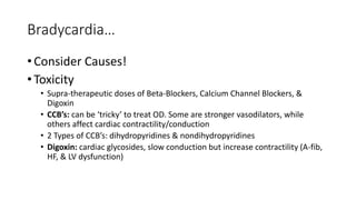
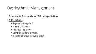
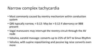
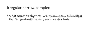
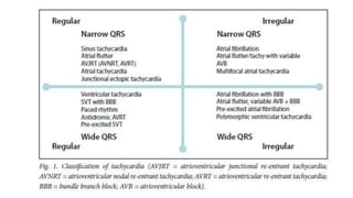
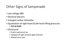
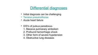
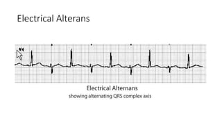
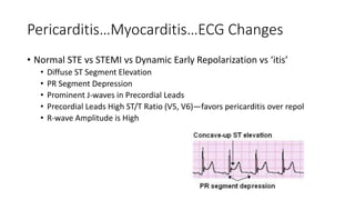
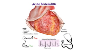
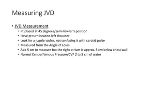
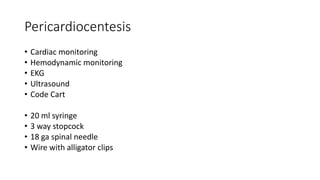
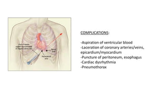
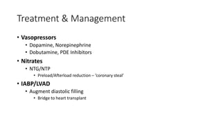
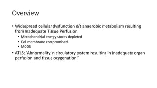
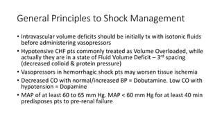
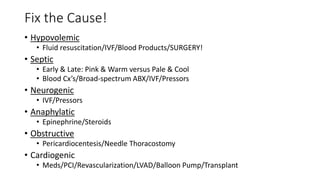
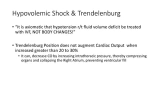
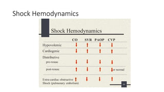
Acute Cardiovascular
Disorders
Initial Stabilization & Management
Presentation Topics
• Sudden Cardiac Arrest (SCA): Current Management Strategies
• Shock: Identification, Stratification, & Treatment
• Heart Failure: 1 Heart, 2 Pumps!
• Cardiopulmonary Hemodynamics of Shock
• Pericardial Tamponade: Identification & Treatment
• Continuous Cardiovascular Pharmacology: Principles & Practice
Hard & Fast!
• What have we learned?
• 250,000 SCA outside the Hospital
• 7.6% (?) ROSC-does not correlate with Survival-to-D/C, OR—Neuro Intact!
• Compressions: Hard and Fast 100 to 120. UNINTERRUPTED!
• 30:2
• Push first? Defib when?
• Differences in Witnessed vs Unwitnessed CA
Limited Progress over 30 Years!
• Access to AED’s & Therapeutic
Hypothermia
• Selected populations
• National Effort Focus: Delivery of
HIGH-QUALITY CPR
• Greatest Improvement/No
Improvement:
Northeast/Midwest
• Biphasic/Double Sequential?
• Role of Mechanical Compression
• Studies reveal 7% increase in
Survival-to-Hospital
• 2 MINUTES! 5 Cycles! No Pulse
Checks!
• Anti-dysrhythmics? Timing
remains unclear…
• Vasopressors?
• Timing is clear, efficacy unclear…
CRM
• Crisis Resource Management
• Leadership & Communication
• Team Leader
• ‘Closed-Loop’
• Team Members
• Delegation of duties
Defibrillation
• Biphasic
• Limit time to defib
• Quicker the better in witnessed OHCA!
• Max energy level
• Increased efficacy at lower energy levels
• 120 to 200 joules
• Different manufacturers deliver different
biphasic shock
Double Sequential Defibrillation?
• Perform after Vfib persists after 5 shocks
• The change in vectors presumed to ‘set up’ for the next shock
• Reduces Vfib/Vtach defib threshold
• Access to Procainamide may be limited
• Considered a ‘Hail Mary’ intervention for refractory/recurrent VF/VT
Double Sequential Defib Pad Placement
VF/Pulseless VT
• Epinephrine, after 2 minutes CPR, defib
• Admin 1 mg every 3 to 5 minutes
• ‘Some’ researchers ‘theorize’ high circulating levels of catecholamines
may be harmful to patients who experience ROSC
• Increased afterload/SVR can decrease Cardiac Output
• ‘Some’ studies have raised doubts about efficacy of Epi
• Perhaps lower doses/longer intervals
Refractory VF/VT
• Anti-dysrhythmics?
• Evidence suggests meds provide little survival benefit
• Timing not specified in protocols
• Evidence suggests administration after 2nd unsuccessful defib, in anticipation
of 3rd shock
• Amiodarone: 300mg IVP, followed by 150mg IVP (post-ROSC hypotension)
• Lidocaine: 1 to 1.5mg IVP, followed by 0.5 to 0.75mg/kg every 5 to 10 minutes
• Magnesium: 2gm IVP, followed by maintenance, consistent for polymorphic
VT/Torsade de Pointes
• Procanimide?
Asystole/PEA
• Addressed together
• Absence of mechanical cardiac contracture
• Both non-perfusing rhythms depending upon EXCELLENT CPR!
• Require Rapid Reversal of underlying causes
• H’s & T’s
• Hypoxia, hypercarbia, hyperkalemia, hypokalemia, hemothorax, hypoglycemia,
hypotension, tension pneumo, toxins, tamponade, thrombosis, thromboembolism
H’s & T’s
• H’s
• Hypoxia
• Hydrogen Ion (acidosis)
• Hypothermia
• Hyper/hypo-kalemia
• Hypovolemia
• Hypoglycemia
• T’s
• Toxins
• Tamponade
• Tension pnx
• Thrombosis
• Cardiac
• PE
Bradycardia
• ‘Generally’ not symptomatic until HR < 50 BPM
• Hypoxemia common cause
• Signs & Symptoms = hypotension, altered LOC, dyspnea, chest pain
• Atropine: 0.5mg IVP, 3 to 5 minutes up to 3mg
• Myocardial Demand: use cautiously in cardiac ischemia/NSTEMI/STEMI
• Ineffective in transplanted hearts
• Ineffective in high-degree AV block (Mobitz II/3rd degree AVB)
• TCP: do not delay
• Chronotropic Infusions: Epinephrine & Norepinephrine
• Role of Calcium in bradycardia/shock?
Bradycardia…
• Consider Causes!
• Toxicity
• Supra-therapeutic doses of Beta-Blockers, Calcium Channel Blockers, &
Digoxin
• CCB’s: can be ‘tricky’ to treat OD. Some are stronger vasodilators, while
others affect cardiac contractility/conduction
• 2 Types of CCB’s: dihydropyridines & nondihydropyridines
• Digoxin: cardiac glycosides, slow conduction but increase contractility (A-fib,
HF, & LV dysfunction)
Tachycardia
• ‘Generally’ not symptomatic until HR > 150 BPM
• No treatment for Sinus Tachycardia: focus on cause, Anemia, Shock,
Fever…
• Approach similar to Bradycardia: assess stability, signs & symptoms of
decreased Cardiac Output (CO)
• Unstable = immediate synchronized cardioversion!
• Stable = Adenosine: 6mg IVP, 12mg IVP X2
Dysrhythmia Management
• Systematic Approach to ECG Interpretation
• 5 Questions
• Regular or Irregular?
• Stable, Unstable?
• Too Fast, Too Slow?
• Complex Narrow or Wide?
• Is there a P wave for every QRS?
Narrow complex tachycardia
• Most commonly caused by reentry mechanism within conduction
system
• QRS typically narrow, < 0.12: May be > 0.12 if aberrancy or BBB
present
• Vagal maneuvers may interrupt the reentry circuit through the AV
node
• Valsalva, carotid massage: converts up to 25% of SVT to Sinus Rhythm
• Valsalva, with supine repositioning and passive leg raise converts even
more
Regular narrow complex
• Adenosine
• Administer into large, proximal vein
• Followed by 20mL flush
• Raise the extremity
• Repeat second/third time
• Warn/prepare the patient: flushing, dyspnea, chest discomfort. Continuous
ECG monitoring
• Larger doses (18mg) for pts on Theophylline/Caffeine
• Smaller doses (3mg) for pts on Tegretol. Transplanted hearts, central line
Conversion failure
• Consider other etiologies for the rhythm
• Atrial Flutter or Non-reentry SVT
• Administer CCB or BB: Cardizem/Verapamil or Metoprolol/Labetalol
Irregular narrow complex
• Most common rhythms: Afib, Multifocal Atrial Tach (MAT), &
Sinus Tachycardia with frequent, premature atrial beats
This page left blank intentionally…
Pericardial Tamponade
• Pericardial sac normally has ‘small’ amount of fluid
• Sac can hold up to 2 liters of fluid if effusion is chronic
• Can take less < 50 ml of blood/fluid to cause an acute tamponade
• Fluid or blood accumulates between visceral & parietal pericardium
• Causes myocardial constriction, resulting in decreased Cardiac Output
• Can lead to Obstructive Shock, decreased ventricular filling, & right
atrial collapse
• If left untreated can lead to PEA
• May occur from Blunt or Penetrating injuries (less common)
• Acute Pericarditis (EKG changes?)
• ESRD/Neoplasm
Beck’s Triad
• Hypotension
• Muffled/distant Heart Sounds
• Jugular Venous Distention
• Decreased CO/Hypotension
• CO = HR x SV
• Patient may exhibit Pulsus Paradoxus
• Pulse intensity Decreases with inspiration
• Decrease in Systolic BP of 10 mm Hg during inspiration
Removal of as little of 5 to
10 ml’s of blood/fluid can
result in an increase of SV
to 25% to 50%!
Other Signs of tamponade
• Low voltage QRS
• Electrical alterans
• Enlarged cardiac silhouette
• Equalization of right-heart & left-heart filling pressures
• CVP & PCWP
• Ultrasound
• Fluid in pericarial sac
• Collapse of right artria & right ventricle
• Dilated vena cava
Electrical Alterans
Pericarditis…Myocarditis…ECG Changes
• Normal STE vs STEMI vs Dynamic Early Repolarization vs ‘itis’
• Diffuse ST Segment Elevation
• PR Segment Depression
• Prominent J-waves in Precordial Leads
• Precordial Leads High ST/T Ratio (V5, V6)—favors pericarditis over repol
• R-wave Amplitude is High
Measuring JVD
• JVD Measurement
• Pt placed at 45 degrees/semi-fowler’s position
• Have pt turn head to left shoulder
• Look for a jugular pulse, not confusing it with carotid pulse
• Measured from the Angle of Louis
• Add 5 cm to measure b/c the right atrium is approx. 5 cm below chest wall
• Normal Central Venous Pressure/CVP 2 to 5 cm of water
Clinical Scenario
• 42 yr old male patient arrives POV to the ED
• Physical examination reveals multiple stab wounds to left & right chest
• Attack occurred approx. 1 hour prior. No pmhx, no meds, no allergies
• Patient is diaphoretic, but speaking in clear, full sentences
• Lung sounds were diminished on left, left chest tube placed/returned 1200cc
blood
• FAST exam was negative for blood/fluid
• BP 90/50
• JVD at 14 cm
• Most likely cause of hypotension/decreased CO?
Pericardiocentesis
• Cardiac monitoring
• Hemodynamic monitoring
• EKG
• Ultrasound
• Code Cart
• 20 ml syringe
• 3 way stopcock
• 18 ga spinal needle
• Wire with alligator clips
Procedure
• Immediate sub-xiphoid approach
• Insert needle with catheter in place, removing the needle once placement is
verified via ultrasound
• Attach stopcock to catheter and advance towards left shoulder while
aspirating continuously
• Monitor with ultrasound and EKG
• IF U/S not available, utilized wire with alligator clip, attach to a precordial
lead.
• If the need is to far, ST segment elevation will be noted on EKG tracing.
Withdraw until it disappears. Aspirate blood/fluid.
• Alternative: PARASTERNAL APPROACH (5 ICS, LSB)
COMPLICATIONS:
-Aspiration of ventricular blood
-Laceration of coronary arteries/veins,
epicardium/myocardium
-Puncture of peritoneum, esophagus
-Cardiac dysrhythmia
-Pneumothorax
Aftercare
• X-ray to rule out pleural effusion, PXT
• Continue to monitor for repeat accumulations in pericardial sac
• Monitor for cardiac dysrhythmias, tamponade, hypotension, etc
This page left blank intentionally…
Heart Failure: 1 Heart, 2 Pumps
• Clinical syndrome of decreased cardiac function
• One or both ventricles are unable to maintain adequate CO
• Typically refers to LV
• Can be bi-ventricular, systolic/diastolic
• Systole: Ejection of blood from the ventricle
• Diastole: Relaxation of the ventricle, ventricular filling
To be ‘Preserved,’ or not to be…that is the ?
Heart Failure
• Systolic Dysfunction: Weak ventricle, difficulty pumping blood
forward
• Diastolic Dysfunction Not enough filling time to maintain stroke
volume, myocardial coronary arteries
Defined
• Sustained hypotension of SBP < 90 mm Hg lasting > 30 minutes with
evidence of tissue hypoperfusion, despite adequate LV filling
pressures
• Insufficient tissue perfusion = oliguria, cool extremities, altered LOC
Mortality/Morbidity
• Most common initiating event in Cardiogenic Shock in AMI, producing
marked decrease in contractility (reduced EF/CO)
• Prevalence 8.6% of pts with STEMI
• Leading COD in AMI
• Overall in-hosp mortality 57%
• Outcomes significantly improve with rapid revascularization
Normal Cardiac Cycle
• Opening/closing of atrio-
ventricular & semi-lunar valves
• When pressure increases &
exceeds, valves open
• Ventricular Filling occurs
• 2 phases:Systole/Diastole
• Isovolumic Contraction
• Ultimately: Pressure to Volume
& Volume to Compliance affect
Cardiac Output
Compensatory Mechanisms
• Increased HR & Contractility
• Arterial constriction
• Venoconstriction
• Retention of salt & water
• Increased blood glucose & lipids
• Release of inflammatory mediators
RAAS
• Renin-Angiotension-Aldosterone System
• Compensation for decreased cardiac output
• Sustained/prolonged compensation results in excessive fluid/CHF
Dysfunction
• Left-sided Heart Failure/Systolic Dysfunction: LV function, decreased
ability to pump blood forward
• Ejection fraction < 60%, normal >65%
• Left-sided Heart Failure/Diastolic Dysfunction: Normal EF
• Compensation:
• Left atrium & pulmonary venous pressure increase, pulmonary congestion &
edema result
• Increased pressure in the pulmonary system leads to right heart failure,
systemic congestion, & edema develops
Treatment & Management
• Vasopressors
• Dopamine, Norepinephrine
• Dobutamine, PDE Inhibitors
• Nitrates
• NTG/NTP
• Preload/Afterload reduction – ‘coronary steal’
• IABP/LVAD
• Augment diastolic filling
• Bridge to heart transplant
Caveats
• Reductions in afterload will increase CO
• High doses of systemic vasoconstrictors will produce increased
afterload/SVR, paradoxically compromising CO further
• In pts with refractory shock on high levels of vasopressors, decrease
dosages incrementally to increase CO & SBP
This page left blank intentionally…
Shock…The Rude Unhinging of the Machinery
of Life…
• Definition
• Inadequate tissue perfusion/oxygenation
• Multiple Forms
• Hypovolemic
• Distributive
• Obstructive
• 3 Stages
• Compensatory
• Decompensated
• Irreversible
Overview
• Widespread cellular dysfunction d/t anaerobic metabolism resulting
from Inadequate Tissue Perfusion
• Mitrochondrial energy stores depleted
• Cell membrane compromised
• MODS
• ATLS: “Abnormality in circulatory system resulting in inadequate organ
perfusion and tissue oxygenation.”
Shock
• Inadequate tissue oxygenation &
perfusion…
• Types & Subtypes:
• Hypovolemic
• Distributive
• Obstructive
• Hypovolemic
• Hemorrhagic
• Pressure Problem!
• Distributive
• Septic
• Anaphylatic
• Neurogenic
• Pipes Problem!
• Obstructive
• Tamponade
• Cardiogenic
• Tension pnx
• Pump Problem!
Caveats!
• Look for an identifiable cause…treatment depends on dx
• Tachycardia is the earliest measurable circulatory sign of shock!
• PEARL: Any pt that is diaphoretic & tachycardic is presumed to be in
shock until proven otherwise
Serum Lactate?
• Normal < 2 mmol; <5 mmol poor prognosis
• What’s the latest on lactate levels?
• Relationship between serum lactate levels and epinephrine…
General Principles to Shock Management
• Intravascular volume deficits should be initially tx with isotonic fluids
before administering vasopressors
• Hypotensive CHF pts commonly treated as Volume Overloaded, while
actually they are in a state of Fluid Volume Deficit – 3rd spacing
(decreased colloid & protein pressure)
• Vasopressors in hemorrhagic shock pts may worsen tissue ischemia
• Decreased CO with normal/increased BP = Dobutamine. Low CO with
hypotension = Dopamine
• MAP of at least 60 to 65 mm Hg. MAP < 60 mm Hg for at least 40 min
predisposes pts to pre-renal failure
Management
• Rapidly identify!
• Treat specifically!
• Treat aggressively!
Any patient diaphoretic & tachycardic is in
shock until proven otherwise!
Fix the Cause!
• Hypovolemic
• Fluid resuscitation/IVF/Blood Products/SURGERY!
• Septic
• Early & Late: Pink & Warm versus Pale & Cool
• Blood Cx’s/Broad-spectrum ABX/IVF/Pressors
• Neurogenic
• IVF/Pressors
• Anaphylatic
• Epinephrine/Steroids
• Obstructive
• Pericardiocentesis/Needle Thoracostomy
• Cardiogenic
• Meds/PCI/Revascularization/LVAD/Balloon Pump/Transplant
Hypovolemic Shock & Trendelenburg
• “It is axiomatic that hypotension r/t fluid volume deficit be treated
with IVF, NOT BODY CHANGES!”
• Trendelenburg Position does not augment Cardiac Output when
increased greater than 20 to 30%
• It can, decrease CO by increasing intrathoracic pressure, thereby compressing
organs and collapsing the Right Atrium, preventing ventricular fill
Dangers of Compensated Shock?
• Value of Vital Signs?
• BP?
• HR?
• RR?
• Skin?
• Mentation?
• Urine output?
This page left blank intentionally…
Shock Hemodynamics
This page left blank intentionally…
Vasoactive Medications
• Indications/Contraindications
• Classes/Effects
• Hemodynamic profile
• Titration
General Principles of Use
• One drug, many receptors!
• Alpha-1
• Beta-1, beta-2
• Not a definitive treatment!
• Supports MAP & tissue & end-
organ perfusion
• Dose-responsive curve
• Dopamine, example…
• Direct versus Reflex Actions
• Example,bradycardia associated
with pure alpha-adrenergic
receptor agonists
• Do not “micromanage” every BP
• Causes lability in vital signs
• Medications specifically with short
half-like, e.g., Sodium Nitroprusside
Vasopressor Caveats
• Fluid resuscitate with 2 liters of Isotonic IVF prior to starting pressors
• Increased afterload/SVR will decrease cardiac output/compromise
perfusion
• Beta-1 agonists can potentiate cardiac dysrhythmias, tachycardia &
worsen myocardial ischemia, etc
• Sometimes ‘less is more…’
• Best if infused via large, antecubital veins/CVC not necessary
• Cardiac output
• 4-8 lpm
• HR x SV = CO
• SV
• Preload
• Afterload
• Contractility
• Viscosity
Vasopressors
• Epinephrine/Adrenalin
• Norepinephrine/Levophed©
“Levophed, Leave ‘em Dead”
• Neosynephrine/Phenylephrine©
• Dopamine/Intropin
• Dobutamine
• Phosphodiesterase Inhibitors
• Systemic vasodilator
• Increased end-organ perfusion
• Decreased afterload/SVR
• Decreased arterial pressure
• may cause hypotension when
initiating infusion
• Cardiopulmonary
• + chronotropic/+dromotropic
• + inotropic/ contractility
• Decreased preload
Vasopressors
• Epinephrine
• 2-20 mcg/min
• Effect on serum lactate levels?
• 8mg in 250 ml’s
• Beta-1 agonist
• Mixed alpha-beta
• More beta than alpha
• Norepinephrine
• 2-20 mcg/min
• 8 mg in 250 ml’s
• Mixed alpha-beta
• More alpha than beta
• Phenylephrine
• Large therapeutic window
• 10 -200 mcg/min
• Reserved for late shock
• Reflex bradycardia
• 100 mg in 250 ml’s
• Pure alpha-agonist
Vasodilators: Mediated via nitric oxide
• Nitroglycerin
• 2-200 mcg/min
• Use low-absorption IV tubing
• Reflex tachycardia!
• Sodium Nitroprusside (Nipride©)
• 0.2 -10 mcg/kg/min
• Coronary Steal Phenom
• Protect from light!
• 10 mcg/kg/min MAX dose..10
minute limit!!
• Breaks down to cyanide! (thiosulfate)
• Dilates
• Veins
• Arteries
• Cardiac Output
• Augments stroke volume
• Decreases afterload
• Ventricular Filling Pressures
• Preload
• CVP (right heart)
• PCWP (left heart)
Vasoactive Management in Heart Failure
• Concomitant Strategies
• Utilizing a vasopressor +
vasodilator
= anytime afterload is decreased,
cardiac output will increase
- Preserved Ejection Fraction
- Dobutamine Gtt + Nipride Gtt
- Sounds counterintuitive
End of presentation…

Acute cardiovascular disorders

  • 1.
  • 2.
    Presentation Topics • SuddenCardiac Arrest (SCA): Current Management Strategies • Shock: Identification, Stratification, & Treatment • Heart Failure: 1 Heart, 2 Pumps! • Cardiopulmonary Hemodynamics of Shock • Pericardial Tamponade: Identification & Treatment • Continuous Cardiovascular Pharmacology: Principles & Practice
  • 3.
    Hard & Fast! •What have we learned? • 250,000 SCA outside the Hospital • 7.6% (?) ROSC-does not correlate with Survival-to-D/C, OR—Neuro Intact! • Compressions: Hard and Fast 100 to 120. UNINTERRUPTED! • 30:2 • Push first? Defib when? • Differences in Witnessed vs Unwitnessed CA
  • 4.
    Limited Progress over30 Years! • Access to AED’s & Therapeutic Hypothermia • Selected populations • National Effort Focus: Delivery of HIGH-QUALITY CPR • Greatest Improvement/No Improvement: Northeast/Midwest • Biphasic/Double Sequential? • Role of Mechanical Compression • Studies reveal 7% increase in Survival-to-Hospital • 2 MINUTES! 5 Cycles! No Pulse Checks! • Anti-dysrhythmics? Timing remains unclear… • Vasopressors? • Timing is clear, efficacy unclear…
  • 5.
    CRM • Crisis ResourceManagement • Leadership & Communication • Team Leader • ‘Closed-Loop’ • Team Members • Delegation of duties
  • 6.
    Defibrillation • Biphasic • Limittime to defib • Quicker the better in witnessed OHCA! • Max energy level • Increased efficacy at lower energy levels • 120 to 200 joules • Different manufacturers deliver different biphasic shock
  • 9.
    Double Sequential Defibrillation? •Perform after Vfib persists after 5 shocks • The change in vectors presumed to ‘set up’ for the next shock • Reduces Vfib/Vtach defib threshold • Access to Procainamide may be limited • Considered a ‘Hail Mary’ intervention for refractory/recurrent VF/VT
  • 10.
  • 11.
    VF/Pulseless VT • Epinephrine,after 2 minutes CPR, defib • Admin 1 mg every 3 to 5 minutes • ‘Some’ researchers ‘theorize’ high circulating levels of catecholamines may be harmful to patients who experience ROSC • Increased afterload/SVR can decrease Cardiac Output • ‘Some’ studies have raised doubts about efficacy of Epi • Perhaps lower doses/longer intervals
  • 12.
    Refractory VF/VT • Anti-dysrhythmics? •Evidence suggests meds provide little survival benefit • Timing not specified in protocols • Evidence suggests administration after 2nd unsuccessful defib, in anticipation of 3rd shock • Amiodarone: 300mg IVP, followed by 150mg IVP (post-ROSC hypotension) • Lidocaine: 1 to 1.5mg IVP, followed by 0.5 to 0.75mg/kg every 5 to 10 minutes • Magnesium: 2gm IVP, followed by maintenance, consistent for polymorphic VT/Torsade de Pointes • Procanimide?
  • 13.
    Asystole/PEA • Addressed together •Absence of mechanical cardiac contracture • Both non-perfusing rhythms depending upon EXCELLENT CPR! • Require Rapid Reversal of underlying causes • H’s & T’s • Hypoxia, hypercarbia, hyperkalemia, hypokalemia, hemothorax, hypoglycemia, hypotension, tension pneumo, toxins, tamponade, thrombosis, thromboembolism
  • 14.
    H’s & T’s •H’s • Hypoxia • Hydrogen Ion (acidosis) • Hypothermia • Hyper/hypo-kalemia • Hypovolemia • Hypoglycemia • T’s • Toxins • Tamponade • Tension pnx • Thrombosis • Cardiac • PE
  • 15.
    Bradycardia • ‘Generally’ notsymptomatic until HR < 50 BPM • Hypoxemia common cause • Signs & Symptoms = hypotension, altered LOC, dyspnea, chest pain • Atropine: 0.5mg IVP, 3 to 5 minutes up to 3mg • Myocardial Demand: use cautiously in cardiac ischemia/NSTEMI/STEMI • Ineffective in transplanted hearts • Ineffective in high-degree AV block (Mobitz II/3rd degree AVB) • TCP: do not delay • Chronotropic Infusions: Epinephrine & Norepinephrine • Role of Calcium in bradycardia/shock?
  • 16.
    Bradycardia… • Consider Causes! •Toxicity • Supra-therapeutic doses of Beta-Blockers, Calcium Channel Blockers, & Digoxin • CCB’s: can be ‘tricky’ to treat OD. Some are stronger vasodilators, while others affect cardiac contractility/conduction • 2 Types of CCB’s: dihydropyridines & nondihydropyridines • Digoxin: cardiac glycosides, slow conduction but increase contractility (A-fib, HF, & LV dysfunction)
  • 17.
    Tachycardia • ‘Generally’ notsymptomatic until HR > 150 BPM • No treatment for Sinus Tachycardia: focus on cause, Anemia, Shock, Fever… • Approach similar to Bradycardia: assess stability, signs & symptoms of decreased Cardiac Output (CO) • Unstable = immediate synchronized cardioversion! • Stable = Adenosine: 6mg IVP, 12mg IVP X2
  • 18.
    Dysrhythmia Management • SystematicApproach to ECG Interpretation • 5 Questions • Regular or Irregular? • Stable, Unstable? • Too Fast, Too Slow? • Complex Narrow or Wide? • Is there a P wave for every QRS?
  • 19.
    Narrow complex tachycardia •Most commonly caused by reentry mechanism within conduction system • QRS typically narrow, < 0.12: May be > 0.12 if aberrancy or BBB present • Vagal maneuvers may interrupt the reentry circuit through the AV node • Valsalva, carotid massage: converts up to 25% of SVT to Sinus Rhythm • Valsalva, with supine repositioning and passive leg raise converts even more
  • 20.
    Regular narrow complex •Adenosine • Administer into large, proximal vein • Followed by 20mL flush • Raise the extremity • Repeat second/third time • Warn/prepare the patient: flushing, dyspnea, chest discomfort. Continuous ECG monitoring • Larger doses (18mg) for pts on Theophylline/Caffeine • Smaller doses (3mg) for pts on Tegretol. Transplanted hearts, central line
  • 21.
    Conversion failure • Considerother etiologies for the rhythm • Atrial Flutter or Non-reentry SVT • Administer CCB or BB: Cardizem/Verapamil or Metoprolol/Labetalol
  • 22.
    Irregular narrow complex •Most common rhythms: Afib, Multifocal Atrial Tach (MAT), & Sinus Tachycardia with frequent, premature atrial beats
  • 25.
    This page leftblank intentionally…
  • 26.
    Pericardial Tamponade • Pericardialsac normally has ‘small’ amount of fluid • Sac can hold up to 2 liters of fluid if effusion is chronic • Can take less < 50 ml of blood/fluid to cause an acute tamponade • Fluid or blood accumulates between visceral & parietal pericardium • Causes myocardial constriction, resulting in decreased Cardiac Output • Can lead to Obstructive Shock, decreased ventricular filling, & right atrial collapse • If left untreated can lead to PEA • May occur from Blunt or Penetrating injuries (less common) • Acute Pericarditis (EKG changes?) • ESRD/Neoplasm
  • 28.
    Beck’s Triad • Hypotension •Muffled/distant Heart Sounds • Jugular Venous Distention • Decreased CO/Hypotension • CO = HR x SV • Patient may exhibit Pulsus Paradoxus • Pulse intensity Decreases with inspiration • Decrease in Systolic BP of 10 mm Hg during inspiration Removal of as little of 5 to 10 ml’s of blood/fluid can result in an increase of SV to 25% to 50%!
  • 29.
    Other Signs oftamponade • Low voltage QRS • Electrical alterans • Enlarged cardiac silhouette • Equalization of right-heart & left-heart filling pressures • CVP & PCWP • Ultrasound • Fluid in pericarial sac • Collapse of right artria & right ventricle • Dilated vena cava
  • 31.
  • 36.
    Pericarditis…Myocarditis…ECG Changes • NormalSTE vs STEMI vs Dynamic Early Repolarization vs ‘itis’ • Diffuse ST Segment Elevation • PR Segment Depression • Prominent J-waves in Precordial Leads • Precordial Leads High ST/T Ratio (V5, V6)—favors pericarditis over repol • R-wave Amplitude is High
  • 40.
    Measuring JVD • JVDMeasurement • Pt placed at 45 degrees/semi-fowler’s position • Have pt turn head to left shoulder • Look for a jugular pulse, not confusing it with carotid pulse • Measured from the Angle of Louis • Add 5 cm to measure b/c the right atrium is approx. 5 cm below chest wall • Normal Central Venous Pressure/CVP 2 to 5 cm of water
  • 41.
    Clinical Scenario • 42yr old male patient arrives POV to the ED • Physical examination reveals multiple stab wounds to left & right chest • Attack occurred approx. 1 hour prior. No pmhx, no meds, no allergies • Patient is diaphoretic, but speaking in clear, full sentences • Lung sounds were diminished on left, left chest tube placed/returned 1200cc blood • FAST exam was negative for blood/fluid • BP 90/50 • JVD at 14 cm • Most likely cause of hypotension/decreased CO?
  • 42.
    Pericardiocentesis • Cardiac monitoring •Hemodynamic monitoring • EKG • Ultrasound • Code Cart • 20 ml syringe • 3 way stopcock • 18 ga spinal needle • Wire with alligator clips
  • 43.
    Procedure • Immediate sub-xiphoidapproach • Insert needle with catheter in place, removing the needle once placement is verified via ultrasound • Attach stopcock to catheter and advance towards left shoulder while aspirating continuously • Monitor with ultrasound and EKG • IF U/S not available, utilized wire with alligator clip, attach to a precordial lead. • If the need is to far, ST segment elevation will be noted on EKG tracing. Withdraw until it disappears. Aspirate blood/fluid. • Alternative: PARASTERNAL APPROACH (5 ICS, LSB)
  • 44.
    COMPLICATIONS: -Aspiration of ventricularblood -Laceration of coronary arteries/veins, epicardium/myocardium -Puncture of peritoneum, esophagus -Cardiac dysrhythmia -Pneumothorax
  • 45.
    Aftercare • X-ray torule out pleural effusion, PXT • Continue to monitor for repeat accumulations in pericardial sac • Monitor for cardiac dysrhythmias, tamponade, hypotension, etc
  • 46.
    This page leftblank intentionally…
  • 47.
    Heart Failure: 1Heart, 2 Pumps • Clinical syndrome of decreased cardiac function • One or both ventricles are unable to maintain adequate CO • Typically refers to LV • Can be bi-ventricular, systolic/diastolic • Systole: Ejection of blood from the ventricle • Diastole: Relaxation of the ventricle, ventricular filling
  • 48.
    To be ‘Preserved,’or not to be…that is the ?
  • 49.
    Heart Failure • SystolicDysfunction: Weak ventricle, difficulty pumping blood forward • Diastolic Dysfunction Not enough filling time to maintain stroke volume, myocardial coronary arteries
  • 55.
    Defined • Sustained hypotensionof SBP < 90 mm Hg lasting > 30 minutes with evidence of tissue hypoperfusion, despite adequate LV filling pressures • Insufficient tissue perfusion = oliguria, cool extremities, altered LOC
  • 56.
    Mortality/Morbidity • Most commoninitiating event in Cardiogenic Shock in AMI, producing marked decrease in contractility (reduced EF/CO) • Prevalence 8.6% of pts with STEMI • Leading COD in AMI • Overall in-hosp mortality 57% • Outcomes significantly improve with rapid revascularization
  • 57.
    Normal Cardiac Cycle •Opening/closing of atrio- ventricular & semi-lunar valves • When pressure increases & exceeds, valves open • Ventricular Filling occurs • 2 phases:Systole/Diastole • Isovolumic Contraction • Ultimately: Pressure to Volume & Volume to Compliance affect Cardiac Output
  • 58.
    Compensatory Mechanisms • IncreasedHR & Contractility • Arterial constriction • Venoconstriction • Retention of salt & water • Increased blood glucose & lipids • Release of inflammatory mediators
  • 59.
    RAAS • Renin-Angiotension-Aldosterone System •Compensation for decreased cardiac output • Sustained/prolonged compensation results in excessive fluid/CHF
  • 61.
    Dysfunction • Left-sided HeartFailure/Systolic Dysfunction: LV function, decreased ability to pump blood forward • Ejection fraction < 60%, normal >65% • Left-sided Heart Failure/Diastolic Dysfunction: Normal EF • Compensation: • Left atrium & pulmonary venous pressure increase, pulmonary congestion & edema result • Increased pressure in the pulmonary system leads to right heart failure, systemic congestion, & edema develops
  • 62.
    Treatment & Management •Vasopressors • Dopamine, Norepinephrine • Dobutamine, PDE Inhibitors • Nitrates • NTG/NTP • Preload/Afterload reduction – ‘coronary steal’ • IABP/LVAD • Augment diastolic filling • Bridge to heart transplant
  • 63.
    Caveats • Reductions inafterload will increase CO • High doses of systemic vasoconstrictors will produce increased afterload/SVR, paradoxically compromising CO further • In pts with refractory shock on high levels of vasopressors, decrease dosages incrementally to increase CO & SBP
  • 64.
    This page leftblank intentionally…
  • 65.
    Shock…The Rude Unhingingof the Machinery of Life… • Definition • Inadequate tissue perfusion/oxygenation • Multiple Forms • Hypovolemic • Distributive • Obstructive • 3 Stages • Compensatory • Decompensated • Irreversible
  • 66.
    Overview • Widespread cellulardysfunction d/t anaerobic metabolism resulting from Inadequate Tissue Perfusion • Mitrochondrial energy stores depleted • Cell membrane compromised • MODS • ATLS: “Abnormality in circulatory system resulting in inadequate organ perfusion and tissue oxygenation.”
  • 67.
    Shock • Inadequate tissueoxygenation & perfusion… • Types & Subtypes: • Hypovolemic • Distributive • Obstructive • Hypovolemic • Hemorrhagic • Pressure Problem! • Distributive • Septic • Anaphylatic • Neurogenic • Pipes Problem! • Obstructive • Tamponade • Cardiogenic • Tension pnx • Pump Problem!
  • 68.
    Caveats! • Look foran identifiable cause…treatment depends on dx • Tachycardia is the earliest measurable circulatory sign of shock! • PEARL: Any pt that is diaphoretic & tachycardic is presumed to be in shock until proven otherwise
  • 69.
    Serum Lactate? • Normal< 2 mmol; <5 mmol poor prognosis • What’s the latest on lactate levels? • Relationship between serum lactate levels and epinephrine…
  • 70.
    General Principles toShock Management • Intravascular volume deficits should be initially tx with isotonic fluids before administering vasopressors • Hypotensive CHF pts commonly treated as Volume Overloaded, while actually they are in a state of Fluid Volume Deficit – 3rd spacing (decreased colloid & protein pressure) • Vasopressors in hemorrhagic shock pts may worsen tissue ischemia • Decreased CO with normal/increased BP = Dobutamine. Low CO with hypotension = Dopamine • MAP of at least 60 to 65 mm Hg. MAP < 60 mm Hg for at least 40 min predisposes pts to pre-renal failure
  • 71.
    Management • Rapidly identify! •Treat specifically! • Treat aggressively!
  • 72.
    Any patient diaphoretic& tachycardic is in shock until proven otherwise!
  • 73.
    Fix the Cause! •Hypovolemic • Fluid resuscitation/IVF/Blood Products/SURGERY! • Septic • Early & Late: Pink & Warm versus Pale & Cool • Blood Cx’s/Broad-spectrum ABX/IVF/Pressors • Neurogenic • IVF/Pressors • Anaphylatic • Epinephrine/Steroids • Obstructive • Pericardiocentesis/Needle Thoracostomy • Cardiogenic • Meds/PCI/Revascularization/LVAD/Balloon Pump/Transplant
  • 74.
    Hypovolemic Shock &Trendelenburg • “It is axiomatic that hypotension r/t fluid volume deficit be treated with IVF, NOT BODY CHANGES!” • Trendelenburg Position does not augment Cardiac Output when increased greater than 20 to 30% • It can, decrease CO by increasing intrathoracic pressure, thereby compressing organs and collapsing the Right Atrium, preventing ventricular fill
  • 75.
    Dangers of CompensatedShock? • Value of Vital Signs? • BP? • HR? • RR? • Skin? • Mentation? • Urine output?
  • 76.
    This page leftblank intentionally…
  • 77.
  • 81.
    This page leftblank intentionally…
  • 82.
    Vasoactive Medications • Indications/Contraindications •Classes/Effects • Hemodynamic profile • Titration
  • 83.
    General Principles ofUse • One drug, many receptors! • Alpha-1 • Beta-1, beta-2 • Not a definitive treatment! • Supports MAP & tissue & end- organ perfusion • Dose-responsive curve • Dopamine, example… • Direct versus Reflex Actions • Example,bradycardia associated with pure alpha-adrenergic receptor agonists • Do not “micromanage” every BP • Causes lability in vital signs • Medications specifically with short half-like, e.g., Sodium Nitroprusside
  • 84.
    Vasopressor Caveats • Fluidresuscitate with 2 liters of Isotonic IVF prior to starting pressors • Increased afterload/SVR will decrease cardiac output/compromise perfusion • Beta-1 agonists can potentiate cardiac dysrhythmias, tachycardia & worsen myocardial ischemia, etc • Sometimes ‘less is more…’ • Best if infused via large, antecubital veins/CVC not necessary
  • 85.
    • Cardiac output •4-8 lpm • HR x SV = CO • SV • Preload • Afterload • Contractility • Viscosity
  • 91.
    Vasopressors • Epinephrine/Adrenalin • Norepinephrine/Levophed© “Levophed,Leave ‘em Dead” • Neosynephrine/Phenylephrine© • Dopamine/Intropin • Dobutamine • Phosphodiesterase Inhibitors • Systemic vasodilator • Increased end-organ perfusion • Decreased afterload/SVR • Decreased arterial pressure • may cause hypotension when initiating infusion • Cardiopulmonary • + chronotropic/+dromotropic • + inotropic/ contractility • Decreased preload
  • 92.
    Vasopressors • Epinephrine • 2-20mcg/min • Effect on serum lactate levels? • 8mg in 250 ml’s • Beta-1 agonist • Mixed alpha-beta • More beta than alpha • Norepinephrine • 2-20 mcg/min • 8 mg in 250 ml’s • Mixed alpha-beta • More alpha than beta • Phenylephrine • Large therapeutic window • 10 -200 mcg/min • Reserved for late shock • Reflex bradycardia • 100 mg in 250 ml’s • Pure alpha-agonist
  • 93.
    Vasodilators: Mediated vianitric oxide • Nitroglycerin • 2-200 mcg/min • Use low-absorption IV tubing • Reflex tachycardia! • Sodium Nitroprusside (Nipride©) • 0.2 -10 mcg/kg/min • Coronary Steal Phenom • Protect from light! • 10 mcg/kg/min MAX dose..10 minute limit!! • Breaks down to cyanide! (thiosulfate) • Dilates • Veins • Arteries • Cardiac Output • Augments stroke volume • Decreases afterload • Ventricular Filling Pressures • Preload • CVP (right heart) • PCWP (left heart)
  • 94.
    Vasoactive Management inHeart Failure • Concomitant Strategies • Utilizing a vasopressor + vasodilator = anytime afterload is decreased, cardiac output will increase - Preserved Ejection Fraction - Dobutamine Gtt + Nipride Gtt - Sounds counterintuitive
  • 95.

Editor's Notes

  • #4 Limited scientific evidence d/t high mortality of SCA. Mostly retrospective or anectdotal. Compressions of 100-119 resulted in only 35% inadequate quality. Over 120 ineffective compressions. Witnessed SCA defib first. If > 4 to 5 minutes, or unwitnessed, 2 minutes of CPR. Lesser energy with biphasic 150 to 200, 360 monophasic. 30:2, in the past we over-ventilated pts, reducing venous return, decreased cardiac output, decreased cerebral perfusion. Intubated patients receive no more than 6 to 10 breaths per minute, 600 ml tidal volume. Lower minute ventilations. Supraglottic airway until ROSC is achieved. All studies proved significant rates of neurologic function w
  • #6 CRM utilized in aviation industry. Team Leader refrains from performing procedures, unless necessary, leadership turned over until done. (Portable Radio Example)
  • #7 In witnessed arrest, perform defib ASAP! Decreased time to defib results in improved rates of successful conversion to a perfusing rhythm and patient survival.
  • #12 Premature Epi results in decreased ROSC/no improvement in survival. Excessive vasoconstriction can result in significant afterload thus reducing cardiac output.
  • #13 Pick one! Prodysrhythmic.
  • #16 Transplanted hearts lack vagal innervation. May be useful in narrow-complex 3rd AVB. Might buy you a few minutes. Block is at level of Node or Bundle of HIS.
  • #17 Massive med doses above may not be easily reversed with Atropine or TCP: May required transvenous pacing & expert cardiology consultation. Use of Dig has decreased substantially, however, the rate of toxicity has remained stable. In 2011, 132 patient toxicities, 27 died. Chance of toxicity increases in hypokalemia.
  • #18 Cardiac monitors reset to unsynchronized defib after every cardioversion, thus must hit ‘Sync’ button EVERYTIME! Sedate, if appropriate.
  • #29 Most patients will exhibit at least 1 of these symptoms, rarely all three, and mostly prior to cardiac arrest.
  • #35 ‘Hour Glass’ appearance
  • #41 Jugular pulse is normally non-pulsatile and consists of 2 beats per cardiac cycle, carotid 1 beat per cycle. If not palpable, push the abdominojugular reflex, or hepatojugular reflex.
  • #43 Relative contraindications: aortic dissection, bleeding disorder, myocardial rupture. A traumatic pericardial effusion may require resuscitative thoracotomy.
  • #44 Blind Approach associated with increased complications.