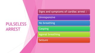
This document outlines cardiac life support and rhythm recognition. It begins with an introduction and then discusses pulseless arrest, including shockable rhythms like ventricular fibrillation and ventricular tachycardia, and non-shockable rhythms like pulseless electrical activity and asystole. It also covers tachyarrhythmias like sinus tachycardia, supraventricular tachycardia, atrial flutter, and atrial fibrillation. Bradyarrhythmias like sinus bradycardia and various types of heart block are also discussed. Medications used in resuscitation like adrenaline, atropine, amiodarone, and antiarrhythmics are outlined. The document concludes with information on transcut
1) Chest injuries can range from fractured ribs to collapsed lungs (pneumothorax) to sections of detached rib cage (flail segment) to open chest wounds.
2) Fractured ribs are the most common chest injury and are managed by positioning the victim in comfort, stabilizing the fracture, seeking medical aid, and monitoring for breathing issues.
3) A collapsed lung (pneumothorax) occurs when air enters the space between the lungs and chest wall, causing pain and breathing difficulty. Immediate medical help is needed along with oxygen support.
This document provides information on cardiac rhythm recognition and management according to advanced cardiac life support protocols. It defines key ECG parameters such as rate, rhythm, P waves, QRS complexes and intervals. It outlines a step-by-step approach to ECG interpretation and describes the treatment of various cardiac arrhythmias and arrests including defibrillation, cardioversion, transcutaneous pacing and management of conditions like supraventricular tachycardia, atrial fibrillation, ventricular tachycardia and bradyarrhythmias. Reversible causes of cardiac arrest are also listed.
Brady and tachyarrythmias diagnosis and managementAlphonse Aswin
This document provides information on interpreting electrocardiograms (ECGs) and diagnosing and treating bradyarrhythmias and tachyarrhythmias. It discusses the conduction pathway, interpreting ECG waveforms, common arrhythmias originating from the sinoatrial node, atria, atrioventricular node and ventricles. Diagnostic criteria and treatment options are provided for sinus bradycardia, sick sinus syndrome, junctional rhythms, ventricular escape rhythms, first-, second- and third-degree heart block, sinus tachycardia, atrial flutter, atrial fibrillation, multifocal atrial tachycardia, ventricular tachycardia, and ventricular fibrillation. The
This document discusses bradyarrhythmias, which are heart rhythms that are slower than normal. It defines key measurements in electrocardiograms including the P wave, PR interval, QRS complex, and QT interval. Common types of bradyarrhythmias include sinus bradycardia, junctional rhythm, and atrioventricular blocks. Treatment depends on whether the patient is stable or unstable and may involve medications to increase heart rate like atropine or transcutaneous pacing. The document provides details on evaluating and treating different types of bradycardic rhythms and their underlying causes.
This presentation describes the emergency department management of sinus tachycardia, supraventricular tachycardia, atrial flutter, atrial fibrillation, ventricular tachycardia and ventricular ectopic
This document discusses the management of peri-arrest arrhythmias. It defines arrhythmias and describes their assessment and general treatment options. It covers the management of specific arrhythmias like bradycardia and tachycardias. It also discusses the pharmacology of common antiarrhythmic drugs like amiodarone, atropine, digoxin. The document provides guidelines on stabilizing patients and restoring normal heart rhythm in peri-arrest settings.
A deep dive into management of cardiac arrhythmia from a Critical Care perspective. Covers brady- and tachyarrhythmias and management of both the stable and unstable patient.
This document discusses the identification and treatment of various cardiac arrhythmias including tachycardias like sinus tachycardia, atrial fibrillation, atrial flutter, multifocal atrial tachycardia, re-entry tachycardia, monomorphic ventricular tachycardia, polymorphic ventricular tachycardia, and ventricular fibrillation. It also discusses bradycardias like premature ventricular contractions, first degree AV block, second degree AV block types 1 and 2, and third degree AV block. For each arrhythmia, the document provides information on characteristics, identification, and treatment approaches.
anti arrhythmic drugs anaesthesiology cardiacNeelkantRaju
1. The document discusses antiarrhythmic drugs and their classification and mechanisms of action. It focuses on Class I drugs that work by blocking sodium channels.
2. Class IA drugs like quinidine and procainamide have a moderate effect on sodium channels and are used for supraventricular and ventricular arrhythmias. They can cause side effects like QT prolongation.
3. Class IB drugs like lidocaine have a weak effect on sodium channels and are the drugs of choice for ventricular arrhythmias. They are used for ventricular arrhythmias due to ischemia or digoxin toxicity.
1. The document discusses antiarrhythmic drugs and their classification and mechanisms of action.
2. Antiarrhythmic drugs are classified based on their effects on the cardiac action potential as Class I-IV. Class I drugs block sodium channels, Class II drugs are beta blockers, Class III drugs prolong the action potential duration, and Class IV drugs slow calcium channels.
3. Examples of different classes of drugs are provided along with their indications, mechanisms, effects, and side effects. The goal of antiarrhythmic therapy is to restore normal sinus rhythm and conduction while preventing more serious arrhythmias.
This presentation provides an overview of ventricular tachycardia (VT). VT is diagnosed based on an electrocardiogram showing three or more consecutive ventricular beats at a rate greater than 120 beats per minute with a wide QRS complex greater than 140 milliseconds. VT can be sustained for over 30 seconds or non-sustained for under 30 seconds. Causes of VT include myocardial infarction, cardiomyopathy, electrolyte abnormalities, and drugs. Treatment depends on hemodynamic stability and includes medications, cardioversion, ablation, or an implantable cardioverter defibrillator. Long term management focuses on underlying heart conditions, antiarrhythmic drugs, and reducing risk of sudden cardiac death.
ADVANCED CARDIAC LIFE SUPPORT (ACLS).pdfezrys54ety5
This document provides information about advanced cardiac life support (ACLS) procedures for treating cardiac emergencies. It outlines the adult chain of survival, including immediate recognition of cardiac arrest, early CPR, defibrillation, and advanced life support. It then describes shockable and non-shockable cardiac rhythms like ventricular fibrillation, asystole, and pulseless electrical activity. It also discusses defibrillation, cardioversion, transcutaneous pacing, and reversible causes and treatment of cardiac arrest.
Supraventricular arrhythmias are tachyarrhythmias with a rate over 90 beats per minute that are usually associated with symptoms. They are classified as narrow or wide complex tachycardias based on the width of the QRS complex on ECG. Narrow complex tachycardias (NCTs) require rapid activation of the ventricles through the His-Purkinje system and depend on AV node conduction. The most common types of NCTs are atrioventricular nodal reentrant tachycardia (AVNRT), atrioventricular reentrant tachycardia (AVRT), and atrial tachycardia. Initial management of stable NCTs involves
This document provides a tutorial on electrocardiography (ECG) including:
- The basics of ECG calibration and electrical impulse propagation.
- Descriptions of the P wave, QRS complex, ST segment, T wave, and other ECG components.
- Identification of abnormalities including hypertrophy, infarction, arrhythmias, and axis deviations.
- Guidance on interpreting ECG findings related to conditions like myocardial infarction, hypertrophy, and conduction blocks.
- Algorithms for managing cardiac arrest and arrhythmia rhythms.
- Recommended resources for further ECG education.
The document discusses tachycardia, including:
1) Tachycardia is an abnormally fast heart rate over 100 beats per minute that can be caused by irregular heart rhythms.
2) Tachycardias are classified based on their origin and heart rhythm characteristics and can be stable or unstable.
3) Unstable tachycardias require prompt treatment such as synchronized cardioversion while stable tachycardias can often be treated with vagal maneuvers or medications like adenosine.
This document provides an overview of cardiac arrhythmias for medical students. It begins by describing the normal conduction pathways in the heart and normal sinus rhythm on an electrocardiogram. It then classifies arrhythmias as rapid and regular, rapid and irregular, slow and regular, or slow and irregular based on heart rate and rhythm. Various types of tachycardias and bradycardias are defined, along with their typical electrocardiogram presentations and common causes. Causes, presentations, and treatments of atrial fibrillation, atrial flutter, supraventricular tachycardia, Wolff-Parkinson-White syndrome, heart block, and ventricular tachycardia are summarized. Catheter ablation techniques
The document describes several types of normal and abnormal cardiac rhythms as identified by an electrocardiogram (EKG or ECG). It provides descriptions and EKG criteria for normal sinus rhythm, sinus bradycardia, sinus tachycardia, premature atrial complexes, atrial fibrillation, atrial flutter, supraventricular tachycardia, premature junctional complexes, junctional rhythm, various degrees of atrioventricular block, premature ventricular contractions, ventricular bigeminy, and ventricular tachycardia.
In patients with acute myocardial infarction (AMI), arrhythmias are common. 90% will develop some form of cardiac arrhythmia, with 25% occurring within the first 24 hours. Arrhythmias can be classified as tachyarrhythmias (ventricular, supraventricular) or bradyarrhythmias (bradycardias, heart blocks). Life-threatening ventricular arrhythmias like ventricular fibrillation and ventricular tachycardia are treated with defibrillation or antiarrhythmic drugs like amiodarone. Other arrhythmias seen in AMI include atrial fibrillation, sinus tachycardia, and various heart blocks. Temporary pacing may be needed for symptomatic bradyarrhythmias
2019 esc guidelines for the management of patientsVijay Yadav
This document discusses guidelines for managing supraventricular tachycardia. It classifies different types of SVT such as atrial tachycardias, AV junctional tachycardias, and AV re-entrant tachycardias. It describes the characteristics, ECG patterns, mechanisms, and treatment recommendations for different SVTs including AV nodal reentrant tachycardia, focal atrial tachycardia, atrial flutter, and AV reciprocating tachycardias. The guidelines provide classifications for acute and chronic therapy for SVTs, with recommendations for electrical or pharmacological cardioversion as well as catheter ablation procedures.
This document provides information on cardiac conduction systems, normal ECG rhythms, cardiac arrhythmias, and arrhythmia management. It defines normal sinus rhythm on ECG and describes the two main types of arrhythmias as bradycardia and tachycardia. Nine common arrhythmias are defined including atrial flutter, atrial fibrillation, junctional rhythm, and various ventricular arrhythmias. Treatment options for arrhythmias include electrical cardioversion, antiarrhythmic medications, and pacemakers.
ARRHYTHMIA - WHAT YOU NEED TO KNOW FOR ACLS.SMSRAZA
This document provides an overview of various arrhythmias that may be encountered in ACLS situations. It discusses pulseless rhythms like ventricular fibrillation and ventricular tachycardia, which require immediate defibrillation. It also covers pulseless electrical activity, asystole, and various bradyarrhythmias and tachyarrhythmias like atrial fibrillation and atrial flutter. For each arrhythmia, it outlines characteristics like heart rate, pulse presence, ECG patterns, potential causes, and recommended treatment approaches. The goal is to help providers quickly recognize rhythms and deliver the appropriate life-saving therapy in cardiac arrest situations.
The document discusses various types of arrhythmias that may occur during anesthesia including narrow and broad complex arrhythmias. It defines arrhythmia and outlines the conduction pathways in the heart. For narrow complex arrhythmias it describes sinus arrhythmias, premature atrial contractions, sinus bradycardia, sinus tachycardia, junctional tachycardia, atrial flutter and fibrillation. For broad complex arrhythmias it covers ventricular ectopy, ventricular tachycardia and fibrillation. Management strategies are provided for selected arrhythmias.
THE SACRIFICE HOW PRO-PALESTINE PROTESTS STUDENTS ARE SACRIFICING TO CHANGE T...indexPub
The recent surge in pro-Palestine student activism has prompted significant responses from universities, ranging from negotiations and divestment commitments to increased transparency about investments in companies supporting the war on Gaza. This activism has led to the cessation of student encampments but also highlighted the substantial sacrifices made by students, including academic disruptions and personal risks. The primary drivers of these protests are poor university administration, lack of transparency, and inadequate communication between officials and students. This study examines the profound emotional, psychological, and professional impacts on students engaged in pro-Palestine protests, focusing on Generation Z's (Gen-Z) activism dynamics. This paper explores the significant sacrifices made by these students and even the professors supporting the pro-Palestine movement, with a focus on recent global movements. Through an in-depth analysis of printed and electronic media, the study examines the impacts of these sacrifices on the academic and personal lives of those involved. The paper highlights examples from various universities, demonstrating student activism's long-term and short-term effects, including disciplinary actions, social backlash, and career implications. The researchers also explore the broader implications of student sacrifices. The findings reveal that these sacrifices are driven by a profound commitment to justice and human rights, and are influenced by the increasing availability of information, peer interactions, and personal convictions. The study also discusses the broader implications of this activism, comparing it to historical precedents and assessing its potential to influence policy and public opinion. The emotional and psychological toll on student activists is significant, but their sense of purpose and community support mitigates some of these challenges. However, the researchers call for acknowledging the broader Impact of these sacrifices on the future global movement of FreePalestine.
Andreas Schleicher presents PISA 2022 Volume III - Creative Thinking - 18 Jun...EduSkills OECD
Andreas Schleicher, Director of Education and Skills at the OECD presents at the launch of PISA 2022 Volume III - Creative Minds, Creative Schools on 18 June 2024.
This document discusses the identification and treatment of various cardiac arrhythmias including tachycardias like sinus tachycardia, atrial fibrillation, atrial flutter, multifocal atrial tachycardia, re-entry tachycardia, monomorphic ventricular tachycardia, polymorphic ventricular tachycardia, and ventricular fibrillation. It also discusses bradycardias like premature ventricular contractions, first degree AV block, second degree AV block types 1 and 2, and third degree AV block. For each arrhythmia, the document provides information on characteristics, identification, and treatment approaches.
anti arrhythmic drugs anaesthesiology cardiacNeelkantRaju
1. The document discusses antiarrhythmic drugs and their classification and mechanisms of action. It focuses on Class I drugs that work by blocking sodium channels.
2. Class IA drugs like quinidine and procainamide have a moderate effect on sodium channels and are used for supraventricular and ventricular arrhythmias. They can cause side effects like QT prolongation.
3. Class IB drugs like lidocaine have a weak effect on sodium channels and are the drugs of choice for ventricular arrhythmias. They are used for ventricular arrhythmias due to ischemia or digoxin toxicity.
1. The document discusses antiarrhythmic drugs and their classification and mechanisms of action.
2. Antiarrhythmic drugs are classified based on their effects on the cardiac action potential as Class I-IV. Class I drugs block sodium channels, Class II drugs are beta blockers, Class III drugs prolong the action potential duration, and Class IV drugs slow calcium channels.
3. Examples of different classes of drugs are provided along with their indications, mechanisms, effects, and side effects. The goal of antiarrhythmic therapy is to restore normal sinus rhythm and conduction while preventing more serious arrhythmias.
This presentation provides an overview of ventricular tachycardia (VT). VT is diagnosed based on an electrocardiogram showing three or more consecutive ventricular beats at a rate greater than 120 beats per minute with a wide QRS complex greater than 140 milliseconds. VT can be sustained for over 30 seconds or non-sustained for under 30 seconds. Causes of VT include myocardial infarction, cardiomyopathy, electrolyte abnormalities, and drugs. Treatment depends on hemodynamic stability and includes medications, cardioversion, ablation, or an implantable cardioverter defibrillator. Long term management focuses on underlying heart conditions, antiarrhythmic drugs, and reducing risk of sudden cardiac death.
ADVANCED CARDIAC LIFE SUPPORT (ACLS).pdfezrys54ety5
This document provides information about advanced cardiac life support (ACLS) procedures for treating cardiac emergencies. It outlines the adult chain of survival, including immediate recognition of cardiac arrest, early CPR, defibrillation, and advanced life support. It then describes shockable and non-shockable cardiac rhythms like ventricular fibrillation, asystole, and pulseless electrical activity. It also discusses defibrillation, cardioversion, transcutaneous pacing, and reversible causes and treatment of cardiac arrest.
Supraventricular arrhythmias are tachyarrhythmias with a rate over 90 beats per minute that are usually associated with symptoms. They are classified as narrow or wide complex tachycardias based on the width of the QRS complex on ECG. Narrow complex tachycardias (NCTs) require rapid activation of the ventricles through the His-Purkinje system and depend on AV node conduction. The most common types of NCTs are atrioventricular nodal reentrant tachycardia (AVNRT), atrioventricular reentrant tachycardia (AVRT), and atrial tachycardia. Initial management of stable NCTs involves
This document provides a tutorial on electrocardiography (ECG) including:
- The basics of ECG calibration and electrical impulse propagation.
- Descriptions of the P wave, QRS complex, ST segment, T wave, and other ECG components.
- Identification of abnormalities including hypertrophy, infarction, arrhythmias, and axis deviations.
- Guidance on interpreting ECG findings related to conditions like myocardial infarction, hypertrophy, and conduction blocks.
- Algorithms for managing cardiac arrest and arrhythmia rhythms.
- Recommended resources for further ECG education.
The document discusses tachycardia, including:
1) Tachycardia is an abnormally fast heart rate over 100 beats per minute that can be caused by irregular heart rhythms.
2) Tachycardias are classified based on their origin and heart rhythm characteristics and can be stable or unstable.
3) Unstable tachycardias require prompt treatment such as synchronized cardioversion while stable tachycardias can often be treated with vagal maneuvers or medications like adenosine.
This document provides an overview of cardiac arrhythmias for medical students. It begins by describing the normal conduction pathways in the heart and normal sinus rhythm on an electrocardiogram. It then classifies arrhythmias as rapid and regular, rapid and irregular, slow and regular, or slow and irregular based on heart rate and rhythm. Various types of tachycardias and bradycardias are defined, along with their typical electrocardiogram presentations and common causes. Causes, presentations, and treatments of atrial fibrillation, atrial flutter, supraventricular tachycardia, Wolff-Parkinson-White syndrome, heart block, and ventricular tachycardia are summarized. Catheter ablation techniques
The document describes several types of normal and abnormal cardiac rhythms as identified by an electrocardiogram (EKG or ECG). It provides descriptions and EKG criteria for normal sinus rhythm, sinus bradycardia, sinus tachycardia, premature atrial complexes, atrial fibrillation, atrial flutter, supraventricular tachycardia, premature junctional complexes, junctional rhythm, various degrees of atrioventricular block, premature ventricular contractions, ventricular bigeminy, and ventricular tachycardia.
In patients with acute myocardial infarction (AMI), arrhythmias are common. 90% will develop some form of cardiac arrhythmia, with 25% occurring within the first 24 hours. Arrhythmias can be classified as tachyarrhythmias (ventricular, supraventricular) or bradyarrhythmias (bradycardias, heart blocks). Life-threatening ventricular arrhythmias like ventricular fibrillation and ventricular tachycardia are treated with defibrillation or antiarrhythmic drugs like amiodarone. Other arrhythmias seen in AMI include atrial fibrillation, sinus tachycardia, and various heart blocks. Temporary pacing may be needed for symptomatic bradyarrhythmias
2019 esc guidelines for the management of patientsVijay Yadav
This document discusses guidelines for managing supraventricular tachycardia. It classifies different types of SVT such as atrial tachycardias, AV junctional tachycardias, and AV re-entrant tachycardias. It describes the characteristics, ECG patterns, mechanisms, and treatment recommendations for different SVTs including AV nodal reentrant tachycardia, focal atrial tachycardia, atrial flutter, and AV reciprocating tachycardias. The guidelines provide classifications for acute and chronic therapy for SVTs, with recommendations for electrical or pharmacological cardioversion as well as catheter ablation procedures.
This document provides information on cardiac conduction systems, normal ECG rhythms, cardiac arrhythmias, and arrhythmia management. It defines normal sinus rhythm on ECG and describes the two main types of arrhythmias as bradycardia and tachycardia. Nine common arrhythmias are defined including atrial flutter, atrial fibrillation, junctional rhythm, and various ventricular arrhythmias. Treatment options for arrhythmias include electrical cardioversion, antiarrhythmic medications, and pacemakers.
ARRHYTHMIA - WHAT YOU NEED TO KNOW FOR ACLS.SMSRAZA
This document provides an overview of various arrhythmias that may be encountered in ACLS situations. It discusses pulseless rhythms like ventricular fibrillation and ventricular tachycardia, which require immediate defibrillation. It also covers pulseless electrical activity, asystole, and various bradyarrhythmias and tachyarrhythmias like atrial fibrillation and atrial flutter. For each arrhythmia, it outlines characteristics like heart rate, pulse presence, ECG patterns, potential causes, and recommended treatment approaches. The goal is to help providers quickly recognize rhythms and deliver the appropriate life-saving therapy in cardiac arrest situations.
The document discusses various types of arrhythmias that may occur during anesthesia including narrow and broad complex arrhythmias. It defines arrhythmia and outlines the conduction pathways in the heart. For narrow complex arrhythmias it describes sinus arrhythmias, premature atrial contractions, sinus bradycardia, sinus tachycardia, junctional tachycardia, atrial flutter and fibrillation. For broad complex arrhythmias it covers ventricular ectopy, ventricular tachycardia and fibrillation. Management strategies are provided for selected arrhythmias.
Similar to CARDIAC LIFE SUPPORT - EDITED.pptx (20)
THE SACRIFICE HOW PRO-PALESTINE PROTESTS STUDENTS ARE SACRIFICING TO CHANGE T...indexPub
The recent surge in pro-Palestine student activism has prompted significant responses from universities, ranging from negotiations and divestment commitments to increased transparency about investments in companies supporting the war on Gaza. This activism has led to the cessation of student encampments but also highlighted the substantial sacrifices made by students, including academic disruptions and personal risks. The primary drivers of these protests are poor university administration, lack of transparency, and inadequate communication between officials and students. This study examines the profound emotional, psychological, and professional impacts on students engaged in pro-Palestine protests, focusing on Generation Z's (Gen-Z) activism dynamics. This paper explores the significant sacrifices made by these students and even the professors supporting the pro-Palestine movement, with a focus on recent global movements. Through an in-depth analysis of printed and electronic media, the study examines the impacts of these sacrifices on the academic and personal lives of those involved. The paper highlights examples from various universities, demonstrating student activism's long-term and short-term effects, including disciplinary actions, social backlash, and career implications. The researchers also explore the broader implications of student sacrifices. The findings reveal that these sacrifices are driven by a profound commitment to justice and human rights, and are influenced by the increasing availability of information, peer interactions, and personal convictions. The study also discusses the broader implications of this activism, comparing it to historical precedents and assessing its potential to influence policy and public opinion. The emotional and psychological toll on student activists is significant, but their sense of purpose and community support mitigates some of these challenges. However, the researchers call for acknowledging the broader Impact of these sacrifices on the future global movement of FreePalestine.
Andreas Schleicher presents PISA 2022 Volume III - Creative Thinking - 18 Jun...EduSkills OECD
Andreas Schleicher, Director of Education and Skills at the OECD presents at the launch of PISA 2022 Volume III - Creative Minds, Creative Schools on 18 June 2024.
Philippine Edukasyong Pantahanan at Pangkabuhayan (EPP) CurriculumMJDuyan
(𝐓𝐋𝐄 𝟏𝟎𝟎) (𝐋𝐞𝐬𝐬𝐨𝐧 𝟏)-𝐏𝐫𝐞𝐥𝐢𝐦𝐬
𝐃𝐢𝐬𝐜𝐮𝐬𝐬 𝐭𝐡𝐞 𝐄𝐏𝐏 𝐂𝐮𝐫𝐫𝐢𝐜𝐮𝐥𝐮𝐦 𝐢𝐧 𝐭𝐡𝐞 𝐏𝐡𝐢𝐥𝐢𝐩𝐩𝐢𝐧𝐞𝐬:
- Understand the goals and objectives of the Edukasyong Pantahanan at Pangkabuhayan (EPP) curriculum, recognizing its importance in fostering practical life skills and values among students. Students will also be able to identify the key components and subjects covered, such as agriculture, home economics, industrial arts, and information and communication technology.
𝐄𝐱𝐩𝐥𝐚𝐢𝐧 𝐭𝐡𝐞 𝐍𝐚𝐭𝐮𝐫𝐞 𝐚𝐧𝐝 𝐒𝐜𝐨𝐩𝐞 𝐨𝐟 𝐚𝐧 𝐄𝐧𝐭𝐫𝐞𝐩𝐫𝐞𝐧𝐞𝐮𝐫:
-Define entrepreneurship, distinguishing it from general business activities by emphasizing its focus on innovation, risk-taking, and value creation. Students will describe the characteristics and traits of successful entrepreneurs, including their roles and responsibilities, and discuss the broader economic and social impacts of entrepreneurial activities on both local and global scales.
This document provides an overview of wound healing, its functions, stages, mechanisms, factors affecting it, and complications.
A wound is a break in the integrity of the skin or tissues, which may be associated with disruption of the structure and function.
Healing is the body’s response to injury in an attempt to restore normal structure and functions.
Healing can occur in two ways: Regeneration and Repair
There are 4 phases of wound healing: hemostasis, inflammation, proliferation, and remodeling. This document also describes the mechanism of wound healing. Factors that affect healing include infection, uncontrolled diabetes, poor nutrition, age, anemia, the presence of foreign bodies, etc.
Complications of wound healing like infection, hyperpigmentation of scar, contractures, and keloid formation.
CapTechTalks Webinar Slides June 2024 Donovan Wright.pptxCapitolTechU
Slides from a Capitol Technology University webinar held June 20, 2024. The webinar featured Dr. Donovan Wright, presenting on the Department of Defense Digital Transformation.
Elevate Your Nonprofit's Online Presence_ A Guide to Effective SEO Strategies...TechSoup
Whether you're new to SEO or looking to refine your existing strategies, this webinar will provide you with actionable insights and practical tips to elevate your nonprofit's online presence.
A Visual Guide to 1 Samuel | A Tale of Two HeartsSteve Thomason
These slides walk through the story of 1 Samuel. Samuel is the last judge of Israel. The people reject God and want a king. Saul is anointed as the first king, but he is not a good king. David, the shepherd boy is anointed and Saul is envious of him. David shows honor while Saul continues to self destruct.
6. SINUS RHYTHM
Regular rhythm.
Normal P wave
morphology and axis
(upright in I and II,
inverted in aVR).
Narrow QRS complexes
(< 100 ms wide).
Each P wave is followed
by a QRS complex.
The PR interval is
constant. ( P-R interval
< 0.2sec )
7. ECG INTERVALS
INTERVALS NORMAL DURATION EVENTS ON HEART
DURING INTERVALS
PR INTERVAL 0.12-0.20s (3-5 small
squares)
Atrial depolarization and
conduction through AV
node
QRS INTERVAL <0.12s Ventricular depolarization
and atrial repolarization
QT INTERVAL 0.38 – 0.42s Ventricular depolarization
plus ventricular
repolarization
ST INTERVAL (QT - QRS) -Measured from J Point to
end of the T Wave,
represents the
repolarisation of the
ventricular tissue
-Usually isoelectric
1. Elevation (>1mm)
2. Horizontal/isoelectric
3. Depression (>0.5mm)
Ventricular repolarization
8. THE J POINT
• J point – where the QRS
complex and ST segment
meet
• ST segment elevation -
evaluated 0.04 seconds
(one small box) after J
point
14. 1. VENTRICULAR FIBRILLATION
Chaotic irregular deflections of varying
amplitude.
No identifiable P waves, QRS complexes,
or T waves.
Rate 150 to 500 per minute.
Amplitude decreases with duration (coarse
VF-> fine VF).
18. 3. PULSELESS ELECTRICAL ACTIVITY
(PEA)
Organized electrical activity other than VT with absence of pulse
Pathophysiology:
Cardiac conduction impulses occurs in organized pattern but this fails to
produce myocardial contraction ; or insufficient ventricular filling during
diastole ; or ineffective contraction.
24. TACHYARRYTHMIAS
Rhythm with heart rate >100 bpm
Has many potential causes
May be symptomatic or asymptomatic
The key management is to determine whether pulses are present or not
If pulses are present, determine whether the patient is stable or unstable
Tachycardia is HR > 100 beats/minute
Tachyarrhythmia normally seen when HR > 150 beats/minute
25. SINUS
TACHYCARDIA
Heart rate > 100 bpm.
Regular rhythm.
P wave for every normal QRS complex
P waves may be hidden within each preceding
T wave at higher rate.
29. ATRIAL FLUTTER
Rhythm can be regular or variable.
Rapid identical undulating waves.
No P waves.
Sawtooth appearance known as flutter waves.
31. ATRIAL FIBRILLATION
Irregularly irregular rhythm.
No P waves.
Absence of an isoelectric baseline.
Variable ventricular rate.
QRS complexes usually < 120 ms.
Fibrillatory waves may mimic P waves leading to misdiagnosis.
40. FIRST-DEGREE AV BLOCK
PR interval >200ms (five small squares).
Sinus rhythm.
Normal P wave followed by normal QRS complex.
‘Marked’ first degree block if PR interval > 300ms.
42. SECOND-DEGREE AV BLOCK MOBITZ TYPE
I (WENKEBACH)
Progressive prolongation of the PR interval culminating in a non-conducted P
wave.
The PR interval is longest immediately before the dropped beat.
The PR interval is shortest immediately after the dropped beat.
44. SECOND-DEGREE AV BLOCK MOBITZ TYPE
II
Intermittent non-conducted P waves without progressive prolongation of
thePR interval (compare this to Mobitz I).
The PR interval in the conducted beats remains constant.
The P waves ‘march through’ at a constant rate.
The RR interval surrounding the dropped beat(s) is an exact multiple of
thepreceding RR interval (e.g. double the preceding RR interval for a
singledropped beat, treble for two dropped beats, etc).
46. THIRD-DEGREE AV BLOCK
In complete heart block, there is complete absence of AV conduction – noneof
the supraventricular impulses are conducted to the ventricles.
Atrial and ventricular rate regular but indipendently dissociated.
The P wave is normal.
47.
48. DRUGS IN RESUSCITATION
DRUG INDICATIONS DOSE AND ADMINISTRATION SIDE EFFECT AND
PRECAUTIONS
ADRENALINE • Cardiac arrest
• Symptomatic bradycardia
<40bpm
• Severe hypotension
• Anaphylaxis
• IV/IO: 1mg ( 1 ml 1:1000 ),
administered every 3-
5minutes followed by 20ml
flush
• For Anaphylactic Shock
• Severe Hypertension
• Tachyarrhythmias
• Tissue necrosis if
extravasation occurs
ATROPINE • First line drug for
symptomatic Bradycardia
• Organophosphate
poisoningSide
• The recommended dose for
bradycardia is 0.5mg IV
every 3 to 5 minutes to
amax total dose of 3mg•
• Use atropine cautiously
in the presenceof acute
coronary ischemia or
MI;increased heart rate
may worsen ischemiaor
increase infarction size.
49. DRUGS IN RESUSCITATION
DRUG INDICATIONS DOSE AND ADMINISTRATION SIDE EFFECT AND
PRECAUTIONS
ADENOSINE • First drug for most form
of stable narrow-
complex PSVT
• Give 6 mg adenosine as a rapid IV
push followed by a 20mL saline flush.
If unsuccessful, this can be followed
with up to two doses each of 12mg
every 1-2 minutes
• Transient unpleasant
side effects, in
particular nausea,
flushing, and chest
discomfort
• Caution if need to be
given in asthmatic
patient
AMIODARONE • Refractory pulseless
VT/VF ( persistent after
at least 3 shock and
adrenaline )
• Unstable
tachyarrhythmias
(failed3x cardioversion )
• Stable tachyarrhythmias
• Refractory pulseless VT /VF ; IV/IO
300mg bolus (dilute in 20mL Dextrose
5% solution) Can repeat after the 5th
shock :150 mg
• Unstable tachyarrhythmias; 300mg IV
over 10-20 minutes
• Stable tachyarrhythmias; 300mg IV
over 20-60 minutes
• Maintenance infusion; 900 mg IV over
24h
• hypotension,
bradycardia and heart
block
50. DRUGS IN RESUSCITATION
DRUG INDICATIONS DOSE AND
ADMINISTRATION
SIDE EFFECT AND
PRECAUTIONS
CALCIUM Only in Pulseless
Electrical Activity caused
by
• hyperkalaemia
• hypocalcaemia
• overdose of calcium
channel blocker
• The initial dose of 10
ml 10% calcium
chloride (6.8 mmol
Ca2+) may be repeated
if necessary
• Calcium can slow heart
rate and precipitate
arrhythmias
• In cardiac arrest,
calcium may be given
by rapid intravenous
injection
LIGNOCAINE Alternative to amiodarone
in cardiac arrest from
VT/VF
Stable monomorphic VT
with preserved ventricular
function
Cardiac arrest from VT/VF
Initial dose:1-1.5mg/kg IV
or IO
For refractory VF: may
give additional dose 0.5-
0.75mg/kg and repeat 5-
10 minutes up to 3 times
or maximal dose of
3mg/kg
In overdose it can cause
slurred speech, altered
consciousness, muscle
twitching and seizure
It also can cause
hypotension, bradycardia,
heart block and
asytoleAdvanced
51. DRUGS IN RESUSCITATION
DRUG INDICATIONS DOSE AND ADMINISTRATION SIDE EFFECT AND
PRECAUTIONS
DOPAMINE • Second-line drug for
symptomatic
bradycardia
• Use for hypotension
• Usual infusion rate is 2-
20μg/kg/minute and dose titrated
according to response
• Can cause tachycardia,
hypertension
• Can precipitate
arrhythmias
MAGNESIUM • Torsades de pointes
• Hypomagnesemia
• Cardiac arrest due to Torsades de
pointes or hypomagnesemia: 1-2g
diluted in 10 mL D5% to be givenover
5-20 minutes
• Torsades de pointes with pulse or
AMI with hypomagnesemia: Loading
dose of 1-2g mixed with 50 mLD5%
over 5-60 minute, followed with 0.5
to 1g/hour (titrate to control
Torsades)
• Occasional fall in blood
pressure with rapid
administration
52. DRUGS IN RESUSCITATION
DRUG INDICATIONS DOSE AND
ADMINISTRATION
SIDE EFFECT AND
PRECAUTIONS
VERAPAMIL • Used for narrow-
complex paroxysymal
SVT (unconverted by
vagal maneuvers or
adenosine )
• Arrhythmias known
with certainty to be of
supraventricular origin
• 2.5– 5 mg IV over 2
min: repeated doses 5-
10 mg every 15-30 min
to a maximum 20 mg
• If given to patient with
ventricular tachycardia
may cause
cardiovascular collapse
SODIUM BICARBONATE • Known prexisting
hyperkalemia
• Known preexisting
bicarbonate responsive
acidosis e.g. : aspirin
overdose, diabetic
ketoacidosis, tricyclic
antidepressant or
cocaine
• 1 mEq/kg IV bolus • May cause tissue
necrosis if
extravasation occurs
• Do not administer
together with IV line
used for vasopressors
or Calcium
53. DRUGS IN RESUSCITATION
DRUG INDICATIONS DOSE AND
ADMINISTRATION
SIDE EFFECT AND
PRECAUTIONS
DOBUTAMINE • In hypotension with
poor output state
• With present of
pulmonary oedema and
hypotension prevents
the use of other
vasodilators
• 5– 20 mcg/kg/min as
continuous infusion
• May worsen
hypotension especially
at the initial
treatment.
• Can increase risk of
arrhythmia, including
fatal arrhythmias
NOREADRENALINE • Used for hypotension
in post resuscitation
period
• Cardiogenic shock
• 0.05– 1mcg/kg/min as
continuous infusion
• Cause tissue necrosis if
extravasation occurs
• Do not administer
Sodium Bicarbonate
• through the same IV
line containing
• Noradrenaline
54. TRANSCUTANEOUS PACING
Explain and consent
IV sedation and analgesia, oxygenation
Attach self adhesive pacing pad
Set pacing mode:
FIXED MODE: deliver a fixed number of beats regardless of patient’s
intrinsic heart rate
DEMAND MODE: only deliver beats when patient’s heart rate falls below
the heart rate
Set heart rate about 60-70 beats/minute
Set current output level to minimum setting
Turn pacer ON
Note presence of pacing spike
Increase current slowly
55. Watch for electrical capture:
Pacing spikes followed by a broad QRS complex with T waves
Check for mechanical capture: palpable pulse with broad QRS complex
56. DEFIBRILLATION VS SYNCHRONIZED
CARDIOVERSION
DEFIBRILLATION
The passage of an electrical
current across the myocardium to
depolarise a critical mass of
myocardium and enable restoration
of coordinated electrical activity
Aims to restore sinus rhythm
Indicated only for VF or pulseless
ventricular tachycardia (pVT)
SYNCHRONIZED
CARDIOVERSION
Deliver shock at the peak of QRS
complex (highest point of R wave)
Deliver lower energy than
defibrillation
Can avoid delivery of shock during
cardiac repolarization (T wave)
which can precipitate VF.
Remember: low-energy shock
should always be delivered as
synchronized shock to avoid
precipitating VF
57. DEFIBRILLATION VS SYNCHRONIZED
CARDIOVERSION
DEFIBRILLATION CARDIOVERSION
Not synchronized Synchronized on the R wave
For cardiac arrest For periarrest tachyarrythmias
(unstable)
Higher energy joules Lower energy joules
No escalating energy for next
shock
Escalate energy for next shock
(100 – 200 – 300 – 360J)
58. REFERENCES
Advanced Life Support – Training Manual by MOH 2017
Guidelines for Resuscitation Training in Ministry of Health Malaysia Hospitals
and Healthcare Facilities
Tintinalli Emergency Medicine (8th edition 2015)