SlideShare a Scribd company logo
Orenburg State Medical University
Surgery Department
Acute appendicitis
Appendicitis is defined as an inflammation of the inner lining
of the vermiform appendix that spreads to its other parts.
Statistics
 The most frequent of the abdominal surgical pathologies.
 4—5 cases out of 1000 persons a year.
 During the whole life 6% of the planet’s population have
an acute appendicitis.
 Lethality – 0,1% (1 person out of 1000 patients).
 At any age, more frequently at 10—30 years, man or
woman.
 The most frequent cause of the generalized peritonitis
Anatomy
Anatomical variants
А – типичное (antecolica);
Б – тазовое;
В – ретроцекальное,
ретроперитонеальное;
Г – мезоцекальное
(медиальное)
 NB! Localisation gives all the symptoms appendicitis
– «monkey» of all the diseases
Appendix vessels
NB! Venous flow:
v. appendicularis
v. ileocolica
v. mesenterica superior
v. portae
Portal way of the infection spread –
pilephlebitis (fatal comlication of
appendicitis)
Innervation
 sympathic system– celiac and upper mesenteric
plexus
 Parasympathic - fibers of the nervus vagus
 NB! Nociceptive (pain)
afferent impulsation goes by
the sympathetic fibers
 NB! Celiac plexus – server
station of the sympathetic
innervation of the abdominal
cavity - frequently the pain
begins in the epigastrium
Classification – on the stage of destructive changes
I. Acute appendicitis
1. Acute simple (catharral) appendicitis.
2. Acute destructive appendicitis:
- Flegmonous;
- Gangrenous;
- Gangreno-perforative.
3. Complications of the acute appendicitis:
- Peritonitis - localized, generalized;
- Appendicular infiltrat;
- Appendicular abscess;
- Pilephlebitis;
II. Chronic appendicitis – result of the not operated resolved
acute appendicitis.
Etiology and pathogenesis
 1. Mechanical reason - obturation of the appendix lumen
(coprolythiase, bending , foreign body)
luminal hypertension
vessels’ compression, interruption of the venous and lymphatic
outflow, oedema of the organ’s wall, vessels’ thrombosis
loss of the barrier function of the mucous
penetration of the intestinal flora into the wall of the appendix
mural destruction
Etiology and pathogenesis
 2. Vascular reason– interruption of the blood flow in the
appendix’ wall (appendix infarction)
Necrosis of the wall of the organ
loss of the mucous’ barrier function
penetration of the intestinal flora into the appendix wall
Causes:
1. atherosclerosis, thrombosis of mesenteric vessels
2. emboly of the mesenteric vessels
3. systemic vasculitis
 NB! primitively-gangrenous (fulminant) appendicitis
Etiology and pathogenesis
 3. Infectious reason – some infections (typhoid
fever, yersiniosis, TB, parasite infections,
amoebiase, salmonellosis) give acute
appendicitis as their complications.
 NB! In other causes (mechanical, vascular)
infection is only secondary
Symptoms
 Can vary a lot, considering anatomical variations
of the appendix position
Symptoms
 Abdominal pain (100%) constant, moderately intensive.
 40-50% epigastric phase: first pain in epigastric or ombilical region
or more rarely all over the abdomen (in children), after a couple of
hours the pain migrates to the right iliac region – symptom of pain
migration Kocher’s-Wolkowitch’s (only almost pathognomonic
symptom of appendicitis).
 50-60% pain right away in the right iliac region.
Symptoms
Pain irradiations
 into the perineum – if pelvic localisation;
 right lumbar region - if retroperitoneal localisation;
 right flank, right hypochondrium – if retrocecal
localisation
 in mesogastrium - if median localisation
Symptoms
Anorexy 100%;
Nausea, vomitting (40-50%) 1-2 times, comes after
pain, reflectory. can be absent;
NB! if nausea or vomitting before pain - not a
appendicitis characteristic;
Tongue firstly wet, then dry.
NB! if dry tongue - sign of dehydration.
Клиника
Stool – no specific signs. But!
 can be liquid if pelvic (rectum’s irritation) or retrocecal
(right colon irritation) localisations.
 If generalized peritonitis, there is no stool because of
the intestinal paralysis.
Urination is not disturbed. But!
If pelvic (irritation of the bladder) and retroperitoneal
(irritation of right kidney and ureter) localisations
frequent painful urination
Symptoms
Tachycardia (100%) и hyperthermia (≈ 40%) - inflammation
process’ consequence
 Catharral appendicitis ≈ up to 90 t ≈ 37,0 – 37,5
 Flegmonous ≈ до 110 t ≈ 37,5 – 38,5
 Gangrenous ≈ > 110 t ≈ 38,5 and >
NB! Tachycardia – intoxication sign
NB! normally if 1 degree elevation, 10 heart beats elevation
Hyperthermia is not constant and can be absent in elderly
patients
NB! if not adequation of heart rate and body temperature (tachycardia
if normal t) – first toxic scissors
Symptoms:
NB! Peritoneal symptoms – universal symptoms of peritoneal irritation.
There is a lesson about peritonitis .
Local symptoms of
appendicitis (more than
100)
Peritoneal symptoms
(4 main symptoms)
Peritoneal symptoms
 1. Woskresensky symptom – pain (skin
hyperestesia) above the pathological source during
fast sliding hand movements on the abdominal
wall.
 Segmentary body innervation
 Lines of Gued
Peritoneal symptoms
2. Razdolsky’s
symptom – pain
during percussion
of the anterior
abdominal wall
Peritoneal symptoms
3. Blumberg’s symptom - The abdominal wall is
compressed slowly and then rapidly released.
A positive sign is indicated by presence of pain
upon removal of pressure on the abdominal
wall.
Peritoneal symptoms
4. Anterior abdominal wall’s
muscular guarding (defense
musculaire)
The main peritoneal symptom
Maximal expression – desk-
like abdomen
can be absent in multipares,
in overwight, in elderly
patients
Local symptoms
 NB! No pathognomonic signs
1. Kokher-Wolkowitch’s sign – see before – only one
almost pathognomonic.
But! Can be simulated if perforative gastroduodenal
ulcer
Local symptoms
2. Rovsing’s symptom
Local symptoms
3. Sitkowsky’s
symptom
Local symptoms
4. Bartomier-Mickelson symptom
Local symptoms
 5. Krymov’s symptom– pain in right iliac region during
finger examination of the external opening of the inguinal
canal;
 6. Dumbadze’s symptom – pain if ombilical palpation.
 7. Yaure-Rosanoff’s symptom — pain if finger pression in
the Petit’s triangle (if retroperitoneal localisation of the
appendix).
 8. Gabay’s symptom – positive Blumberg’s symptom in
the Petit’s triangle (if retroperitoneal localisation).
Local psoas symptoms
9. Obrastsov’s
symptom – pain
increasing during
pressure on the
caecum and in
the same time
raising
straightened in
the knee leg
Local psoas symptoms
10. Ostrovsky’s symptom – the patient raises his straighned
right leg. The surgeon quickly unbends it and puts it
horizontally. Pain in the right iliac region.
11. Koul’s symptom 1 – presence of pain in ileocecal region
if unbending of right hip.
12. Koul’s symptom 2 – increasing of the right iliac pain if
right hip rotation (if pelvic localisation).
Laboratory diagnosis
 Leucocytosis with neutrophilic shift (PNN) increase.
Increases if process’ progression
NB! Leucocytosis doesn’t have its own meaning.
NB! normal or decreased leucocytes count with PNN – 2
toxic scissors (if hypoallergic immune response,
inclusing elderly patients; if toxic and terminal stages of
peritonitis)
Always look at the leucocytes formula
laboratory diagnosis
 Urine analysis – no specific changes But!
Important meaning:
1. if retroperitoneal or pelvic localisation of appendix
(hematurea, leucocyturea);
2. in differential diagnosis of the urinary tract
pathology.
Instrumental diagnosis
1. Palpation, percussion, auscutation.
2. PR if men PV if women – «Douglasses cry»
(effusion), differential diagnosis with internal sexual
organs pathology in women
Instrumental diagnosis
3. US – effective and not expensive method that doesn’t have a big
popularity in our region yet
local mural lesioins of the appendix (local loss of
the sub-mucous echogenous layer), that
shows the transmural lesion.
difficulty in determination of the top of the
appendix (blurred contours and loss of
echogenous sub-mucous layer
Instrumental diagnosis
4. Computer tomography -
highly effective method,
but expensive and
though not highly popular
in our region.
Thickened appendix
Instrumental diagnosis
5. Laparoscopy – invasive method, that allows to
visualise the abdominal cavity, evaluate the
appendix condition and to treat the
appendicitis surgically.
Differential diagnosis
 Appendicitis – monkey of all diseases.
NB! Well constructed anamnesis – 50% of diagnosis.
1. pathology of right kidney and ureter;
2. intestinal infection;
3. genital pathology (in women);
4. acute pancreatitis;
5. covered gastroduodenal perforation;
6. Crohn’s disease;
7. Meckel’s diverticulitis;
8. Acute gastritis.
Differential diagnosis
Pathology of right kidney and ureter (kidney colic,
pyelonephritis):
 pain in the right lumbar region with the irradiation to the
right half of the abdomen;
 Sometimes nausea, vomiting;
 Changes in the urine: hematurie if kidney colic,
leucocytes and bacteries if pyelonephritis;
 Intoxication in pyelonephritis;
 Not treated and not resolved kidney colic obturated
pyelonephritis.
 US!
Differential diagnosis
Intestinal infection (alimentary toxicoinfection):
 Flagrant onset;
 Multiple vomiting;
 Frequent liquid stool;
 Quickly intoxication (hyperthermia!) and
dehydration;
 Pain, sometimes contractory (salmonellous
triangle)
 Anamnesis!
Differential diagnosis
Genital pathology in women (very difficult):
1. Adnexitis – inflammatory signs, simulaiton of the appendicitis
symptoms + vaginal discharge.
2. disturbed tubal pregnancy, ovary apoplexy (hemorragic form),
broken ovary cyst – and also blood loss signs.
3. Ovary apoplexy (painful form) – middl of the cycle, only pain
without blood loss signs.
NB! every woman with appendicitis suspicion should be seen by a
gynecologist.
Differential diagnosis
Acute pancreatitis – strong, frequently belting pain in
superior regions of the abdomen, multiple vomiting.
Sometimes epigastric phase of acute appendicitis is
falsely taken for an acute pancreatitis
Covered gastroduodenal perforation – a little quantity
of effusion in the superior regions give epigastric
pain, then goes to the right iliac region – simulation
of the Kokher-Wolkowitch symptom.
Differential diagnosis
Crohn’s disease in its acute form is almost
identical to the acute appendicitis
symptomatology.
Meckel’s diverticulitis – identical symptoms
Acute gastritis – can simulate the epigastric
phase of acute appendicitis
NB! Epigastric phase’s lenght - 2-3 hours, max
1 day.
Retrocecal (retroperitoneal) acute appendicitis (5-7%)
 Pain in the right lumbar region.
 Dysuria (irritation of the ureter,
lidney).
 liquid stool (irritation of the
caecum, right colon).
 Urine analysis – hematuria,
leucocyturia.
 Blood analysis – lucocytosis with
PNN increased
 Psoas-symptoms, s. of Gabay, s.
Yaure-Rosanoff’s
Pelvic acute appendicitis (16-30%)
 Pubic pain with perineum
irradiation
 Dysuria (bladder’s irritation)
 Liquid stool (rectum irritation)
 Urine analysis – hematuria,
leucocyturia.
 Blood analysis – leucocytosis
with PNN increase
 Koup’s symptom 2, PR, PV
Sub hepatic localisation (rare)
 right hypochondrium pain
simulating acute
cholecystitis pain
 US
Left appendix localisation (rare)
1. situs viscerum inversus;
2. caecum mobile.
Acute appendicitis symptoms on the left side
Acute appendicitis in pregnant women (1%)
 pregnant uterus moves the
caecum and appendix up;
 Pain is not expressed;
 no expression of local
symptoms (uterus covers the
caecum);
 No expression of the
peritoneal symptoms (over
extension of the anterior
abdominal wall).
Acute appendicitis in pregnant women (1%)
 one of the difficult diagnostic problems:
1. Possibility of the physiological pain beкcase of the ligament
system of the uterus extension and its hypertonus;
2.possibility of the pregnancy dyspepsia (nausea, vomiting, stool
problems);
3. possibility of the physiological luecocytosis;
4. atypical symptoms, especially during the second half of the
pregnancy.
5. impossibility of the laparoscopy during late pregnancy period.
6. anamnesis, US, exclude pyelonephritis and the risk of the
pregnancy interruption.
Acute appendicitis in children
 Particularities of children organism
1. Hyperergic response – fulminant symptoms;
2. Short big omentum – no limitations;
3. the child can not sometimes well relate the
anamnesis.
Acute appendicitis in children
 Symptom’s particularities
1. pain can be not localized, contraction-like;
2. Multiple vomiting;
3. Frequent stool;
4. Quickly febrile hyperthermia;
5. Quickly fast leucocytosis;
6. Symptom of the leg lifting;
7. Symptom of the surgeon’s hand repulsion;
Acute appendicitis in elderly patients
 Particularities of the elderly patients’ organism
1. Hypoergic response, reduced symptoms;
2. Atherosclerosis of the vessels – fast ischemia and
necrosis (primarily-gangrenous appendicitis);
3. Diabetes mellitus – fast destruction.
Abundance of the complicated appendicitis forms
Acute appendicitis in elderly patients
 Symptoms particularities
1. Reduced pain syndrom;
2. Reduces local and peritoneal symptoms;
3. First and second toxic scissors.
Treatment
 Hospitalisation in surgery department.
 If diagnosis not clear (table 0, IV, once intramuscular
spasmolytic (но-шпа, папаверин).
 Without positive dynamics during 2 hours (laparoscopy or
laparotomy).
 If diagnosis acute appendicitis is established, it is an absolute
surgery indication.
 contraindication –agonic patient’s condition.
 pain-killers – general anesthesia
Operation stages
Operations stages
 Standard
(anterograde)
appendectomy
Operation stages
 Retrograde appendectomy
Operation features
 In pregnant women - incision higher with the
pregnancy length.
 In children– ligature method without purse-string
and Z-stitches (thin wall of the caecum, riskof the
ileocaecalis involvement in the stitches).
 If laparoscopy – only ligature method.
Open appendectomy
Laparoscopic appendectomy
 Conditions
1. Presence of necessary mechanisms
(endosurgical stand);
2. Surgeon’s preparation;
3. Possibility of the laparoscopy (not in every case)
 Russian surgeon L.I. Rogozov have done an appendectomy on himself in Antarctic in 1961
Complications’ classification
I. Early:
1. Before the operation:
- Appendicular infiltat;
- Appendicular abscess;
- Peritonitis - local, generalized;
- retroperitoneal flegmona (if retroperitoneal localisation)
- pylephlebitis.
2. Post-surgical complications:
- Intraperitoneal hemorrage;
- failure of the appendix stump;
- Local infiltrat and abscess of the peritoneal cavity;
- suoouration of the wound;
- post-surgical peritonitis (tertiary peritonitis);
- Intestinal paralysis;
- early adhesive intestinal blockage.
II. Late:
- Ligature fistulas;
- Adhesive intestinal blockage;
- Post-surgical ventral hernia.
III. Involving other systems and organs:
- Pneumonia, pleuritis, lung abscess;
- Myocardial infarction, acute failure of brain circulation.
- Thrombosis of the deep veines of the shin, lung emboly
Progression of not-operated appendicitis
Appendicular infiltrat
peritonitis
Resolution destruction
(abscesses)
Chronic
appendicitis
local
generalized
Break into the
intestinal
lumen
Break into the
retroperitoneal
space
break through the
anterior abdominal
wall
break into the abdominal
cavity
Appendicular infiltrat (1-3%)
 Inflammatory tumor, Inflammatory tumor, conglomerate of losely fixed to
one another tissues around the appendix with participation of parietal
peritoneum, big omentum, caecum, small intestin.
• Infiltrat – limitation, protection reaction in order to limitate the inflammation
1. Formation (loose) of infiltrat – 3-4 days;
2. Formed (dense) infiltrat – after 5 days.
Appendicular infiltrat
Symptoms:
• Less abdominal pain;
• Better general patient’s condition;
• Dense, not painful, fixed formation in the right iliac
region,
• Infiltrate’s size can vary a lot, can occupy all the right
iliac region;
• There can be positive local symptoms and peritoneal
symptoms - negative;
• Mild leucocytosis with PNN, sub febrile body
temperature
Appendicular infiltrat
NB! it’s localisaiton (abscess)
depends on the initial
appendix’ localisation, it can be
localized in the pelvis, in
mesogastrium, in sub hepatic
space.
Appendicular infiltrat
Diagnosis:
1. Disease’s anamnesis (reduced symptoms in
elderly);
2. US;
3. CT
Differential diagnosis: cecal tumor (especially in
elderly)
US diagnosis
Appendicular infiltrat Appendicular abscess
Appendicular infiltrat
Conservative treatment
1. Table 0 with IV infusions 30 ml/kg, then 1А;
2. Cold on abdomen;
3. Bed regimen;
4. Antibacterial therapy;
5. Physical treatment on the infiltrat region
6. No surgical indication (risk of organs participating
in infiltrat damage)
Appendicular infiltrat
Results:
1. Resolution: pain decreases, infiltrat resorbs,
temperature normalizes discharge
with planned hospitalisation in 3 month for a
planned appendectomy
2. abscess operation
Pain increase
Hyperthermia even if one of these sign
Leucocytosis
Interventions variants
 Wolkowitch-Diakonov’s Laparotomy
 Pirogov’s method (extraperitoneal);
 Punction drainage under US control;
 Laparoscopy (risk!).
 No obligation of appendectomy, appendix can be
removed if well visualised Risk of organ damage
(organs that participate in the pyogenic capsule)
Pirogov’s method
Laparoscopy
 video
Pilephlebitis
Septic thrombophlebitis of the portal veine –
rare, but fatal.
 Necrotic prosess that invades appendix
mesentery and its vessels.
 then invasion of the vessels of the ileocecal
angle.
 in 2-3 days goes to the portal veine and
hepatic veines
 then retrograde invasion of the splenic and
other mesenteric veines.
Пилефлебит
Клиника:
1. Бурно развивающаяся картина системной
воспалительной реакции (фебрильная
гипертермия);
2.Усиление боли в животе (правая половина);
3.Желтуха;
4.Гепатоспленомегалия; портальная гипертензия
5.Асцит;
6.Прогрессирующая печеночно-почечная
недостаточность.
Pilephlebitis
 Treatment:
1. Maximal section of the appendix
mesentery;
2. ligature v. ileocolica;
3. resection of the ileocecal angle;
4. Massive antibacterial therapy;
5. Masive infusion-detox therapy;
Abscesses of the abdominal cavity
 Appendicitis complication after surgery;
 Universal complication;
 Reasons:
1. Not adequate sanation of the abdominal
cavity;
2. suppuration of hematomas after not
adequate hemostasis;
 Typical localisation:
- pelvic (Douglass’ space);
- iliac;
- between loops;
- sub hepatic;
- subphrenic.
General signs of the abdominal abscesses
 Clear laps of time – better general condition after operation then
progressive symptoms with 7-8 day pic:
1. Growing hyperthermia
Appendectomy abscess break
ССВР
SIRS
2. Progressive dehydration;
3. Leucocytosis with PNN growth;
4. Infiltrat’s palpation (not always);
5. Local abscess symptoms.
Local clinic of the abdominal abscess
Douglass’ abscess:
1. Pubic pain with perineum irradiation;
2. Dysuria (bladder’s irritation);
3. tenesms (rectum’s irritation).
Intestinal abscess:
1. pain in mesogastrium;
2. frequent liquid stool (small intestins irritation);
Subhepatic abscess:
1. Pain in right hypochondrium;
2. Cholecystitis symptoms (Кера, Ортнера, Мерфи, Мюсси)
3. Frequent liquid stool (colon’s irritation);
Local clinic of the abdominal abscess
Subhepatic abscess – the most difficult
diagnostically and for the therapy
Classificaiton:
1.right-side, left-side, median;
2.one side, both side;
3.anterior, posterior;
4.superior, inferior.
Local symptoms of the abdominal abscess
Symptoms of the sb phrenic abscess
1.inferior thoracic region pain with shoulder irradiation (с.
Мюсси, Элекера);
2.Senator’s syndrom – body’s rigidness;
3.thorax is late for the breathing;
4.pleural effusion signs;
5.inferior lobe pneumonia
6.Signs of SIRS (sometimes the only symptom).
One of the reasons – sucking action of the
diaphragm.
Law: after operation the patient must be with raised
head position (Fowler’s position). Douglass’
abscess is less difficult to treat than sub phrenic.
Retrouperitoneal flegmona
1. Right lumbar pain;
2. Irradiation to the right half of the abdomen;
3. Positive psoas symptoms;
4. softness of the tissues in the lumbar region
(possible);
5. dysuria;
6. Changes in urine analysis;
7. Signs of SIRS – hyperthermy, leucocytosis, etc.
Diagnosis
 US – method of choice;
 CT – method of choice;
 X-rays (for sub phrenic abscess);
 PR! (Douglass);
 White blood (leucocytes elevation).
NB! abscess’ localisation depends on the appendix
localisation.
Surgical treatment
 Laparotomy;
 laparoscopy (risk);
 Punction drainage under US control (method of
выбchoiceора);
 Pelvic abscess – transvaginally or transrectally;
 Sub phrenic abscess – Clermond’s method;
transthoracic acces (transpleural, extrapleural) –
anachronism.
Punction drainage under US control
 Video
 Features:
1. Effectiveness 60-70%;
2. if no positive dynamics for 2-3 days
laparotomy;
3. Risk of organs damage.
Punction drainage under US control
Clermond’s method
Accessible for the anterior
abscesses
Transthoracic methods
Variants
1.Transpleural acces in one or two times (VIII-IX costs);
2.Extrapleural acces (X-XI cost).
Possible during posterior abscesses
Section of the pelvic abscess
vaginal access rectal access
intraperitoneal hemorrage
 Causes:
1. ligature’s slipping
2. abdominal organ’s damage during revision (omentum, intestinal mesentery, liver,
spleen).
 symptoms (during the first hours and depends on blood loss abundance):
1. General blood loss signs (skin palour, hypotony, tachycardia, diziness,
weakness);
2. frequent signs of the intraabdominal hemorrage (increasing pain in the abdomen
during palpation, с. Куленкампфа).
 Diagnosis:
1. Blood analysis – Hb decrease, Ht, erythrocytes;
2. US – free liquid in the abdominal cavity.
 Strategy – urgent median relaparotomy, blood loss stop.
 Prophylaxy – attentive hemostase and its control during the primary operation
Insufficiency of the appendix stump
 causes:
1. defects of the operative technique;
2. Tiphlitis (inflammation of the caecum’s wall).
 Symptoms (from 3-4 days), process can be limited
(abscess formaiton) and generalized (peritonitis):
1. Increasing pain and its generalization;
2. Peritoneal symptoms;
3. SIRS.
 Strategy – relaparotomy, punction drainage (if abscess).
Suppuration of the wound
 cause:
1. operation technique violation (trauma, unsufficient hemostase, pouches left):
hematoma suppuration.
cavity then suppuration.
2. Appendectomy – «dirty» operation risk of infection of operative wound
.
Mechanical protection necessary (limitation of the wound, wound wash).
Perioperational antibiotherapy obligatory.
symptoms: (tumor, rubor, calor, dolor, functio laesa):
painful wound infiltrat, skin hyperhemia, SIRS.
NB! If source is very deep, local symptoms can be absent.
Treatment: revision and drainage of the wound.
 Peritonitis - theme of another lesson
 Intestinal peralysis and intestinal adhesive
blockage - theme of another lesson.

More Related Content

What's hot

Gemtel 1
Gemtel 1Gemtel 1
256 tushaal minister's order of check list buyanjargal
256 tushaal minister's order of check list buyanjargal256 tushaal minister's order of check list buyanjargal
256 tushaal minister's order of check list buyanjargal
Tumuruu Shiileg
 
Mental retardation - mn
Mental retardation - mnMental retardation - mn
Mental retardation - mn
Saibo Boldsaikhan
 
төвөнхийн архаг үрэвсэл
төвөнхийн архаг үрэвсэлтөвөнхийн архаг үрэвсэл
төвөнхийн архаг үрэвсэл
Халиун
 
Durs sudlal
Durs sudlalDurs sudlal
Durs sudlal
Gantulga Nyamdorj
 
Haldvart 1
Haldvart 1Haldvart 1
Haldvart 1
Gantulga Nyamdorj
 
зүрх том судасны дүрс оношлогоо
зүрх том судасны дүрс оношлогоозүрх том судасны дүрс оношлогоо
зүрх том судасны дүрс оношлогоо
mscoder999
 
плексопати , невропати
плексопати , невропати плексопати , невропати
плексопати , невропати Doko Dolgorsuren
 
Zoonoziin ovchin sudlal
Zoonoziin ovchin sudlalZoonoziin ovchin sudlal
Zoonoziin ovchin sudlal
Gantulga Nyamdorj
 
чихний гаралтай гавал тархины эмгэг
чихний гаралтай гавал тархины эмгэгчихний гаралтай гавал тархины эмгэг
чихний гаралтай гавал тархины эмгэг
mscoder999
 
Сэдэв_ Полинноз, Атопийн дерматит.docx
Сэдэв_ Полинноз, Атопийн дерматит.docxСэдэв_ Полинноз, Атопийн дерматит.docx
Сэдэв_ Полинноз, Атопийн дерматит.docx
ZayaBatkhuyag
 
гавал тархины гэмтэл
гавал тархины гэмтэлгавал тархины гэмтэл
гавал тархины гэмтэл
Lkhagvajav Munkhtumur
 
хүний дархлал хомсдлын вирус (Hiv)
хүний дархлал хомсдлын вирус (Hiv)хүний дархлал хомсдлын вирус (Hiv)
хүний дархлал хомсдлын вирус (Hiv)
Altanzul Bayarsaikhan
 
Medrel sudlal
Medrel sudlalMedrel sudlal
Medrel sudlal
Gantulga Nyamdorj
 

What's hot (20)

Gemtel 1
Gemtel 1Gemtel 1
Gemtel 1
 
256 tushaal minister's order of check list buyanjargal
256 tushaal minister's order of check list buyanjargal256 tushaal minister's order of check list buyanjargal
256 tushaal minister's order of check list buyanjargal
 
төрөлтийн 3- р үеийн физиологи, төрлөгийн гэмтэл
төрөлтийн 3- р үеийн физиологи, төрлөгийн гэмтэлтөрөлтийн 3- р үеийн физиологи, төрлөгийн гэмтэл
төрөлтийн 3- р үеийн физиологи, төрлөгийн гэмтэл
 
Mental retardation - mn
Mental retardation - mnMental retardation - mn
Mental retardation - mn
 
төвөнхийн архаг үрэвсэл
төвөнхийн архаг үрэвсэлтөвөнхийн архаг үрэвсэл
төвөнхийн архаг үрэвсэл
 
хуурамч үе
хуурамч үехуурамч үе
хуурамч үе
 
Durs sudlal
Durs sudlalDurs sudlal
Durs sudlal
 
Haldvart 1
Haldvart 1Haldvart 1
Haldvart 1
 
зүрх том судасны дүрс оношлогоо
зүрх том судасны дүрс оношлогоозүрх том судасны дүрс оношлогоо
зүрх том судасны дүрс оношлогоо
 
Tugjrel
TugjrelTugjrel
Tugjrel
 
плексопати , невропати
плексопати , невропати плексопати , невропати
плексопати , невропати
 
Зүрхний цахилгаан бичлэг
Зүрхний цахилгаан бичлэг Зүрхний цахилгаан бичлэг
Зүрхний цахилгаан бичлэг
 
Zoonoziin ovchin sudlal
Zoonoziin ovchin sudlalZoonoziin ovchin sudlal
Zoonoziin ovchin sudlal
 
чихний гаралтай гавал тархины эмгэг
чихний гаралтай гавал тархины эмгэгчихний гаралтай гавал тархины эмгэг
чихний гаралтай гавал тархины эмгэг
 
Сэдэв_ Полинноз, Атопийн дерматит.docx
Сэдэв_ Полинноз, Атопийн дерматит.docxСэдэв_ Полинноз, Атопийн дерматит.docx
Сэдэв_ Полинноз, Атопийн дерматит.docx
 
Mb l7
Mb l7Mb l7
Mb l7
 
гавал тархины гэмтэл
гавал тархины гэмтэлгавал тархины гэмтэл
гавал тархины гэмтэл
 
хүний дархлал хомсдлын вирус (Hiv)
хүний дархлал хомсдлын вирус (Hiv)хүний дархлал хомсдлын вирус (Hiv)
хүний дархлал хомсдлын вирус (Hiv)
 
Gemtel
GemtelGemtel
Gemtel
 
Medrel sudlal
Medrel sudlalMedrel sudlal
Medrel sudlal
 

Similar to Acute appendicitis.pdf

M Hussnain Raza, Acute appendicitis.pptx
M Hussnain Raza, Acute appendicitis.pptxM Hussnain Raza, Acute appendicitis.pptx
M Hussnain Raza, Acute appendicitis.pptx
draadii305
 
Acute appendicitis
Acute appendicitisAcute appendicitis
Acute appendicitisAman Baloch
 
Appendicitis - disease, etiology, treatment
Appendicitis - disease, etiology, treatmentAppendicitis - disease, etiology, treatment
Appendicitis - disease, etiology, treatment
RishikRana3
 
Wasim,Shreeja Symptomatology of acute surgery disease of the abdominal cavity...
Wasim,Shreeja Symptomatology of acute surgery disease of the abdominal cavity...Wasim,Shreeja Symptomatology of acute surgery disease of the abdominal cavity...
Wasim,Shreeja Symptomatology of acute surgery disease of the abdominal cavity...
BHARGAVKINI
 
Appendix Pp For Online
Appendix Pp For OnlineAppendix Pp For Online
Appendix Pp For Onlinesashehri
 
Acute appendicitis(lecture vasilevsky v.p.)
Acute appendicitis(lecture vasilevsky v.p.)Acute appendicitis(lecture vasilevsky v.p.)
Acute appendicitis(lecture vasilevsky v.p.)
Сяржук Батаеў
 
Appendicitis
AppendicitisAppendicitis
Appendicitis
MonitoshPaul
 
Acute.pdf
Acute.pdfAcute.pdf
Acute.pdf
Hays12
 
Appendicitis DELV.ppt
Appendicitis  DELV.pptAppendicitis  DELV.ppt
Appendicitis DELV.ppt
afzal mohd
 
ENGappendicite4.pdf
ENGappendicite4.pdfENGappendicite4.pdf
ENGappendicite4.pdf
EvgenyKhoronko
 
appendicite4.pptx
appendicite4.pptxappendicite4.pptx
appendicite4.pptx
EvgenyKhoronko
 
Seminar Presentation On Appendicitis (1).pptx
Seminar Presentation On Appendicitis (1).pptxSeminar Presentation On Appendicitis (1).pptx
Seminar Presentation On Appendicitis (1).pptx
RebiraWorkineh
 
ACUTE ABDOMEN SLIDES PRESENTATION ABDOMEN
ACUTE ABDOMEN SLIDES PRESENTATION ABDOMENACUTE ABDOMEN SLIDES PRESENTATION ABDOMEN
ACUTE ABDOMEN SLIDES PRESENTATION ABDOMEN
SubbashEkambaram2
 
Bohomolets Surgery 4th year Lecture #3
Bohomolets Surgery 4th year Lecture #3Bohomolets Surgery 4th year Lecture #3
Bohomolets Surgery 4th year Lecture #3
Dr. Rubz
 
appendicite4 — копия.pptx
appendicite4 — копия.pptxappendicite4 — копия.pptx
appendicite4 — копия.pptx
EvgenyKhoronko
 
02. Purulent-inflammatory diseases of abdominal cavity_d38fe14abb13872ba8f006...
02. Purulent-inflammatory diseases of abdominal cavity_d38fe14abb13872ba8f006...02. Purulent-inflammatory diseases of abdominal cavity_d38fe14abb13872ba8f006...
02. Purulent-inflammatory diseases of abdominal cavity_d38fe14abb13872ba8f006...
ssuserca828a
 
01.Acute appendicitis and chronic appendicitis.pdf
01.Acute appendicitis and chronic appendicitis.pdf01.Acute appendicitis and chronic appendicitis.pdf
01.Acute appendicitis and chronic appendicitis.pdf
ravananusmf
 
DDx ACUTE APPENDICITIS AND KIDNEY STONE.ru.en.pptx
DDx ACUTE APPENDICITIS AND KIDNEY STONE.ru.en.pptxDDx ACUTE APPENDICITIS AND KIDNEY STONE.ru.en.pptx
DDx ACUTE APPENDICITIS AND KIDNEY STONE.ru.en.pptx
PRIYANKAPAL468275
 
Acute Appendicitis.ppt
Acute Appendicitis.pptAcute Appendicitis.ppt
Acute Appendicitis.ppt
BarentuShemsu
 
Acute Appendicitis-١.pptx
Acute Appendicitis-١.pptxAcute Appendicitis-١.pptx
Acute Appendicitis-١.pptx
NawrsHasan
 

Similar to Acute appendicitis.pdf (20)

M Hussnain Raza, Acute appendicitis.pptx
M Hussnain Raza, Acute appendicitis.pptxM Hussnain Raza, Acute appendicitis.pptx
M Hussnain Raza, Acute appendicitis.pptx
 
Acute appendicitis
Acute appendicitisAcute appendicitis
Acute appendicitis
 
Appendicitis - disease, etiology, treatment
Appendicitis - disease, etiology, treatmentAppendicitis - disease, etiology, treatment
Appendicitis - disease, etiology, treatment
 
Wasim,Shreeja Symptomatology of acute surgery disease of the abdominal cavity...
Wasim,Shreeja Symptomatology of acute surgery disease of the abdominal cavity...Wasim,Shreeja Symptomatology of acute surgery disease of the abdominal cavity...
Wasim,Shreeja Symptomatology of acute surgery disease of the abdominal cavity...
 
Appendix Pp For Online
Appendix Pp For OnlineAppendix Pp For Online
Appendix Pp For Online
 
Acute appendicitis(lecture vasilevsky v.p.)
Acute appendicitis(lecture vasilevsky v.p.)Acute appendicitis(lecture vasilevsky v.p.)
Acute appendicitis(lecture vasilevsky v.p.)
 
Appendicitis
AppendicitisAppendicitis
Appendicitis
 
Acute.pdf
Acute.pdfAcute.pdf
Acute.pdf
 
Appendicitis DELV.ppt
Appendicitis  DELV.pptAppendicitis  DELV.ppt
Appendicitis DELV.ppt
 
ENGappendicite4.pdf
ENGappendicite4.pdfENGappendicite4.pdf
ENGappendicite4.pdf
 
appendicite4.pptx
appendicite4.pptxappendicite4.pptx
appendicite4.pptx
 
Seminar Presentation On Appendicitis (1).pptx
Seminar Presentation On Appendicitis (1).pptxSeminar Presentation On Appendicitis (1).pptx
Seminar Presentation On Appendicitis (1).pptx
 
ACUTE ABDOMEN SLIDES PRESENTATION ABDOMEN
ACUTE ABDOMEN SLIDES PRESENTATION ABDOMENACUTE ABDOMEN SLIDES PRESENTATION ABDOMEN
ACUTE ABDOMEN SLIDES PRESENTATION ABDOMEN
 
Bohomolets Surgery 4th year Lecture #3
Bohomolets Surgery 4th year Lecture #3Bohomolets Surgery 4th year Lecture #3
Bohomolets Surgery 4th year Lecture #3
 
appendicite4 — копия.pptx
appendicite4 — копия.pptxappendicite4 — копия.pptx
appendicite4 — копия.pptx
 
02. Purulent-inflammatory diseases of abdominal cavity_d38fe14abb13872ba8f006...
02. Purulent-inflammatory diseases of abdominal cavity_d38fe14abb13872ba8f006...02. Purulent-inflammatory diseases of abdominal cavity_d38fe14abb13872ba8f006...
02. Purulent-inflammatory diseases of abdominal cavity_d38fe14abb13872ba8f006...
 
01.Acute appendicitis and chronic appendicitis.pdf
01.Acute appendicitis and chronic appendicitis.pdf01.Acute appendicitis and chronic appendicitis.pdf
01.Acute appendicitis and chronic appendicitis.pdf
 
DDx ACUTE APPENDICITIS AND KIDNEY STONE.ru.en.pptx
DDx ACUTE APPENDICITIS AND KIDNEY STONE.ru.en.pptxDDx ACUTE APPENDICITIS AND KIDNEY STONE.ru.en.pptx
DDx ACUTE APPENDICITIS AND KIDNEY STONE.ru.en.pptx
 
Acute Appendicitis.ppt
Acute Appendicitis.pptAcute Appendicitis.ppt
Acute Appendicitis.ppt
 
Acute Appendicitis-١.pptx
Acute Appendicitis-١.pptxAcute Appendicitis-١.pptx
Acute Appendicitis-١.pptx
 

More from AbhishekKumar671692

cystic fibrosis_Abhishek kumar_2nd group.pptx
cystic fibrosis_Abhishek kumar_2nd group.pptxcystic fibrosis_Abhishek kumar_2nd group.pptx
cystic fibrosis_Abhishek kumar_2nd group.pptx
AbhishekKumar671692
 
Intervertebral disc herniation trauma.pptx
Intervertebral disc herniation trauma.pptxIntervertebral disc herniation trauma.pptx
Intervertebral disc herniation trauma.pptx
AbhishekKumar671692
 
introduction of infectious disease microbiology
introduction of infectious disease microbiologyintroduction of infectious disease microbiology
introduction of infectious disease microbiology
AbhishekKumar671692
 
Bone Cancer ONCOLOGY BONE CANCER IN THE ONCOLOGY
Bone Cancer ONCOLOGY BONE CANCER IN THE ONCOLOGYBone Cancer ONCOLOGY BONE CANCER IN THE ONCOLOGY
Bone Cancer ONCOLOGY BONE CANCER IN THE ONCOLOGY
AbhishekKumar671692
 
Urinary system disease in children presentation
Urinary system disease in children presentationUrinary system disease in children presentation
Urinary system disease in children presentation
AbhishekKumar671692
 
chest trauma no orthopedist presentation
chest trauma no orthopedist presentationchest trauma no orthopedist presentation
chest trauma no orthopedist presentation
AbhishekKumar671692
 
754_Respiratory_Tract_Infection lungs disease
754_Respiratory_Tract_Infection lungs disease754_Respiratory_Tract_Infection lungs disease
754_Respiratory_Tract_Infection lungs disease
AbhishekKumar671692
 
3 Bacterial infections derma lecture bacteria
3 Bacterial infections derma lecture bacteria3 Bacterial infections derma lecture bacteria
3 Bacterial infections derma lecture bacteria
AbhishekKumar671692
 
Abhishek_grp2_dmtype2 diabetic melitus type2
Abhishek_grp2_dmtype2 diabetic melitus type2Abhishek_grp2_dmtype2 diabetic melitus type2
Abhishek_grp2_dmtype2 diabetic melitus type2
AbhishekKumar671692
 
lewy_body_dementia of psycitarit disease
lewy_body_dementia of psycitarit diseaselewy_body_dementia of psycitarit disease
lewy_body_dementia of psycitarit disease
AbhishekKumar671692
 
ROTAVIRAL INFECTION.pptx
ROTAVIRAL INFECTION.pptxROTAVIRAL INFECTION.pptx
ROTAVIRAL INFECTION.pptx
AbhishekKumar671692
 
vaccines-191201151736.pptx
vaccines-191201151736.pptxvaccines-191201151736.pptx
vaccines-191201151736.pptx
AbhishekKumar671692
 
lec1(1).pptx
lec1(1).pptxlec1(1).pptx
lec1(1).pptx
AbhishekKumar671692
 
Traumatic Brain Injury.pptx
Traumatic Brain Injury.pptxTraumatic Brain Injury.pptx
Traumatic Brain Injury.pptx
AbhishekKumar671692
 
INFECTIOUS DIARRHEA..pdf
INFECTIOUS  DIARRHEA..pdfINFECTIOUS  DIARRHEA..pdf
INFECTIOUS DIARRHEA..pdf
AbhishekKumar671692
 
proteinuriapresentation-161220141802.pdf
proteinuriapresentation-161220141802.pdfproteinuriapresentation-161220141802.pdf
proteinuriapresentation-161220141802.pdf
AbhishekKumar671692
 
examination of patient.pdf
examination of patient.pdfexamination of patient.pdf
examination of patient.pdf
AbhishekKumar671692
 
ABHISHEK KUMAR-2ND GRP.pptx
ABHISHEK KUMAR-2ND GRP.pptxABHISHEK KUMAR-2ND GRP.pptx
ABHISHEK KUMAR-2ND GRP.pptx
AbhishekKumar671692
 

More from AbhishekKumar671692 (18)

cystic fibrosis_Abhishek kumar_2nd group.pptx
cystic fibrosis_Abhishek kumar_2nd group.pptxcystic fibrosis_Abhishek kumar_2nd group.pptx
cystic fibrosis_Abhishek kumar_2nd group.pptx
 
Intervertebral disc herniation trauma.pptx
Intervertebral disc herniation trauma.pptxIntervertebral disc herniation trauma.pptx
Intervertebral disc herniation trauma.pptx
 
introduction of infectious disease microbiology
introduction of infectious disease microbiologyintroduction of infectious disease microbiology
introduction of infectious disease microbiology
 
Bone Cancer ONCOLOGY BONE CANCER IN THE ONCOLOGY
Bone Cancer ONCOLOGY BONE CANCER IN THE ONCOLOGYBone Cancer ONCOLOGY BONE CANCER IN THE ONCOLOGY
Bone Cancer ONCOLOGY BONE CANCER IN THE ONCOLOGY
 
Urinary system disease in children presentation
Urinary system disease in children presentationUrinary system disease in children presentation
Urinary system disease in children presentation
 
chest trauma no orthopedist presentation
chest trauma no orthopedist presentationchest trauma no orthopedist presentation
chest trauma no orthopedist presentation
 
754_Respiratory_Tract_Infection lungs disease
754_Respiratory_Tract_Infection lungs disease754_Respiratory_Tract_Infection lungs disease
754_Respiratory_Tract_Infection lungs disease
 
3 Bacterial infections derma lecture bacteria
3 Bacterial infections derma lecture bacteria3 Bacterial infections derma lecture bacteria
3 Bacterial infections derma lecture bacteria
 
Abhishek_grp2_dmtype2 diabetic melitus type2
Abhishek_grp2_dmtype2 diabetic melitus type2Abhishek_grp2_dmtype2 diabetic melitus type2
Abhishek_grp2_dmtype2 diabetic melitus type2
 
lewy_body_dementia of psycitarit disease
lewy_body_dementia of psycitarit diseaselewy_body_dementia of psycitarit disease
lewy_body_dementia of psycitarit disease
 
ROTAVIRAL INFECTION.pptx
ROTAVIRAL INFECTION.pptxROTAVIRAL INFECTION.pptx
ROTAVIRAL INFECTION.pptx
 
vaccines-191201151736.pptx
vaccines-191201151736.pptxvaccines-191201151736.pptx
vaccines-191201151736.pptx
 
lec1(1).pptx
lec1(1).pptxlec1(1).pptx
lec1(1).pptx
 
Traumatic Brain Injury.pptx
Traumatic Brain Injury.pptxTraumatic Brain Injury.pptx
Traumatic Brain Injury.pptx
 
INFECTIOUS DIARRHEA..pdf
INFECTIOUS  DIARRHEA..pdfINFECTIOUS  DIARRHEA..pdf
INFECTIOUS DIARRHEA..pdf
 
proteinuriapresentation-161220141802.pdf
proteinuriapresentation-161220141802.pdfproteinuriapresentation-161220141802.pdf
proteinuriapresentation-161220141802.pdf
 
examination of patient.pdf
examination of patient.pdfexamination of patient.pdf
examination of patient.pdf
 
ABHISHEK KUMAR-2ND GRP.pptx
ABHISHEK KUMAR-2ND GRP.pptxABHISHEK KUMAR-2ND GRP.pptx
ABHISHEK KUMAR-2ND GRP.pptx
 

Recently uploaded

Report Back from SGO 2024: What’s the Latest in Cervical Cancer?
Report Back from SGO 2024: What’s the Latest in Cervical Cancer?Report Back from SGO 2024: What’s the Latest in Cervical Cancer?
Report Back from SGO 2024: What’s the Latest in Cervical Cancer?
bkling
 
263778731218 Abortion Clinic /Pills In Harare ,
263778731218 Abortion Clinic /Pills In Harare ,263778731218 Abortion Clinic /Pills In Harare ,
263778731218 Abortion Clinic /Pills In Harare ,
sisternakatoto
 
Maxilla, Mandible & Hyoid Bone & Clinical Correlations by Dr. RIG.pptx
Maxilla, Mandible & Hyoid Bone & Clinical Correlations by Dr. RIG.pptxMaxilla, Mandible & Hyoid Bone & Clinical Correlations by Dr. RIG.pptx
Maxilla, Mandible & Hyoid Bone & Clinical Correlations by Dr. RIG.pptx
Dr. Rabia Inam Gandapore
 
Surat @ℂall @Girls ꧁❤8527049040❤꧂@ℂall @Girls Service Vip Top Model Safe
Surat @ℂall @Girls ꧁❤8527049040❤꧂@ℂall @Girls Service Vip Top Model SafeSurat @ℂall @Girls ꧁❤8527049040❤꧂@ℂall @Girls Service Vip Top Model Safe
Surat @ℂall @Girls ꧁❤8527049040❤꧂@ℂall @Girls Service Vip Top Model Safe
Savita Shen $i11
 
Lung Cancer: Artificial Intelligence, Synergetics, Complex System Analysis, S...
Lung Cancer: Artificial Intelligence, Synergetics, Complex System Analysis, S...Lung Cancer: Artificial Intelligence, Synergetics, Complex System Analysis, S...
Lung Cancer: Artificial Intelligence, Synergetics, Complex System Analysis, S...
Oleg Kshivets
 
Couples presenting to the infertility clinic- Do they really have infertility...
Couples presenting to the infertility clinic- Do they really have infertility...Couples presenting to the infertility clinic- Do they really have infertility...
Couples presenting to the infertility clinic- Do they really have infertility...
Sujoy Dasgupta
 
New Directions in Targeted Therapeutic Approaches for Older Adults With Mantl...
New Directions in Targeted Therapeutic Approaches for Older Adults With Mantl...New Directions in Targeted Therapeutic Approaches for Older Adults With Mantl...
New Directions in Targeted Therapeutic Approaches for Older Adults With Mantl...
i3 Health
 
micro teaching on communication m.sc nursing.pdf
micro teaching on communication m.sc nursing.pdfmicro teaching on communication m.sc nursing.pdf
micro teaching on communication m.sc nursing.pdf
Anurag Sharma
 
Phone Us ❤85270-49040❤ #ℂall #gIRLS In Surat By Surat @ℂall @Girls Hotel With...
Phone Us ❤85270-49040❤ #ℂall #gIRLS In Surat By Surat @ℂall @Girls Hotel With...Phone Us ❤85270-49040❤ #ℂall #gIRLS In Surat By Surat @ℂall @Girls Hotel With...
Phone Us ❤85270-49040❤ #ℂall #gIRLS In Surat By Surat @ℂall @Girls Hotel With...
Savita Shen $i11
 
BRACHYTHERAPY OVERVIEW AND APPLICATORS
BRACHYTHERAPY OVERVIEW  AND  APPLICATORSBRACHYTHERAPY OVERVIEW  AND  APPLICATORS
BRACHYTHERAPY OVERVIEW AND APPLICATORS
Krishan Murari
 
Novas diretrizes da OMS para os cuidados perinatais de mais qualidade
Novas diretrizes da OMS para os cuidados perinatais de mais qualidadeNovas diretrizes da OMS para os cuidados perinatais de mais qualidade
Novas diretrizes da OMS para os cuidados perinatais de mais qualidade
Prof. Marcus Renato de Carvalho
 
The Normal Electrocardiogram - Part I of II
The Normal Electrocardiogram - Part I of IIThe Normal Electrocardiogram - Part I of II
The Normal Electrocardiogram - Part I of II
MedicoseAcademics
 
Flu Vaccine Alert in Bangalore Karnataka
Flu Vaccine Alert in Bangalore KarnatakaFlu Vaccine Alert in Bangalore Karnataka
Flu Vaccine Alert in Bangalore Karnataka
addon Scans
 
ARTHROLOGY PPT NCISM SYLLABUS AYURVEDA STUDENTS
ARTHROLOGY PPT NCISM SYLLABUS AYURVEDA STUDENTSARTHROLOGY PPT NCISM SYLLABUS AYURVEDA STUDENTS
ARTHROLOGY PPT NCISM SYLLABUS AYURVEDA STUDENTS
Dr. Vinay Pareek
 
Ophthalmology Clinical Tests for OSCE exam
Ophthalmology Clinical Tests for OSCE examOphthalmology Clinical Tests for OSCE exam
Ophthalmology Clinical Tests for OSCE exam
KafrELShiekh University
 
Alcohol_Dr. Jeenal Mistry MD Pharmacology.pdf
Alcohol_Dr. Jeenal Mistry MD Pharmacology.pdfAlcohol_Dr. Jeenal Mistry MD Pharmacology.pdf
Alcohol_Dr. Jeenal Mistry MD Pharmacology.pdf
Dr Jeenal Mistry
 
Cervical & Brachial Plexus By Dr. RIG.pptx
Cervical & Brachial Plexus By Dr. RIG.pptxCervical & Brachial Plexus By Dr. RIG.pptx
Cervical & Brachial Plexus By Dr. RIG.pptx
Dr. Rabia Inam Gandapore
 
BENIGN PROSTATIC HYPERPLASIA.BPH. BPHpdf
BENIGN PROSTATIC HYPERPLASIA.BPH. BPHpdfBENIGN PROSTATIC HYPERPLASIA.BPH. BPHpdf
BENIGN PROSTATIC HYPERPLASIA.BPH. BPHpdf
DR SETH JOTHAM
 
Pulmonary Thromboembolism - etilogy, types, medical- Surgical and nursing man...
Pulmonary Thromboembolism - etilogy, types, medical- Surgical and nursing man...Pulmonary Thromboembolism - etilogy, types, medical- Surgical and nursing man...
Pulmonary Thromboembolism - etilogy, types, medical- Surgical and nursing man...
VarunMahajani
 
Ozempic: Preoperative Management of Patients on GLP-1 Receptor Agonists
Ozempic: Preoperative Management of Patients on GLP-1 Receptor Agonists  Ozempic: Preoperative Management of Patients on GLP-1 Receptor Agonists
Ozempic: Preoperative Management of Patients on GLP-1 Receptor Agonists
Saeid Safari
 

Recently uploaded (20)

Report Back from SGO 2024: What’s the Latest in Cervical Cancer?
Report Back from SGO 2024: What’s the Latest in Cervical Cancer?Report Back from SGO 2024: What’s the Latest in Cervical Cancer?
Report Back from SGO 2024: What’s the Latest in Cervical Cancer?
 
263778731218 Abortion Clinic /Pills In Harare ,
263778731218 Abortion Clinic /Pills In Harare ,263778731218 Abortion Clinic /Pills In Harare ,
263778731218 Abortion Clinic /Pills In Harare ,
 
Maxilla, Mandible & Hyoid Bone & Clinical Correlations by Dr. RIG.pptx
Maxilla, Mandible & Hyoid Bone & Clinical Correlations by Dr. RIG.pptxMaxilla, Mandible & Hyoid Bone & Clinical Correlations by Dr. RIG.pptx
Maxilla, Mandible & Hyoid Bone & Clinical Correlations by Dr. RIG.pptx
 
Surat @ℂall @Girls ꧁❤8527049040❤꧂@ℂall @Girls Service Vip Top Model Safe
Surat @ℂall @Girls ꧁❤8527049040❤꧂@ℂall @Girls Service Vip Top Model SafeSurat @ℂall @Girls ꧁❤8527049040❤꧂@ℂall @Girls Service Vip Top Model Safe
Surat @ℂall @Girls ꧁❤8527049040❤꧂@ℂall @Girls Service Vip Top Model Safe
 
Lung Cancer: Artificial Intelligence, Synergetics, Complex System Analysis, S...
Lung Cancer: Artificial Intelligence, Synergetics, Complex System Analysis, S...Lung Cancer: Artificial Intelligence, Synergetics, Complex System Analysis, S...
Lung Cancer: Artificial Intelligence, Synergetics, Complex System Analysis, S...
 
Couples presenting to the infertility clinic- Do they really have infertility...
Couples presenting to the infertility clinic- Do they really have infertility...Couples presenting to the infertility clinic- Do they really have infertility...
Couples presenting to the infertility clinic- Do they really have infertility...
 
New Directions in Targeted Therapeutic Approaches for Older Adults With Mantl...
New Directions in Targeted Therapeutic Approaches for Older Adults With Mantl...New Directions in Targeted Therapeutic Approaches for Older Adults With Mantl...
New Directions in Targeted Therapeutic Approaches for Older Adults With Mantl...
 
micro teaching on communication m.sc nursing.pdf
micro teaching on communication m.sc nursing.pdfmicro teaching on communication m.sc nursing.pdf
micro teaching on communication m.sc nursing.pdf
 
Phone Us ❤85270-49040❤ #ℂall #gIRLS In Surat By Surat @ℂall @Girls Hotel With...
Phone Us ❤85270-49040❤ #ℂall #gIRLS In Surat By Surat @ℂall @Girls Hotel With...Phone Us ❤85270-49040❤ #ℂall #gIRLS In Surat By Surat @ℂall @Girls Hotel With...
Phone Us ❤85270-49040❤ #ℂall #gIRLS In Surat By Surat @ℂall @Girls Hotel With...
 
BRACHYTHERAPY OVERVIEW AND APPLICATORS
BRACHYTHERAPY OVERVIEW  AND  APPLICATORSBRACHYTHERAPY OVERVIEW  AND  APPLICATORS
BRACHYTHERAPY OVERVIEW AND APPLICATORS
 
Novas diretrizes da OMS para os cuidados perinatais de mais qualidade
Novas diretrizes da OMS para os cuidados perinatais de mais qualidadeNovas diretrizes da OMS para os cuidados perinatais de mais qualidade
Novas diretrizes da OMS para os cuidados perinatais de mais qualidade
 
The Normal Electrocardiogram - Part I of II
The Normal Electrocardiogram - Part I of IIThe Normal Electrocardiogram - Part I of II
The Normal Electrocardiogram - Part I of II
 
Flu Vaccine Alert in Bangalore Karnataka
Flu Vaccine Alert in Bangalore KarnatakaFlu Vaccine Alert in Bangalore Karnataka
Flu Vaccine Alert in Bangalore Karnataka
 
ARTHROLOGY PPT NCISM SYLLABUS AYURVEDA STUDENTS
ARTHROLOGY PPT NCISM SYLLABUS AYURVEDA STUDENTSARTHROLOGY PPT NCISM SYLLABUS AYURVEDA STUDENTS
ARTHROLOGY PPT NCISM SYLLABUS AYURVEDA STUDENTS
 
Ophthalmology Clinical Tests for OSCE exam
Ophthalmology Clinical Tests for OSCE examOphthalmology Clinical Tests for OSCE exam
Ophthalmology Clinical Tests for OSCE exam
 
Alcohol_Dr. Jeenal Mistry MD Pharmacology.pdf
Alcohol_Dr. Jeenal Mistry MD Pharmacology.pdfAlcohol_Dr. Jeenal Mistry MD Pharmacology.pdf
Alcohol_Dr. Jeenal Mistry MD Pharmacology.pdf
 
Cervical & Brachial Plexus By Dr. RIG.pptx
Cervical & Brachial Plexus By Dr. RIG.pptxCervical & Brachial Plexus By Dr. RIG.pptx
Cervical & Brachial Plexus By Dr. RIG.pptx
 
BENIGN PROSTATIC HYPERPLASIA.BPH. BPHpdf
BENIGN PROSTATIC HYPERPLASIA.BPH. BPHpdfBENIGN PROSTATIC HYPERPLASIA.BPH. BPHpdf
BENIGN PROSTATIC HYPERPLASIA.BPH. BPHpdf
 
Pulmonary Thromboembolism - etilogy, types, medical- Surgical and nursing man...
Pulmonary Thromboembolism - etilogy, types, medical- Surgical and nursing man...Pulmonary Thromboembolism - etilogy, types, medical- Surgical and nursing man...
Pulmonary Thromboembolism - etilogy, types, medical- Surgical and nursing man...
 
Ozempic: Preoperative Management of Patients on GLP-1 Receptor Agonists
Ozempic: Preoperative Management of Patients on GLP-1 Receptor Agonists  Ozempic: Preoperative Management of Patients on GLP-1 Receptor Agonists
Ozempic: Preoperative Management of Patients on GLP-1 Receptor Agonists
 

Acute appendicitis.pdf

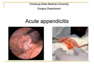
  • 1. Orenburg State Medical University Surgery Department Acute appendicitis
  • 2. Appendicitis is defined as an inflammation of the inner lining of the vermiform appendix that spreads to its other parts. Statistics  The most frequent of the abdominal surgical pathologies.  4—5 cases out of 1000 persons a year.  During the whole life 6% of the planet’s population have an acute appendicitis.  Lethality – 0,1% (1 person out of 1000 patients).  At any age, more frequently at 10—30 years, man or woman.  The most frequent cause of the generalized peritonitis
  • 4. Anatomical variants А – типичное (antecolica); Б – тазовое; В – ретроцекальное, ретроперитонеальное; Г – мезоцекальное (медиальное)  NB! Localisation gives all the symptoms appendicitis – «monkey» of all the diseases
  • 5. Appendix vessels NB! Venous flow: v. appendicularis v. ileocolica v. mesenterica superior v. portae Portal way of the infection spread – pilephlebitis (fatal comlication of appendicitis)
  • 6. Innervation  sympathic system– celiac and upper mesenteric plexus  Parasympathic - fibers of the nervus vagus  NB! Nociceptive (pain) afferent impulsation goes by the sympathetic fibers  NB! Celiac plexus – server station of the sympathetic innervation of the abdominal cavity - frequently the pain begins in the epigastrium
  • 7. Classification – on the stage of destructive changes I. Acute appendicitis 1. Acute simple (catharral) appendicitis. 2. Acute destructive appendicitis: - Flegmonous; - Gangrenous; - Gangreno-perforative. 3. Complications of the acute appendicitis: - Peritonitis - localized, generalized; - Appendicular infiltrat; - Appendicular abscess; - Pilephlebitis; II. Chronic appendicitis – result of the not operated resolved acute appendicitis.
  • 8. Etiology and pathogenesis  1. Mechanical reason - obturation of the appendix lumen (coprolythiase, bending , foreign body) luminal hypertension vessels’ compression, interruption of the venous and lymphatic outflow, oedema of the organ’s wall, vessels’ thrombosis loss of the barrier function of the mucous penetration of the intestinal flora into the wall of the appendix mural destruction
  • 9. Etiology and pathogenesis  2. Vascular reason– interruption of the blood flow in the appendix’ wall (appendix infarction) Necrosis of the wall of the organ loss of the mucous’ barrier function penetration of the intestinal flora into the appendix wall Causes: 1. atherosclerosis, thrombosis of mesenteric vessels 2. emboly of the mesenteric vessels 3. systemic vasculitis  NB! primitively-gangrenous (fulminant) appendicitis
  • 10. Etiology and pathogenesis  3. Infectious reason – some infections (typhoid fever, yersiniosis, TB, parasite infections, amoebiase, salmonellosis) give acute appendicitis as their complications.  NB! In other causes (mechanical, vascular) infection is only secondary
  • 11. Symptoms  Can vary a lot, considering anatomical variations of the appendix position
  • 12. Symptoms  Abdominal pain (100%) constant, moderately intensive.  40-50% epigastric phase: first pain in epigastric or ombilical region or more rarely all over the abdomen (in children), after a couple of hours the pain migrates to the right iliac region – symptom of pain migration Kocher’s-Wolkowitch’s (only almost pathognomonic symptom of appendicitis).  50-60% pain right away in the right iliac region.
  • 13. Symptoms Pain irradiations  into the perineum – if pelvic localisation;  right lumbar region - if retroperitoneal localisation;  right flank, right hypochondrium – if retrocecal localisation  in mesogastrium - if median localisation
  • 14. Symptoms Anorexy 100%; Nausea, vomitting (40-50%) 1-2 times, comes after pain, reflectory. can be absent; NB! if nausea or vomitting before pain - not a appendicitis characteristic; Tongue firstly wet, then dry. NB! if dry tongue - sign of dehydration.
  • 15. Клиника Stool – no specific signs. But!  can be liquid if pelvic (rectum’s irritation) or retrocecal (right colon irritation) localisations.  If generalized peritonitis, there is no stool because of the intestinal paralysis. Urination is not disturbed. But! If pelvic (irritation of the bladder) and retroperitoneal (irritation of right kidney and ureter) localisations frequent painful urination
  • 16. Symptoms Tachycardia (100%) и hyperthermia (≈ 40%) - inflammation process’ consequence  Catharral appendicitis ≈ up to 90 t ≈ 37,0 – 37,5  Flegmonous ≈ до 110 t ≈ 37,5 – 38,5  Gangrenous ≈ > 110 t ≈ 38,5 and > NB! Tachycardia – intoxication sign NB! normally if 1 degree elevation, 10 heart beats elevation Hyperthermia is not constant and can be absent in elderly patients NB! if not adequation of heart rate and body temperature (tachycardia if normal t) – first toxic scissors
  • 17. Symptoms: NB! Peritoneal symptoms – universal symptoms of peritoneal irritation. There is a lesson about peritonitis . Local symptoms of appendicitis (more than 100) Peritoneal symptoms (4 main symptoms)
  • 18. Peritoneal symptoms  1. Woskresensky symptom – pain (skin hyperestesia) above the pathological source during fast sliding hand movements on the abdominal wall.  Segmentary body innervation  Lines of Gued
  • 19. Peritoneal symptoms 2. Razdolsky’s symptom – pain during percussion of the anterior abdominal wall
  • 20. Peritoneal symptoms 3. Blumberg’s symptom - The abdominal wall is compressed slowly and then rapidly released. A positive sign is indicated by presence of pain upon removal of pressure on the abdominal wall.
  • 21. Peritoneal symptoms 4. Anterior abdominal wall’s muscular guarding (defense musculaire) The main peritoneal symptom Maximal expression – desk- like abdomen can be absent in multipares, in overwight, in elderly patients
  • 22. Local symptoms  NB! No pathognomonic signs 1. Kokher-Wolkowitch’s sign – see before – only one almost pathognomonic. But! Can be simulated if perforative gastroduodenal ulcer
  • 26. Local symptoms  5. Krymov’s symptom– pain in right iliac region during finger examination of the external opening of the inguinal canal;  6. Dumbadze’s symptom – pain if ombilical palpation.  7. Yaure-Rosanoff’s symptom — pain if finger pression in the Petit’s triangle (if retroperitoneal localisation of the appendix).  8. Gabay’s symptom – positive Blumberg’s symptom in the Petit’s triangle (if retroperitoneal localisation).
  • 27. Local psoas symptoms 9. Obrastsov’s symptom – pain increasing during pressure on the caecum and in the same time raising straightened in the knee leg
  • 28. Local psoas symptoms 10. Ostrovsky’s symptom – the patient raises his straighned right leg. The surgeon quickly unbends it and puts it horizontally. Pain in the right iliac region. 11. Koul’s symptom 1 – presence of pain in ileocecal region if unbending of right hip. 12. Koul’s symptom 2 – increasing of the right iliac pain if right hip rotation (if pelvic localisation).
  • 29. Laboratory diagnosis  Leucocytosis with neutrophilic shift (PNN) increase. Increases if process’ progression NB! Leucocytosis doesn’t have its own meaning. NB! normal or decreased leucocytes count with PNN – 2 toxic scissors (if hypoallergic immune response, inclusing elderly patients; if toxic and terminal stages of peritonitis) Always look at the leucocytes formula
  • 30. laboratory diagnosis  Urine analysis – no specific changes But! Important meaning: 1. if retroperitoneal or pelvic localisation of appendix (hematurea, leucocyturea); 2. in differential diagnosis of the urinary tract pathology.
  • 31. Instrumental diagnosis 1. Palpation, percussion, auscutation. 2. PR if men PV if women – «Douglasses cry» (effusion), differential diagnosis with internal sexual organs pathology in women
  • 32. Instrumental diagnosis 3. US – effective and not expensive method that doesn’t have a big popularity in our region yet local mural lesioins of the appendix (local loss of the sub-mucous echogenous layer), that shows the transmural lesion. difficulty in determination of the top of the appendix (blurred contours and loss of echogenous sub-mucous layer
  • 33. Instrumental diagnosis 4. Computer tomography - highly effective method, but expensive and though not highly popular in our region. Thickened appendix
  • 34. Instrumental diagnosis 5. Laparoscopy – invasive method, that allows to visualise the abdominal cavity, evaluate the appendix condition and to treat the appendicitis surgically.
  • 35. Differential diagnosis  Appendicitis – monkey of all diseases. NB! Well constructed anamnesis – 50% of diagnosis. 1. pathology of right kidney and ureter; 2. intestinal infection; 3. genital pathology (in women); 4. acute pancreatitis; 5. covered gastroduodenal perforation; 6. Crohn’s disease; 7. Meckel’s diverticulitis; 8. Acute gastritis.
  • 36. Differential diagnosis Pathology of right kidney and ureter (kidney colic, pyelonephritis):  pain in the right lumbar region with the irradiation to the right half of the abdomen;  Sometimes nausea, vomiting;  Changes in the urine: hematurie if kidney colic, leucocytes and bacteries if pyelonephritis;  Intoxication in pyelonephritis;  Not treated and not resolved kidney colic obturated pyelonephritis.  US!
  • 37. Differential diagnosis Intestinal infection (alimentary toxicoinfection):  Flagrant onset;  Multiple vomiting;  Frequent liquid stool;  Quickly intoxication (hyperthermia!) and dehydration;  Pain, sometimes contractory (salmonellous triangle)  Anamnesis!
  • 38. Differential diagnosis Genital pathology in women (very difficult): 1. Adnexitis – inflammatory signs, simulaiton of the appendicitis symptoms + vaginal discharge. 2. disturbed tubal pregnancy, ovary apoplexy (hemorragic form), broken ovary cyst – and also blood loss signs. 3. Ovary apoplexy (painful form) – middl of the cycle, only pain without blood loss signs. NB! every woman with appendicitis suspicion should be seen by a gynecologist.
  • 39. Differential diagnosis Acute pancreatitis – strong, frequently belting pain in superior regions of the abdomen, multiple vomiting. Sometimes epigastric phase of acute appendicitis is falsely taken for an acute pancreatitis Covered gastroduodenal perforation – a little quantity of effusion in the superior regions give epigastric pain, then goes to the right iliac region – simulation of the Kokher-Wolkowitch symptom.
  • 40. Differential diagnosis Crohn’s disease in its acute form is almost identical to the acute appendicitis symptomatology. Meckel’s diverticulitis – identical symptoms Acute gastritis – can simulate the epigastric phase of acute appendicitis NB! Epigastric phase’s lenght - 2-3 hours, max 1 day.
  • 41. Retrocecal (retroperitoneal) acute appendicitis (5-7%)  Pain in the right lumbar region.  Dysuria (irritation of the ureter, lidney).  liquid stool (irritation of the caecum, right colon).  Urine analysis – hematuria, leucocyturia.  Blood analysis – lucocytosis with PNN increased  Psoas-symptoms, s. of Gabay, s. Yaure-Rosanoff’s
  • 42. Pelvic acute appendicitis (16-30%)  Pubic pain with perineum irradiation  Dysuria (bladder’s irritation)  Liquid stool (rectum irritation)  Urine analysis – hematuria, leucocyturia.  Blood analysis – leucocytosis with PNN increase  Koup’s symptom 2, PR, PV
  • 43. Sub hepatic localisation (rare)  right hypochondrium pain simulating acute cholecystitis pain  US
  • 44. Left appendix localisation (rare) 1. situs viscerum inversus; 2. caecum mobile. Acute appendicitis symptoms on the left side
  • 45. Acute appendicitis in pregnant women (1%)  pregnant uterus moves the caecum and appendix up;  Pain is not expressed;  no expression of local symptoms (uterus covers the caecum);  No expression of the peritoneal symptoms (over extension of the anterior abdominal wall).
  • 46. Acute appendicitis in pregnant women (1%)  one of the difficult diagnostic problems: 1. Possibility of the physiological pain beкcase of the ligament system of the uterus extension and its hypertonus; 2.possibility of the pregnancy dyspepsia (nausea, vomiting, stool problems); 3. possibility of the physiological luecocytosis; 4. atypical symptoms, especially during the second half of the pregnancy. 5. impossibility of the laparoscopy during late pregnancy period. 6. anamnesis, US, exclude pyelonephritis and the risk of the pregnancy interruption.
  • 47. Acute appendicitis in children  Particularities of children organism 1. Hyperergic response – fulminant symptoms; 2. Short big omentum – no limitations; 3. the child can not sometimes well relate the anamnesis.
  • 48. Acute appendicitis in children  Symptom’s particularities 1. pain can be not localized, contraction-like; 2. Multiple vomiting; 3. Frequent stool; 4. Quickly febrile hyperthermia; 5. Quickly fast leucocytosis; 6. Symptom of the leg lifting; 7. Symptom of the surgeon’s hand repulsion;
  • 49. Acute appendicitis in elderly patients  Particularities of the elderly patients’ organism 1. Hypoergic response, reduced symptoms; 2. Atherosclerosis of the vessels – fast ischemia and necrosis (primarily-gangrenous appendicitis); 3. Diabetes mellitus – fast destruction. Abundance of the complicated appendicitis forms
  • 50. Acute appendicitis in elderly patients  Symptoms particularities 1. Reduced pain syndrom; 2. Reduces local and peritoneal symptoms; 3. First and second toxic scissors.
  • 51. Treatment  Hospitalisation in surgery department.  If diagnosis not clear (table 0, IV, once intramuscular spasmolytic (но-шпа, папаверин).  Without positive dynamics during 2 hours (laparoscopy or laparotomy).  If diagnosis acute appendicitis is established, it is an absolute surgery indication.  contraindication –agonic patient’s condition.  pain-killers – general anesthesia
  • 55. Operation features  In pregnant women - incision higher with the pregnancy length.  In children– ligature method without purse-string and Z-stitches (thin wall of the caecum, riskof the ileocaecalis involvement in the stitches).  If laparoscopy – only ligature method.
  • 57. Laparoscopic appendectomy  Conditions 1. Presence of necessary mechanisms (endosurgical stand); 2. Surgeon’s preparation; 3. Possibility of the laparoscopy (not in every case)
  • 58.  Russian surgeon L.I. Rogozov have done an appendectomy on himself in Antarctic in 1961
  • 59. Complications’ classification I. Early: 1. Before the operation: - Appendicular infiltat; - Appendicular abscess; - Peritonitis - local, generalized; - retroperitoneal flegmona (if retroperitoneal localisation) - pylephlebitis. 2. Post-surgical complications: - Intraperitoneal hemorrage; - failure of the appendix stump; - Local infiltrat and abscess of the peritoneal cavity; - suoouration of the wound; - post-surgical peritonitis (tertiary peritonitis); - Intestinal paralysis; - early adhesive intestinal blockage. II. Late: - Ligature fistulas; - Adhesive intestinal blockage; - Post-surgical ventral hernia. III. Involving other systems and organs: - Pneumonia, pleuritis, lung abscess; - Myocardial infarction, acute failure of brain circulation. - Thrombosis of the deep veines of the shin, lung emboly
  • 60. Progression of not-operated appendicitis Appendicular infiltrat peritonitis Resolution destruction (abscesses) Chronic appendicitis local generalized Break into the intestinal lumen Break into the retroperitoneal space break through the anterior abdominal wall break into the abdominal cavity
  • 61. Appendicular infiltrat (1-3%)  Inflammatory tumor, Inflammatory tumor, conglomerate of losely fixed to one another tissues around the appendix with participation of parietal peritoneum, big omentum, caecum, small intestin. • Infiltrat – limitation, protection reaction in order to limitate the inflammation 1. Formation (loose) of infiltrat – 3-4 days; 2. Formed (dense) infiltrat – after 5 days.
  • 62. Appendicular infiltrat Symptoms: • Less abdominal pain; • Better general patient’s condition; • Dense, not painful, fixed formation in the right iliac region, • Infiltrate’s size can vary a lot, can occupy all the right iliac region; • There can be positive local symptoms and peritoneal symptoms - negative; • Mild leucocytosis with PNN, sub febrile body temperature
  • 63. Appendicular infiltrat NB! it’s localisaiton (abscess) depends on the initial appendix’ localisation, it can be localized in the pelvis, in mesogastrium, in sub hepatic space.
  • 64. Appendicular infiltrat Diagnosis: 1. Disease’s anamnesis (reduced symptoms in elderly); 2. US; 3. CT Differential diagnosis: cecal tumor (especially in elderly)
  • 65. US diagnosis Appendicular infiltrat Appendicular abscess
  • 66. Appendicular infiltrat Conservative treatment 1. Table 0 with IV infusions 30 ml/kg, then 1А; 2. Cold on abdomen; 3. Bed regimen; 4. Antibacterial therapy; 5. Physical treatment on the infiltrat region 6. No surgical indication (risk of organs participating in infiltrat damage)
  • 67. Appendicular infiltrat Results: 1. Resolution: pain decreases, infiltrat resorbs, temperature normalizes discharge with planned hospitalisation in 3 month for a planned appendectomy 2. abscess operation Pain increase Hyperthermia even if one of these sign Leucocytosis
  • 68. Interventions variants  Wolkowitch-Diakonov’s Laparotomy  Pirogov’s method (extraperitoneal);  Punction drainage under US control;  Laparoscopy (risk!).  No obligation of appendectomy, appendix can be removed if well visualised Risk of organ damage (organs that participate in the pyogenic capsule)
  • 71. Pilephlebitis Septic thrombophlebitis of the portal veine – rare, but fatal.  Necrotic prosess that invades appendix mesentery and its vessels.  then invasion of the vessels of the ileocecal angle.  in 2-3 days goes to the portal veine and hepatic veines  then retrograde invasion of the splenic and other mesenteric veines.
  • 72. Пилефлебит Клиника: 1. Бурно развивающаяся картина системной воспалительной реакции (фебрильная гипертермия); 2.Усиление боли в животе (правая половина); 3.Желтуха; 4.Гепатоспленомегалия; портальная гипертензия 5.Асцит; 6.Прогрессирующая печеночно-почечная недостаточность.
  • 73. Pilephlebitis  Treatment: 1. Maximal section of the appendix mesentery; 2. ligature v. ileocolica; 3. resection of the ileocecal angle; 4. Massive antibacterial therapy; 5. Masive infusion-detox therapy;
  • 74. Abscesses of the abdominal cavity  Appendicitis complication after surgery;  Universal complication;  Reasons: 1. Not adequate sanation of the abdominal cavity; 2. suppuration of hematomas after not adequate hemostasis;  Typical localisation: - pelvic (Douglass’ space); - iliac; - between loops; - sub hepatic; - subphrenic.
  • 75. General signs of the abdominal abscesses  Clear laps of time – better general condition after operation then progressive symptoms with 7-8 day pic: 1. Growing hyperthermia Appendectomy abscess break ССВР SIRS 2. Progressive dehydration; 3. Leucocytosis with PNN growth; 4. Infiltrat’s palpation (not always); 5. Local abscess symptoms.
  • 76. Local clinic of the abdominal abscess Douglass’ abscess: 1. Pubic pain with perineum irradiation; 2. Dysuria (bladder’s irritation); 3. tenesms (rectum’s irritation). Intestinal abscess: 1. pain in mesogastrium; 2. frequent liquid stool (small intestins irritation); Subhepatic abscess: 1. Pain in right hypochondrium; 2. Cholecystitis symptoms (Кера, Ортнера, Мерфи, Мюсси) 3. Frequent liquid stool (colon’s irritation);
  • 77. Local clinic of the abdominal abscess Subhepatic abscess – the most difficult diagnostically and for the therapy Classificaiton: 1.right-side, left-side, median; 2.one side, both side; 3.anterior, posterior; 4.superior, inferior.
  • 78. Local symptoms of the abdominal abscess Symptoms of the sb phrenic abscess 1.inferior thoracic region pain with shoulder irradiation (с. Мюсси, Элекера); 2.Senator’s syndrom – body’s rigidness; 3.thorax is late for the breathing; 4.pleural effusion signs; 5.inferior lobe pneumonia 6.Signs of SIRS (sometimes the only symptom).
  • 79. One of the reasons – sucking action of the diaphragm. Law: after operation the patient must be with raised head position (Fowler’s position). Douglass’ abscess is less difficult to treat than sub phrenic.
  • 80. Retrouperitoneal flegmona 1. Right lumbar pain; 2. Irradiation to the right half of the abdomen; 3. Positive psoas symptoms; 4. softness of the tissues in the lumbar region (possible); 5. dysuria; 6. Changes in urine analysis; 7. Signs of SIRS – hyperthermy, leucocytosis, etc.
  • 81. Diagnosis  US – method of choice;  CT – method of choice;  X-rays (for sub phrenic abscess);  PR! (Douglass);  White blood (leucocytes elevation). NB! abscess’ localisation depends on the appendix localisation.
  • 82. Surgical treatment  Laparotomy;  laparoscopy (risk);  Punction drainage under US control (method of выбchoiceора);  Pelvic abscess – transvaginally or transrectally;  Sub phrenic abscess – Clermond’s method; transthoracic acces (transpleural, extrapleural) – anachronism.
  • 83. Punction drainage under US control  Video  Features: 1. Effectiveness 60-70%; 2. if no positive dynamics for 2-3 days laparotomy; 3. Risk of organs damage.
  • 86. Clermond’s method Accessible for the anterior abscesses
  • 87. Transthoracic methods Variants 1.Transpleural acces in one or two times (VIII-IX costs); 2.Extrapleural acces (X-XI cost). Possible during posterior abscesses
  • 88. Section of the pelvic abscess vaginal access rectal access
  • 89. intraperitoneal hemorrage  Causes: 1. ligature’s slipping 2. abdominal organ’s damage during revision (omentum, intestinal mesentery, liver, spleen).  symptoms (during the first hours and depends on blood loss abundance): 1. General blood loss signs (skin palour, hypotony, tachycardia, diziness, weakness); 2. frequent signs of the intraabdominal hemorrage (increasing pain in the abdomen during palpation, с. Куленкампфа).  Diagnosis: 1. Blood analysis – Hb decrease, Ht, erythrocytes; 2. US – free liquid in the abdominal cavity.  Strategy – urgent median relaparotomy, blood loss stop.  Prophylaxy – attentive hemostase and its control during the primary operation
  • 90. Insufficiency of the appendix stump  causes: 1. defects of the operative technique; 2. Tiphlitis (inflammation of the caecum’s wall).  Symptoms (from 3-4 days), process can be limited (abscess formaiton) and generalized (peritonitis): 1. Increasing pain and its generalization; 2. Peritoneal symptoms; 3. SIRS.  Strategy – relaparotomy, punction drainage (if abscess).
  • 91. Suppuration of the wound  cause: 1. operation technique violation (trauma, unsufficient hemostase, pouches left): hematoma suppuration. cavity then suppuration. 2. Appendectomy – «dirty» operation risk of infection of operative wound . Mechanical protection necessary (limitation of the wound, wound wash). Perioperational antibiotherapy obligatory. symptoms: (tumor, rubor, calor, dolor, functio laesa): painful wound infiltrat, skin hyperhemia, SIRS. NB! If source is very deep, local symptoms can be absent. Treatment: revision and drainage of the wound.
  • 92.  Peritonitis - theme of another lesson  Intestinal peralysis and intestinal adhesive blockage - theme of another lesson.