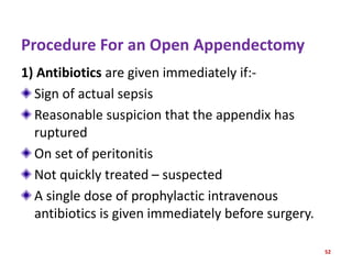
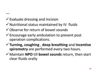
APPENDICITIS
SEMINAR PRESENTATION
ON
APPENDICITIS
1
OUTLINES
Anatomy & Physiology of Appendix
Introduction To Appendicitis
Epidemiology
Risk Factors
Etiologies
Types Of Appendicitis
PP Of Appendicitis
2
OUTLINES…
CXNS Of Appendicitis
Assessment Of Acute Appendicitis
Investigations
Differential Diagnosis
Management Of Appendicitis
References/ Sources
3
ANATOMY
 Appendix is a small, finger-like tube about 10
cm (4 in) long
That is attached to the cecum just below the
ileocecal valve.
The appendix fills with food and empties
regularly into the cecum.
 Because it empties inefficiently and its lumen
is small, the appendix is prone to obstruction
&
4
…
Is particularly vulnerable to infection
Position :- is variable, but it is usually retrocecal
Arterial Supply:- Appendicular branch of iliocolic
artery
Innervation :- From sympathetic elements
contributed by superior mesenteric plexus ( T10-
L1)
Afferents :- From parasympathetic elements via
vagus nerves.
5
Physiology
• Is an immunologic organ
• Secretion of immunoglobulins ( igA)
• May serve as a reservoir for beneficial gut
bacteria
• I,e maintaining gut flora.
6
Appendix
Figure 1.1 location of the appendix.
7
INTRODUCTION TO APPENDICITIS
Def of Appendicitis
• Is a condition where the appendix becomes
swollen, inflamed, and filled with pus.
• Is inflammation of the vermiform appendix.
• Is an inflammation of appendix that develops
most commonly in adolescents and adults.
• Is acute inflammation of the appendix and is the
most common cause for acute, severe abdominal
pain.
8
…
• Is the most common reason for emergency
abdominal surgery.
• Anyone can get it, but occurs most often
between ages 10-30
• The abdomen is most tender at McBurney’s
point-1/3 from the RASIS to the umbilicus
• This corresponds to the location of the base
of the appendix.
9
EPIDEMIOLOGY
World population-7%- 12%,Men - 9% &
Women-7%
Appendicitis is more common in:
Males (aged 21-30 years ) &
Females( aged 11-20 years/10-30years)
Ap. 20% of all patients have evidence of
perforation at presentation , but the
percentage risk is much higher in patients
under 5 or over 65 years of age.
10
Risk Factors
Infection-Stomach infection
Age:20-30 years( peak incidence in early 20s)
Socio-economic condition: High profile (Low
fiber)
Lymphoid hyperplasia of the appendix
Fibrosis of the appendix cos of previous
damage
11
…
• Extreme of age
• Previous abdominal surgery
• Position of Appendix (Retrocecal 56.5%)
• Sex- more in males than females
• Familial- people who have family history of
appendicitis are at high risk of developing it.
12
Risk Factors For Perforation Of Appendicitis
1) Extremes of age
2) Immunosuppression
3) Diabetes mellitus
4) Pelvic appendix
5) Previous abdominal surgery
13
Etiologies
Obstruction of the lumen with Faecolith
Is the predominant etiologic factor in acute
appendicitis
Bacterial proliferation-streptococci & E. coli
Worm infestations
Neoplasms, ca caecum (elderly& middle age)
Viral
14
…
Low dietary fiber
Foreign bodies
King- King of appendix ( Twisting)
Swelling of the bowel wall
Inspissated barium
Vegetable & fruit seeds
15
…
Appendicitis is caused by a blockage of the hollow
portion of the appendix.
This is most commonly due to a calcified stone
made of faeces.
Inflamed lymphoid tissue from a viral infection,
parasites, gallstone or tumors may also cause the
blockage.
Mechanism of luminal obstruction varies
depending upon the pt's age & other factors.
In the young--lymphoid follicular hyperplasia
due to infection is the main cause .
16
…
In older patients more likely to be caused by
• Fibrosis
• Fecaliths or
• Neoplasia (carcinoid, adenocarcinoma or
mucocele)
In endemic areas—parasites can cause
obstruction at any age & groups.
Examples: Schistosomes species ,Pinworms,
Strongyloide & Stercoralis
17
Types of Appendicitis
1) Acute Appendicitis
Develops very fast
Usually in a span of several days or hours
Mostly it is diagnosed within 24 to 48 hours.
It is easier to detect
Requires prompt medical treatment, usually surgery.
occurs when the vermiform appendix is completely
obstructed, either because of a bacterial infection,
feces or other types of blockage
18
2) Chronic Appendicitis
Is a Rare Condition
Is an inflammation that can last for a long time.
It only occurs in only 1.5% of recorded acute
appendicitis cases.
Appendiceal lumen is only partially obstructed,
causing inflammation.
The inflammation worsens over time, causing internal
pressure to buildup.
Until the infection get worsened the appendicitis
undiagnosed for several weeks, months, or years .
19
Can also be of four types
1)Acute simple appendicitis
• Is defined as an inflamed appendix without
any signs of gangrene or perforation.
2) Acute purulent appendicitis
• Purulent, usually already perforated,
appendicitis is the most common and
dangerous differential diagnosis for acute
infectious enteritis, in children as well as in
adults.
20
…
3) Perforation and gangrenous
• Is defined as an inflamed appendix with signs of
grossly necrotic tissue but no frank perforation or
abscess.
4) Appendiceal abscess
• Is a condition in which an abscess is formed
around the appendix as a result of appendiceal
perforation or extension of inflammation to the
adjacent tissues due to aggravation of
appendicitis.
• Occurs in 2-6% of patients with appendicitis.
21
PP OF APPENDICITIS
Obstruction
Build up of mucous
Increased luminal pressure
Decreased blood flow
Decreased oxygen delivery ( hypoxia)
Ulceration or lesion formation
Invasion by bacteria severe inflammation and swelling Appendicitis
22
If appendicitis not get treated / Etiology
Ischemia
Necrosis
Gangrene
Perforation
Peritonitis
23
SIGNS OF PERFORATION
If fever > 102*F
WBC > 18,000
24
COMPLICATIONS OF APPENDICITIS
1) Gangrenous Appendicitis
Thrombosis of the appendiceal artery and veins
2) Perforation
Complication rates 58 %
Perforation rate increases at both ends of the
age spectrum
3) Peri-appendiceal abscess
Most frequent complication
Peri-appendiceal fibrinous adhesions
25
…
4) Peritonitis
Bacterial peritonitis in absence of fibrinous
adhesions usually E. coli
5) Bowel Obstruction
6) Septic seeding of mesenteric vessels
Infection along the mesenteric–portal venous
system
Pylephlebitis , pylethrombosis , or hepatic
abscess
26
Assessment Of Acute appendicitis
Subjective Data
Abdominal Pain
1st noticed in the peri-umblical area-then shifts to the
RIF.
Mechanism of pain--As the appendix becomes
engorged, the visceral afferent nerve fibers entering
the spinal cord at T8-T10 are stimulated, leading to
vague central or periumbilical abdominal pain.
Well-localized pain occurs later in the course when
inflammation involves the adjacent parietal
peritoneum
27
Associated with:-
o Anorexia
o Nausea &
o Vomiting
May be the pt present with constipation &
diarrhea
Cough & sudden movement exacerbate the pain
NB The symptoms of appendicitis vary depending
upon the location of the tip of the appendix
28
Objective Data
 G/A – Acutely Sick Looking (in pain)
 V/S--Low grade fever
Tachycardia
Tachypnoea
 Abdomen
 Direct & rebound tenderness
 Mc Burney’s point…point of maximum tenderness
 1/3 from anterior superior iliac spine ( ASIS) & 2/3
from umbilicus
29
Rovsing’s sign
 Palpating in the left lower quadrant causes pain in the
right lower quadrant
 Psoas sign
o RLQ pain with passive right hip extension
o Associated with a retrocecal appendix.
 Obturator sign
 Flexing the patient's right hip and knee followed by
internal rotation of the right hip elicits RLQ Pain
 Associated with a pelvic appendex
 Dunphy’s sign:- Increased pain in the RLQ with
coughing.
30
IVESTIGATIONSN
1) History collection
2) Physical examination
3) TLC- Raised: 10000 - 18000 ( Neutrophils >75%).
If TLC >18000 (suspect perforation)
4) Urinalysis—To rule out Urinary Tract Infection
5) Abdominal X-Ray
6) Abdominal USG Are the most commonly used tests
7) CT Scan
31
…
Abdominal x-ray films, ultrasound studies, and
CT scans may reveal a right lower quadrant
density or localized distention of the bowel.
NB
8) Urine HCG:- should be done for all women of
childbearing age to exclude pregnancy related
causes of acute abdomen.
32
9) Advanced Diagnosing Technology
Is neutroSpec imaging
Is a new technique to diagnose appendicitis
Uses a technetium
Labeled anti-CD15 monoclonal antibody
That selectively binds to neutrophils
When injected into the blood, neutroSpec
binds to neutrophils present at the infection
site
33
…
Labeling these cells with technetium
As a result, physicians can rapidly detect an
infection using a gamma camera that records
radioactivity
Advantage over the current standard of care
• Is in vivo labeling of WBCs &
• Diagnosis in less than 1 hour
34
10) The Alvarado scoring System
Is a clinical scoring system used in the
diagnosis of appendicitis
This scoring system has 6 clinical items and 2
laboratory measurements with a total of 10
points.
35
The Alvarado Score
SN Symptoms Score
01 Migratory RIF pain 1
02 Anorexia / Loss of appetite) or ketenes in
the urine
1
03 Nausea / Vomiting 1
Signs
04 Tenderness in the RIF 2
05 Rebound tenderness in the RIF 1
06 Elevated temperature 1
Laboratory Findings
07 Leucocytosis / increased in no but
immature)
2
08 Shift to the left of neutrophils 1
09 Total 10 36
Interpretation Of Alvarado Score
Alvarado score Interpretation
A score of 5 or 6 Compatible with dx of acute
appendicitis
A score of 7 or 8 Indicates a probable appendicitis
A score of 9 or 10 Indicates a very probable acute
appendicitis
37
Differential Diagnosis
1) Children
Gastroenteritis
Mesenteric adenitis
Meckel’s diverticulitis
Intussusception
Purpura
Lobar pneumonia
38
2) Adults
Regional enteritis
Ureteric colic
Perforated peptic ulcer
Torsion of testis
Pancreatitis
Rectus sheath haematoma
39
3) Adult Female
Mittelschmerz
PID
Pyelonephritis
Ectopic pregnancy
Torsion / Rupture of ovarian cyst
Endometriosis
40
4) Elderly
Diverticulitis
Intestinal obstruction
Ca colon
Mesenteric infarction
Torsion of appendix epiploicae
Leaking aortic aneurysm
41
5) Rare
Tabetic crisis
Spinal condition
Porphyria
Diabetes
Typhilitis
42
MANAGEMENT
A) MEDICAL MANAGEMENT
1. Absolute bed rest & NPO
2. IV Fluids Supplements
To avoid dehydration & electrolyte imbalance
3. Analgesics
can be administered after the diagnosis is made
Morphine is a common & effective analgesics for
this purpose
43
4.Antibiotics
Ruptured appendix
Evidence of peritonitis or abscess
Medication is given for 6 to 8 hours prior to
appendectomy to prevent sepsis
Examples Cefotan ( cefotetan) & cefotaxime
(claforan, mefotoxin) help prevent wound
infection after surgery.
Levofloxacin ,Metronidazole
or
Ofloxacine + Orinidazole
44
B) SURGICAL MANAGEMENT
APPENDECTOMY
Is surgical removal of the appendix
Is a surgery to remove the appendix which is
usually found in the right lower side of the
abdomen.
Usually carried out on an emergency basis to
treat appendicitis.
This may occur as a result of an obstruction in
part of the appendex.
45
…
Another name for this operation is
appendicectomy.
Performed as soon as possible to decrease the
risk of perforation.
May be performed under general or spinal
anesthesia
46
INDICATION OF APPENDECTOMY
Acute , chronic, recurrent & Perforated
appendicitis
As interval appendectomy after drainage of
abscess or appendicial mass
Carcinoma confined to the mucosa
Mucocele of the appendix
Appendicular graft, ileal conduit
On table colonic lavage
47
Types Of Appendectomy
Laparoscopic appendectomy
Open appendectomy /Traditional surgery
Natural orifice surgery (no incision
appendectomy)
The choice of method is made by the surgeon
on a case-by –case basis.
General anesthesia is used in both
procedures.
48
1) Laparoscopic Appendectomy
Also referred to as lap appendectomy
Is a minimally invasive surgery to remove the
appendix through several small incisions
Recovery time from the lap appendectomy is short.
Under GA, pt is in supine position
Use three ports,10-12 mm port at umbilicus & 5 mm
port at suprapubic & in LLQ
Appendix should be identified by tracking taeniae
libera/ coli
Appendix is removed through infraumbilical trocar in a
retrieval bag.
49
Laparoscopic Procedure is :-
Lower risk for postoperative infection
A smaller scar
A shorter hospital stay
More expensive
Resource intensive
Keyhole” surgery
Lower complication rate
Faster recovery time
50
2) Open Appendectomy
Is the traditional method and the standard treatment
for appendicitis.
Typically performed under general anesthesia pt is in
supine position
Abdomen is prepared & draped
Incision is made at McBurney ‘s point
If appendix is not easily identified , caecum should be
located.
Tracing the taenia libera, the most visible of three
taeniae coli, distally, the base of the appendix can be
identified.
51
Procedure For an Open Appendectomy
1) Antibiotics are given immediately if:-
Sign of actual sepsis
Reasonable suspicion that the appendix has
ruptured
On set of peritonitis
Not quickly treated – suspected
A single dose of prophylactic intravenous
antibiotics is given immediately before surgery.
52
Antibiotics Prophylaxis in Surgery
Usually a single dose is sufficient
A second dose may be required in the following
situations
i ) In prolonged operations
ii ) When there is contamination during surgery
Giving more than 1 or 2 doses is generally not advised
The practice of continuing prophylactic antibiotics until
surgical drains have been removed is not
recommended.
Should be given 1hour prior to surgery preferably with
induction of anesthesia, 30min?
53
2) General anesthesia
Is induced with endotrachcheal intubation &
full muscle relaxation
The pt is placed in supine position
3) The abdomen is prepared and draped & is
examined under anaesthesia.
4) The incision is made over Mc Burney’s point
The most common position of the base of the
appendix
If mass is present, the incision is made over
the mass
54
…
5) The various layers of the abdominal wall are opened.
6) On entering the peritoneum, the appendix is identified,
mobilized, and then ligated and divided at its base.
7) The stump of the appendix is buried by surgeon by inverting it
so it points into the caecum.
8) Each layer of the abdominal wall is then closed in turn.
9) The skin may be closed with staples or stitches
10) The wound is dressed
11) The patient is bought to the recovery room.
55
3) Natural Orifice Surgery
Is an experimental surgical technique whereby
scarless abdominal operations can be
performed
With an endoscope passed through a natural
orifice like mouth, urethra, anus & vagina
Then through an internal incision in the
stomach, vagina, bladder, or colon, thus
avoiding any external incisions or scars.
56
…
No incision appendectomy
Use the natural orifices
Anal canal endoscopically or
Trans-vaginally
Less pain
No scar
Less hospital stay
Fewer complications
Take about 50 minutes
57
COMPLICATION OF APPENDECTOMY
Wound infection 5-10%
,4-5th day
Intra- abdominal
abscess 8%
Hemorrhage
Paralysis ileus
Generalized peritonitis
Aspiration pneumonia
Urinary tract infection
Deep Vein Cut
thrombosis & embolism
Portal pyemia-
 Adhesion intestinal
obstruction
Fecal Fistula
 Ritcher’s hernia
58
C) NURSING MANAGEMENT
Pre-op preparative Care
Intra-operative Care
Post-operative Care
Pt Education
59
Pre-Op Preparative Care
Assessment- History taking & physical
examination
Monitor vital signs BP, To ,PR & RR for
baseline data
NPO ( 6-8 hrs) and IV Fluids should be started
Investigations:- U/A, CT Scan, Ultrasound &
CBC
Analgesics-- to relieve pain as per prescribed
60
…
Administer antibiotics as prescribed :-Second
generation Cephalosporin with Metronidazole
is given
Naso-gastric aspiration
Monitor for signs of ruptured appendix
&peritonitis
Position right-side lying or low to semi fowler
position to promote comfort.
Auscultate Bowel Sounds
61
…
Preparation for surgery i.e. physically & psychologically
Remove anxiety & fears
Maintaining skin integrity
Informed ( Written) consent form must be signed
knowing that the pt understands the procedure, the
potential risks, and that he /she receives certain
medications
Prepare and send the patient for surgery without delay
OT clothes and pre medications to be given 45
minutes before operation
62
Intra-Operative Care
Position the patient on the OR table
Skin preparation
Induction of anesthesia
Procedure is performed aseptically
Closing of the incision
Dressing of the site
63
Post-Operative Care
Following surgery, the patient is taken to the
post anesthesia care unit ( PACU) until the
anesthesia wears off.
During this time, the nurse check the ff:-
Temperature
Heart rate &
Breathing at frequent intervals
64
…
 When the anesthesia wears off & vital signs stabilize, the
pt is transferred to their hospital room.
 Place the pt in a semi-fowler position. This position
reduces the tension on the incision and abdominal
organs, helping to reduce pain.
 Clear airway
 Proper breathing and adequate tissue perfusion by IVF
 Monitor vital signs for signs of infection and shock.
Fever
Hypotension
Tachycardia
65
…
Monitor input & output for signs of fluid
imbalance, dehydration & shock.
Naso-gastric suction to be done regularly to
relieve tension on sutures
Provide safety & effective care environment to
the patient
Care of all drainage tubes
Care of surgical wounds. Watch for soapage
66
…
Evaluate dressing and incision
Nutritional status maintained by IV fluids
Observe for return of bowel sounds
Encourage early ambulation to prevent post
operation complications.
Turning, coughing , deep breathing and incentive
spirometry are performed every two hours.
Maintain NPO till bowel sounds return, then start
clear fluids orally
67
…
Assess abdomen for increased pain,
distension, rigidity, and rebound tenderness
b/c these may indicate post operative
complications
Monitor for nausea & vomiting
Administer & analgesics as per prescribed
Evaluate the passing of flatus or faeces.
68
Pt Education
The pt should avoid heavy lifting for 4 to 6 wks
The pt must report the ff symptoms if there
are:-
Anorexia
Nausea
Vomiting
Fever
Abdominal pain
Redness around incision area & drainage
69
…
If drains from incision, patient may be kept in
the hospital for several days &
Monitored carefully for signs of intestinal
obstruction or 2O hemorrhage
Diet and how to take ordered medication
Returning to health facility on appointment
70
NB
1. Avoid Laxatives and enemas as they increase
peristalsis that may cause perforation of the
appendix.
2. Apply Cold , this may decrease the flow of
blood to RLQ and help reduce the
inflammatory process.
3. Heat is never used because it may cause the
appendix to rupture.
71
References / Sources
1) Medical- surgical Nursing vol 1, 13th edition
2) Schwartz’s principles of Surgery 9th edition
3) Emergency Medicine Clinics of North America.
4) Black M. JOYCE, Medical Surgical Nursing, published
by Elsevier, 8th edition volume 2
5) Brunner and Suddarth’s , Textbook of Medical
Surgical Nursing ,published by Lippincott Williams and
Wilkins , 11th edition
6) Smeltzer C. Suzane, Textbook of Medical Surgical
Nursing ,published by Lippincott, 9th edition.
72
References / Sources…
7) Schwartz’s principles of Surgery 10th edition
8) Baley and Love’s Short Practice of Surgery,
26th edition
9) Schwartz Book of General Surgery
10) Medical- surgical Nursing vol 2, 13th edition
11) Medical- surgical Nursing 10th edition
12) Wikipedia , Google
73
THANKS
74

Seminar Presentation On Appendicitis (1).pptx

  • 1.
  • 2.
    OUTLINES Anatomy & Physiologyof Appendix Introduction To Appendicitis Epidemiology Risk Factors Etiologies Types Of Appendicitis PP Of Appendicitis 2
  • 3.
    OUTLINES… CXNS Of Appendicitis AssessmentOf Acute Appendicitis Investigations Differential Diagnosis Management Of Appendicitis References/ Sources 3
  • 4.
    ANATOMY  Appendix isa small, finger-like tube about 10 cm (4 in) long That is attached to the cecum just below the ileocecal valve. The appendix fills with food and empties regularly into the cecum.  Because it empties inefficiently and its lumen is small, the appendix is prone to obstruction & 4
  • 5.
    … Is particularly vulnerableto infection Position :- is variable, but it is usually retrocecal Arterial Supply:- Appendicular branch of iliocolic artery Innervation :- From sympathetic elements contributed by superior mesenteric plexus ( T10- L1) Afferents :- From parasympathetic elements via vagus nerves. 5
  • 6.
    Physiology • Is animmunologic organ • Secretion of immunoglobulins ( igA) • May serve as a reservoir for beneficial gut bacteria • I,e maintaining gut flora. 6
  • 7.
    Appendix Figure 1.1 locationof the appendix. 7
  • 8.
    INTRODUCTION TO APPENDICITIS Defof Appendicitis • Is a condition where the appendix becomes swollen, inflamed, and filled with pus. • Is inflammation of the vermiform appendix. • Is an inflammation of appendix that develops most commonly in adolescents and adults. • Is acute inflammation of the appendix and is the most common cause for acute, severe abdominal pain. 8
  • 9.
    … • Is themost common reason for emergency abdominal surgery. • Anyone can get it, but occurs most often between ages 10-30 • The abdomen is most tender at McBurney’s point-1/3 from the RASIS to the umbilicus • This corresponds to the location of the base of the appendix. 9
  • 10.
    EPIDEMIOLOGY World population-7%- 12%,Men- 9% & Women-7% Appendicitis is more common in: Males (aged 21-30 years ) & Females( aged 11-20 years/10-30years) Ap. 20% of all patients have evidence of perforation at presentation , but the percentage risk is much higher in patients under 5 or over 65 years of age. 10
  • 11.
    Risk Factors Infection-Stomach infection Age:20-30years( peak incidence in early 20s) Socio-economic condition: High profile (Low fiber) Lymphoid hyperplasia of the appendix Fibrosis of the appendix cos of previous damage 11
  • 12.
    … • Extreme ofage • Previous abdominal surgery • Position of Appendix (Retrocecal 56.5%) • Sex- more in males than females • Familial- people who have family history of appendicitis are at high risk of developing it. 12
  • 13.
    Risk Factors ForPerforation Of Appendicitis 1) Extremes of age 2) Immunosuppression 3) Diabetes mellitus 4) Pelvic appendix 5) Previous abdominal surgery 13
  • 14.
    Etiologies Obstruction of thelumen with Faecolith Is the predominant etiologic factor in acute appendicitis Bacterial proliferation-streptococci & E. coli Worm infestations Neoplasms, ca caecum (elderly& middle age) Viral 14
  • 15.
    … Low dietary fiber Foreignbodies King- King of appendix ( Twisting) Swelling of the bowel wall Inspissated barium Vegetable & fruit seeds 15
  • 16.
    … Appendicitis is causedby a blockage of the hollow portion of the appendix. This is most commonly due to a calcified stone made of faeces. Inflamed lymphoid tissue from a viral infection, parasites, gallstone or tumors may also cause the blockage. Mechanism of luminal obstruction varies depending upon the pt's age & other factors. In the young--lymphoid follicular hyperplasia due to infection is the main cause . 16
  • 17.
    … In older patientsmore likely to be caused by • Fibrosis • Fecaliths or • Neoplasia (carcinoid, adenocarcinoma or mucocele) In endemic areas—parasites can cause obstruction at any age & groups. Examples: Schistosomes species ,Pinworms, Strongyloide & Stercoralis 17
  • 18.
    Types of Appendicitis 1)Acute Appendicitis Develops very fast Usually in a span of several days or hours Mostly it is diagnosed within 24 to 48 hours. It is easier to detect Requires prompt medical treatment, usually surgery. occurs when the vermiform appendix is completely obstructed, either because of a bacterial infection, feces or other types of blockage 18
  • 19.
    2) Chronic Appendicitis Isa Rare Condition Is an inflammation that can last for a long time. It only occurs in only 1.5% of recorded acute appendicitis cases. Appendiceal lumen is only partially obstructed, causing inflammation. The inflammation worsens over time, causing internal pressure to buildup. Until the infection get worsened the appendicitis undiagnosed for several weeks, months, or years . 19
  • 20.
    Can also beof four types 1)Acute simple appendicitis • Is defined as an inflamed appendix without any signs of gangrene or perforation. 2) Acute purulent appendicitis • Purulent, usually already perforated, appendicitis is the most common and dangerous differential diagnosis for acute infectious enteritis, in children as well as in adults. 20
  • 21.
    … 3) Perforation andgangrenous • Is defined as an inflamed appendix with signs of grossly necrotic tissue but no frank perforation or abscess. 4) Appendiceal abscess • Is a condition in which an abscess is formed around the appendix as a result of appendiceal perforation or extension of inflammation to the adjacent tissues due to aggravation of appendicitis. • Occurs in 2-6% of patients with appendicitis. 21
  • 22.
    PP OF APPENDICITIS Obstruction Buildup of mucous Increased luminal pressure Decreased blood flow Decreased oxygen delivery ( hypoxia) Ulceration or lesion formation Invasion by bacteria severe inflammation and swelling Appendicitis 22
  • 23.
    If appendicitis notget treated / Etiology Ischemia Necrosis Gangrene Perforation Peritonitis 23
  • 24.
    SIGNS OF PERFORATION Iffever > 102*F WBC > 18,000 24
  • 25.
    COMPLICATIONS OF APPENDICITIS 1)Gangrenous Appendicitis Thrombosis of the appendiceal artery and veins 2) Perforation Complication rates 58 % Perforation rate increases at both ends of the age spectrum 3) Peri-appendiceal abscess Most frequent complication Peri-appendiceal fibrinous adhesions 25
  • 26.
    … 4) Peritonitis Bacterial peritonitisin absence of fibrinous adhesions usually E. coli 5) Bowel Obstruction 6) Septic seeding of mesenteric vessels Infection along the mesenteric–portal venous system Pylephlebitis , pylethrombosis , or hepatic abscess 26
  • 27.
    Assessment Of Acuteappendicitis Subjective Data Abdominal Pain 1st noticed in the peri-umblical area-then shifts to the RIF. Mechanism of pain--As the appendix becomes engorged, the visceral afferent nerve fibers entering the spinal cord at T8-T10 are stimulated, leading to vague central or periumbilical abdominal pain. Well-localized pain occurs later in the course when inflammation involves the adjacent parietal peritoneum 27
  • 28.
    Associated with:- o Anorexia oNausea & o Vomiting May be the pt present with constipation & diarrhea Cough & sudden movement exacerbate the pain NB The symptoms of appendicitis vary depending upon the location of the tip of the appendix 28
  • 29.
    Objective Data  G/A– Acutely Sick Looking (in pain)  V/S--Low grade fever Tachycardia Tachypnoea  Abdomen  Direct & rebound tenderness  Mc Burney’s point…point of maximum tenderness  1/3 from anterior superior iliac spine ( ASIS) & 2/3 from umbilicus 29
  • 30.
    Rovsing’s sign  Palpatingin the left lower quadrant causes pain in the right lower quadrant  Psoas sign o RLQ pain with passive right hip extension o Associated with a retrocecal appendix.  Obturator sign  Flexing the patient's right hip and knee followed by internal rotation of the right hip elicits RLQ Pain  Associated with a pelvic appendex  Dunphy’s sign:- Increased pain in the RLQ with coughing. 30
  • 31.
    IVESTIGATIONSN 1) History collection 2)Physical examination 3) TLC- Raised: 10000 - 18000 ( Neutrophils >75%). If TLC >18000 (suspect perforation) 4) Urinalysis—To rule out Urinary Tract Infection 5) Abdominal X-Ray 6) Abdominal USG Are the most commonly used tests 7) CT Scan 31
  • 32.
    … Abdominal x-ray films,ultrasound studies, and CT scans may reveal a right lower quadrant density or localized distention of the bowel. NB 8) Urine HCG:- should be done for all women of childbearing age to exclude pregnancy related causes of acute abdomen. 32
  • 33.
    9) Advanced DiagnosingTechnology Is neutroSpec imaging Is a new technique to diagnose appendicitis Uses a technetium Labeled anti-CD15 monoclonal antibody That selectively binds to neutrophils When injected into the blood, neutroSpec binds to neutrophils present at the infection site 33
  • 34.
    … Labeling these cellswith technetium As a result, physicians can rapidly detect an infection using a gamma camera that records radioactivity Advantage over the current standard of care • Is in vivo labeling of WBCs & • Diagnosis in less than 1 hour 34
  • 35.
    10) The Alvaradoscoring System Is a clinical scoring system used in the diagnosis of appendicitis This scoring system has 6 clinical items and 2 laboratory measurements with a total of 10 points. 35
  • 36.
    The Alvarado Score SNSymptoms Score 01 Migratory RIF pain 1 02 Anorexia / Loss of appetite) or ketenes in the urine 1 03 Nausea / Vomiting 1 Signs 04 Tenderness in the RIF 2 05 Rebound tenderness in the RIF 1 06 Elevated temperature 1 Laboratory Findings 07 Leucocytosis / increased in no but immature) 2 08 Shift to the left of neutrophils 1 09 Total 10 36
  • 37.
    Interpretation Of AlvaradoScore Alvarado score Interpretation A score of 5 or 6 Compatible with dx of acute appendicitis A score of 7 or 8 Indicates a probable appendicitis A score of 9 or 10 Indicates a very probable acute appendicitis 37
  • 38.
    Differential Diagnosis 1) Children Gastroenteritis Mesentericadenitis Meckel’s diverticulitis Intussusception Purpura Lobar pneumonia 38
  • 39.
    2) Adults Regional enteritis Uretericcolic Perforated peptic ulcer Torsion of testis Pancreatitis Rectus sheath haematoma 39
  • 40.
    3) Adult Female Mittelschmerz PID Pyelonephritis Ectopicpregnancy Torsion / Rupture of ovarian cyst Endometriosis 40
  • 41.
    4) Elderly Diverticulitis Intestinal obstruction Cacolon Mesenteric infarction Torsion of appendix epiploicae Leaking aortic aneurysm 41
  • 42.
    5) Rare Tabetic crisis Spinalcondition Porphyria Diabetes Typhilitis 42
  • 43.
    MANAGEMENT A) MEDICAL MANAGEMENT 1.Absolute bed rest & NPO 2. IV Fluids Supplements To avoid dehydration & electrolyte imbalance 3. Analgesics can be administered after the diagnosis is made Morphine is a common & effective analgesics for this purpose 43
  • 44.
    4.Antibiotics Ruptured appendix Evidence ofperitonitis or abscess Medication is given for 6 to 8 hours prior to appendectomy to prevent sepsis Examples Cefotan ( cefotetan) & cefotaxime (claforan, mefotoxin) help prevent wound infection after surgery. Levofloxacin ,Metronidazole or Ofloxacine + Orinidazole 44
  • 45.
    B) SURGICAL MANAGEMENT APPENDECTOMY Issurgical removal of the appendix Is a surgery to remove the appendix which is usually found in the right lower side of the abdomen. Usually carried out on an emergency basis to treat appendicitis. This may occur as a result of an obstruction in part of the appendex. 45
  • 46.
    … Another name forthis operation is appendicectomy. Performed as soon as possible to decrease the risk of perforation. May be performed under general or spinal anesthesia 46
  • 47.
    INDICATION OF APPENDECTOMY Acute, chronic, recurrent & Perforated appendicitis As interval appendectomy after drainage of abscess or appendicial mass Carcinoma confined to the mucosa Mucocele of the appendix Appendicular graft, ileal conduit On table colonic lavage 47
  • 48.
    Types Of Appendectomy Laparoscopicappendectomy Open appendectomy /Traditional surgery Natural orifice surgery (no incision appendectomy) The choice of method is made by the surgeon on a case-by –case basis. General anesthesia is used in both procedures. 48
  • 49.
    1) Laparoscopic Appendectomy Alsoreferred to as lap appendectomy Is a minimally invasive surgery to remove the appendix through several small incisions Recovery time from the lap appendectomy is short. Under GA, pt is in supine position Use three ports,10-12 mm port at umbilicus & 5 mm port at suprapubic & in LLQ Appendix should be identified by tracking taeniae libera/ coli Appendix is removed through infraumbilical trocar in a retrieval bag. 49
  • 50.
    Laparoscopic Procedure is:- Lower risk for postoperative infection A smaller scar A shorter hospital stay More expensive Resource intensive Keyhole” surgery Lower complication rate Faster recovery time 50
  • 51.
    2) Open Appendectomy Isthe traditional method and the standard treatment for appendicitis. Typically performed under general anesthesia pt is in supine position Abdomen is prepared & draped Incision is made at McBurney ‘s point If appendix is not easily identified , caecum should be located. Tracing the taenia libera, the most visible of three taeniae coli, distally, the base of the appendix can be identified. 51
  • 52.
    Procedure For anOpen Appendectomy 1) Antibiotics are given immediately if:- Sign of actual sepsis Reasonable suspicion that the appendix has ruptured On set of peritonitis Not quickly treated – suspected A single dose of prophylactic intravenous antibiotics is given immediately before surgery. 52
  • 53.
    Antibiotics Prophylaxis inSurgery Usually a single dose is sufficient A second dose may be required in the following situations i ) In prolonged operations ii ) When there is contamination during surgery Giving more than 1 or 2 doses is generally not advised The practice of continuing prophylactic antibiotics until surgical drains have been removed is not recommended. Should be given 1hour prior to surgery preferably with induction of anesthesia, 30min? 53
  • 54.
    2) General anesthesia Isinduced with endotrachcheal intubation & full muscle relaxation The pt is placed in supine position 3) The abdomen is prepared and draped & is examined under anaesthesia. 4) The incision is made over Mc Burney’s point The most common position of the base of the appendix If mass is present, the incision is made over the mass 54
  • 55.
    … 5) The variouslayers of the abdominal wall are opened. 6) On entering the peritoneum, the appendix is identified, mobilized, and then ligated and divided at its base. 7) The stump of the appendix is buried by surgeon by inverting it so it points into the caecum. 8) Each layer of the abdominal wall is then closed in turn. 9) The skin may be closed with staples or stitches 10) The wound is dressed 11) The patient is bought to the recovery room. 55
  • 56.
    3) Natural OrificeSurgery Is an experimental surgical technique whereby scarless abdominal operations can be performed With an endoscope passed through a natural orifice like mouth, urethra, anus & vagina Then through an internal incision in the stomach, vagina, bladder, or colon, thus avoiding any external incisions or scars. 56
  • 57.
    … No incision appendectomy Usethe natural orifices Anal canal endoscopically or Trans-vaginally Less pain No scar Less hospital stay Fewer complications Take about 50 minutes 57
  • 58.
    COMPLICATION OF APPENDECTOMY Woundinfection 5-10% ,4-5th day Intra- abdominal abscess 8% Hemorrhage Paralysis ileus Generalized peritonitis Aspiration pneumonia Urinary tract infection Deep Vein Cut thrombosis & embolism Portal pyemia-  Adhesion intestinal obstruction Fecal Fistula  Ritcher’s hernia 58
  • 59.
    C) NURSING MANAGEMENT Pre-oppreparative Care Intra-operative Care Post-operative Care Pt Education 59
  • 60.
    Pre-Op Preparative Care Assessment-History taking & physical examination Monitor vital signs BP, To ,PR & RR for baseline data NPO ( 6-8 hrs) and IV Fluids should be started Investigations:- U/A, CT Scan, Ultrasound & CBC Analgesics-- to relieve pain as per prescribed 60
  • 61.
    … Administer antibiotics asprescribed :-Second generation Cephalosporin with Metronidazole is given Naso-gastric aspiration Monitor for signs of ruptured appendix &peritonitis Position right-side lying or low to semi fowler position to promote comfort. Auscultate Bowel Sounds 61
  • 62.
    … Preparation for surgeryi.e. physically & psychologically Remove anxiety & fears Maintaining skin integrity Informed ( Written) consent form must be signed knowing that the pt understands the procedure, the potential risks, and that he /she receives certain medications Prepare and send the patient for surgery without delay OT clothes and pre medications to be given 45 minutes before operation 62
  • 63.
    Intra-Operative Care Position thepatient on the OR table Skin preparation Induction of anesthesia Procedure is performed aseptically Closing of the incision Dressing of the site 63
  • 64.
    Post-Operative Care Following surgery,the patient is taken to the post anesthesia care unit ( PACU) until the anesthesia wears off. During this time, the nurse check the ff:- Temperature Heart rate & Breathing at frequent intervals 64
  • 65.
    …  When theanesthesia wears off & vital signs stabilize, the pt is transferred to their hospital room.  Place the pt in a semi-fowler position. This position reduces the tension on the incision and abdominal organs, helping to reduce pain.  Clear airway  Proper breathing and adequate tissue perfusion by IVF  Monitor vital signs for signs of infection and shock. Fever Hypotension Tachycardia 65
  • 66.
    … Monitor input &output for signs of fluid imbalance, dehydration & shock. Naso-gastric suction to be done regularly to relieve tension on sutures Provide safety & effective care environment to the patient Care of all drainage tubes Care of surgical wounds. Watch for soapage 66
  • 67.
    … Evaluate dressing andincision Nutritional status maintained by IV fluids Observe for return of bowel sounds Encourage early ambulation to prevent post operation complications. Turning, coughing , deep breathing and incentive spirometry are performed every two hours. Maintain NPO till bowel sounds return, then start clear fluids orally 67
  • 68.
    … Assess abdomen forincreased pain, distension, rigidity, and rebound tenderness b/c these may indicate post operative complications Monitor for nausea & vomiting Administer & analgesics as per prescribed Evaluate the passing of flatus or faeces. 68
  • 69.
    Pt Education The ptshould avoid heavy lifting for 4 to 6 wks The pt must report the ff symptoms if there are:- Anorexia Nausea Vomiting Fever Abdominal pain Redness around incision area & drainage 69
  • 70.
    … If drains fromincision, patient may be kept in the hospital for several days & Monitored carefully for signs of intestinal obstruction or 2O hemorrhage Diet and how to take ordered medication Returning to health facility on appointment 70
  • 71.
    NB 1. Avoid Laxativesand enemas as they increase peristalsis that may cause perforation of the appendix. 2. Apply Cold , this may decrease the flow of blood to RLQ and help reduce the inflammatory process. 3. Heat is never used because it may cause the appendix to rupture. 71
  • 72.
    References / Sources 1)Medical- surgical Nursing vol 1, 13th edition 2) Schwartz’s principles of Surgery 9th edition 3) Emergency Medicine Clinics of North America. 4) Black M. JOYCE, Medical Surgical Nursing, published by Elsevier, 8th edition volume 2 5) Brunner and Suddarth’s , Textbook of Medical Surgical Nursing ,published by Lippincott Williams and Wilkins , 11th edition 6) Smeltzer C. Suzane, Textbook of Medical Surgical Nursing ,published by Lippincott, 9th edition. 72
  • 73.
    References / Sources… 7)Schwartz’s principles of Surgery 10th edition 8) Baley and Love’s Short Practice of Surgery, 26th edition 9) Schwartz Book of General Surgery 10) Medical- surgical Nursing vol 2, 13th edition 11) Medical- surgical Nursing 10th edition 12) Wikipedia , Google 73
  • 74.