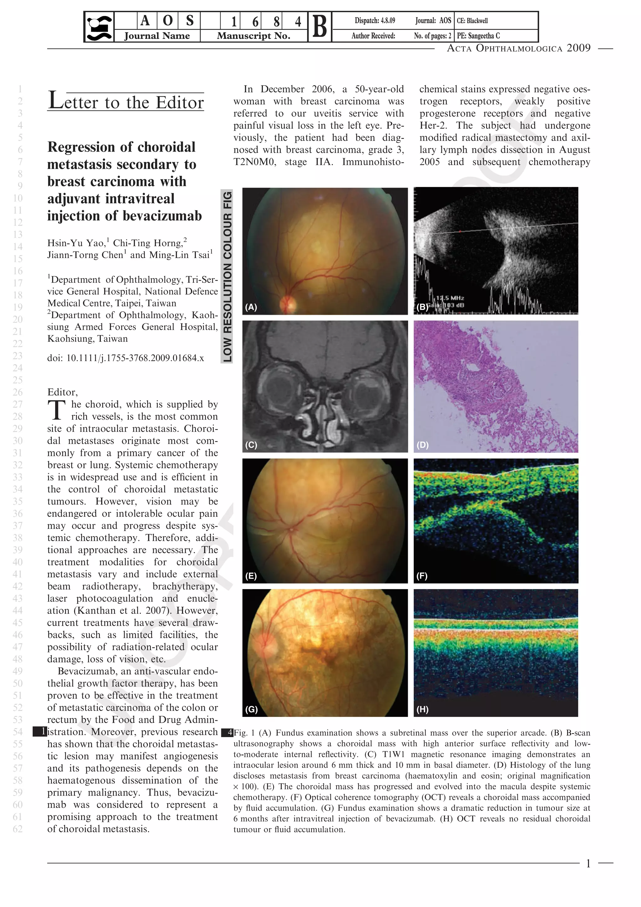
A O S
                         Journal Name          Manuscript No.
                                                                        1 6 8 4
                                                                                               B          Dispatch: 4.8.09
                                                                                                         Author Received:
                                                                                                                             Journal: AOS CE: Blackwell
                                                                                                                             No. of pages: 2 PE: Sangeetha C
                                                                                                                                        Acta Ophthalmologica 2009


 1                                                                              In December 2006, a 50-year-old               chemical stains expressed negative oes-
 2
 3
      Letter to the Editor                                                   woman with breast carcinoma was
                                                                             referred to our uveitis service with
                                                                                                                              trogen receptors, weakly positive
                                                                                                                              progesterone receptors and negative
 4                                                                           painful visual loss in the left eye. Pre-        Her-2. The subject had undergone
 5                                                                           viously, the patient had been diag-              modified radical mastectomy and axil-
 6    Regression of choroidal                                                nosed with breast carcinoma, grade 3,            lary lymph nodes dissection in August
 7    metastasis secondary to                                                T2N0M0, stage IIA. Immunohisto-                  2005 and subsequent chemotherapy
 8
 9    breast carcinoma with


                                                 LOW RESOLUTION COLOUR FIG
10    adjuvant intravitreal
11
12
      injection of bevacizumab
13
14    Hsin-Yu Yao,1 Chi-Ting Horng,2
15    Jiann-Torng Chen1 and Ming-Lin Tsai1
16    1
17      Department of Ophthalmology, Tri-Ser-
18    vice General Hospital, National Defence
19    Medical Centre, Taipei, Taiwan                                           (A)                                           (B)
      2
20      Department of Ophthalmology, Kaoh-
      siung Armed Forces General Hospital,
21
      Kaohsiung, Taiwan
22
23    doi: 10.1111/j.1755-3768.2009.01684.x
24
25
26     Editor,
27
28
29
      T       he choroid, which is supplied by
              rich vessels, is the most common
       site of intraocular metastasis. Choroi-
30     dal metastases originate most com-          (C)                                        (D)
31     monly from a primary cancer of the
32     breast or lung. Systemic chemotherapy
33     is in widespread use and is efficient in
34     the control of choroidal metastatic
35     tumours. However, vision may be
36     endangered or intolerable ocular pain
37     may occur and progress despite sys-
38     temic chemotherapy. Therefore, addi-
39     tional approaches are necessary. The
40     treatment modalities for choroidal
41     metastasis vary and include external        (E)                                        (F)
42     beam radiotherapy, brachytherapy,
43     laser photocoagulation and enucle-
44     ation (Kanthan et al. 2007). However,
45     current treatments have several draw-
46     backs, such as limited facilities, the
47     possibility of radiation-related ocular
48     damage, loss of vision, etc.
49        Bevacizumab, an anti-vascular endo-
50     thelial growth factor therapy, has been
51     proven to be effective in the treatment
52     of metastatic carcinoma of the colon or     (G)                                        (H)
53     rectum by the Food and Drug Admin-
54   1 istration. Moreover, previous research 4 Fig. 1 (A) Fundus examination shows a subretinal mass over the superior arcade. (B) B-scan
55     has shown that the choroidal metastas- ultrasonography shows a choroidal mass with high anterior surface reflectivity and low-
56     tic lesion may manifest angiogenesis     to-moderate internal reflectivity. (C) T1W1 magnetic resonance imaging demonstrates an
57     and its pathogenesis depends on the intraocular lesion around 6 mm thick and 10 mm in basal diameter. (D) Histology of the lung
58     haematogenous dissemination of the discloses metastasis from breast carcinoma (haematoxylin and eosin; original magnification
                                                · 100). (E) The choroidal mass has progressed and evolved into the macula despite systemic
59     primary malignancy. Thus, bevacizu- chemotherapy. (F) Optical coherence tomography (OCT) reveals a choroidal mass accompanied
60     mab was considered to represent a by fluid accumulation. (G) Fundus examination shows a dramatic reduction in tumour size at
61     promising approach to the treatment 6 months after intravitreal injection of bevacizumab. (H) OCT reveals no residual choroidal
62     of choroidal metastasis.                 tumour or fluid accumulation.


                                                                                                                                                                   1
Acta Ophthalmologica 2009


 1   with cyclophosphamide, doxorubicin residual lesion or fluid accumulation              Further studies are necessary to eluci-
 2   and 5-fluorouracil. On ophthalmic           were observed on optical coherence        date its optimal uses.
 3   examination, her best corrected visual     tomography (Fig. 1H). Two years
 4   acuity (BCVA) was 20 ⁄ 25 in the right     later, the patient remained stable and
 5   eye and 20 ⁄ 400 in the left. Fundus there was no recurrent choroidal mass.          References
 6   examination revealed a subretinal             Survival time in choroidal metasta-
 7   mass over the superior arcade (Fig. 1      sis is reported to be poor because cho- Amselem L, Cervera E, Dı´ az-Llopis M,
                                                                                            Montero J, Garcia-Pous M, Udaondo P,
 8   A). B-scan ultrasonography showed a        roidal metastasis represents the            Garcia-Delpech S & Salom D (2007): Intravi-
 9   dome-shaped choroidal lesion (Fig. 1       disseminated stage of cancer. In a ran-     treal bevacizumab (Avastin) for choroidal
10   B). Magnetic resonance imaging dem- domized trial, bevacizumab was                     metastasis secondary to breast carcinoma:
11   onstrated an intraocular lesion around     approved in combination with paclit-        short-term follow-up. Eye 21: 566–567.
12   6 mm thick and measuring 10 mm             axel for the treatment of patients with Kanthan GL, Jayamohan J, Yip D & Con-
13   across its base (Fig. 1C). Optical metastatic breast carcinoma (Michel                 way RM (2007): Management of metastatic
14   coherence tomography indicated a         2 & Xavier 2008). Amselem et al. (2007)       carcinoma of the uveal tract: an evidence-
                                                                                            based analysis. Clin Experiment Ophthal-
15   choroidal mass accompanied by fluid         reported that off-label use of intravi-
                                                                                            mol 35: 553–565.
16   accumulation (Fig. 1F). The patient treal bevacizumab might achieve                  Kuo IC, Haller JA, Maffrand R, Sambuelli
17   underwent a positron emission tomog-       short-term regression of choroidal          RH & Reviglio VE (2008): Regression of a
18   raphy (PET) scan which showed hyper- metastasis secondary to breast carci-             subfoveal choroidal metastasis of colorectal
19   dense lesions in the lung and mediasti-    noma for 3 weeks. Kuo et al. (2008)         carcinoma after intravitreous bevacizumab
20   nal lymph nodes. Biopsy guided by declared a similar efficacy in the treat-             treatment. Arch Ophthalmol 126: 1311–
21   computed tomography was performed ment of choroidal metastasis from                    1313.
22   and confirmed the diagnosis of multi- colorectal carcinoma. In our case, Marty M & Pivot X (2008): The potential of
                                                                                            anti-vascular endothelial growth factor
23   ple metastases of breast carcinoma intravitreal injection of 2.5 mg bev-               therapy in metastatic breast cancer: clinical
24   (Fig. 1D). The patient therefore under-    acizumab led to a significant regres-        experience with anti-angiogenic agents,
25   went systemic chemotherapy with pac- sion of the choroidal lesion. In                  focusing on bevacizumab. Eur J Cancer 44:
26   litaxel and gemcitabine. However, a        addition, there was no recurrent cho- 3 912–920.
27   progressive choroidal mass was noted.      roidal mass during the 24-month fol-
28   Best corrected VA deteriorated to low-up. The possible mechanisms of
29   counting fingers and the choroidal          intravitreal bevacizumab may involve
30   mass evolved into the macular region anti-angiogenic and anti-permeability           Correspondence:
31   (Fig. 1E). An adjuvant intravitreal        effects on angiogenesis in choroidal      Ming-Ling Tsai MD, PhD
32   injection of bevacizumab (2.5 mg in        metastasis. Synergistic effects of intra- Department of Ophthalmology
33   0.1 ml) was administered and a signifi- vitreal bevacizumab combined with Tri-Service General Hospital
34   cant reduction in mass was achieved.       concurrent chemotherapy, such as 325, Section 2, Cheng-Kung Road
35   Six months after the injection, the sub-   paclitaxel, may also play an important Nei-Hu District 114
36   ject’s BCVA improved to 20 ⁄ 30 and role.                                            Taipei
                                                                                          Taiwan
37   fundus examination demonstrated the           Intravitreal bevacizumab provides a
                                                                                          Tel: + 886 9 6839 2245
38   almost entire regression of the choroi-    potential option for adjuvant therapy Fax: + 886 2 2938 8737
39   dal mass (Fig. 1G). Furthermore, no        in patients with choroidal metastasis.    Email: doc30845@yahoo.com.tw
40
41
42
43
44
45
46
47
48
49
50
51
52
53
54
55
56
57
58
59
60
61
62


     2
Author Query Form
Journal:           AOS

Article:           1684
Dear Author,
During the copy-editing of your paper, the following queries arose. Please respond to these by marking up your
proofs with the necessary changes/additions. Please write your answers on the query sheet if there is insufficient
space on the page proofs. Please write clearly and follow the conventions shown on the attached corrections sheet.
If returning the proof by fax do not write too close to the paper’s edge. Please remember that illegible mark-ups
may delay publication.

Many thanks for your assistance.


Query reference       Query                                                                   Remarks
Q1                    AUTHOR: please insert reference for this statement
Q2                    AUTHOR: Michel & Xavier, 2008 has not been included in the Refer-
                      ence List, please supply full publication details.
Q3                    AUTHOR: Marty & Pivot (2008) has not been cited in the text. Please
                      indicate where it should be cited; or delete from the Reference List.
Q4                    AUTHOR: Figure 1 has been saved at a low resolution of 140 dpi.
                      Please resupply at 300 dpi. Check required artwork specifications at
                      http://www.blackwellpublishing.com/authors/digill.asp.
USING E-ANNOTATION TOOLS FOR ELECTRONIC PROOF CORRECTION
Required Software
Adobe Acrobat Professional or Acrobat Reader (version 7.0 or above) is required to e-annotate PDFs.
Acrobat 8 Reader is a free download: http://www.adobe.com/products/acrobat/readstep2.html
Once you have Acrobat Reader 8 on your PC and open the proof, you will see the Commenting Toolbar (if it
does not appear automatically go to Tools>Commenting>Commenting Toolbar). The Commenting Toolbar
looks like this:


If you experience problems annotating files in Adobe Acrobat Reader 9 then you may need to change a
preference setting in order to edit.
In the “Documents” category under “Edit – Preferences”, please select the category ‘Documents’ and
change the setting “PDF/A mode:” to “Never”.


Note Tool — For making notes at specific points in the text
Marks a point on the paper where a note or question needs to be addressed.

                                              How to use it:
                                               1. Right click into area of either inserted
                                                   text or relevance to note
                                               2. Select Add Note and a yellow speech
                                                   bubble symbol and text box will appear
                                               3. Type comment into the text box
                                               4. Click the X in the top right hand corner
                                                   of the note box to close.

Replacement text tool — For deleting one word/section of text and replacing it
Strikes red line through text and opens up a replacement text box.

                                                        How   to use it:
                                                         1.    Select cursor from toolbar
                                                         2.    Highlight word or sentence
                                                         3.    Right click
                                                         4.    Select Replace Text (Comment) option
                                                         5.    Type replacement text in blue box
                                                         6.    Click outside of the blue box to close


Cross out text tool — For deleting text when there is nothing to replace selection
Strikes through text in a red line.
                                        How   to use it:
                                         1.    Select cursor from toolbar
                                         2.    Highlight word or sentence
                                         3.    Right click
                                         4.    Select Cross Out Text


Page 1 of 3
Approved tool — For approving a proof and that no corrections at all are required.

                                                     How to use it:
                                                      1. Click on the Stamp Tool in the toolbar
                                                      2. Select the Approved rubber stamp from
                                                          the ‘standard business’ selection
                                                      3. Click on the text where you want to rubber
                                                          stamp to appear (usually first page)

Highlight tool — For highlighting selection that should be changed to bold or italic.
Highlights text in yellow and opens up a text box.

                                                  How to use it:
                                                  1. Select Highlighter Tool from the
                                                     commenting toolbar
                                                  2. Highlight the desired text
                                                  3. Add a note detailing the required change


Attach File Tool — For inserting large amounts of text or replacement figures as a files.
Inserts symbol and speech bubble where a file has been inserted.

                                       How   to use it:
                                        1.    Click on paperclip icon in the commenting toolbar
                                        2.    Click where you want to insert the attachment
                                        3.    Select the saved file from your PC/network
                                        4.    Select appearance of icon (paperclip, graph, attachment or
                                              tag) and close

Pencil tool — For circling parts of figures or making freeform marks
Creates freeform shapes with a pencil tool. Particularly with graphics within the proof it may be useful to use
the Drawing Markups toolbar. These tools allow you to draw circles, lines and comment on these marks.


  How  to use it:
   1.   Select Tools > Drawing Markups > Pencil Tool
   2.   Draw with the cursor
   3.   Multiple pieces of pencil annotation can be grouped together
   4.   Once finished, move the cursor over the shape until an arrowhead appears
        and right click
    5. Select Open Pop-Up Note and type in a details of required change
    6. Click the X in the top right hand corner of the note box to close.


Page 2 of 3
Acta

Acta

  • 1.
    A O S Journal Name Manuscript No. 1 6 8 4 B Dispatch: 4.8.09 Author Received: Journal: AOS CE: Blackwell No. of pages: 2 PE: Sangeetha C Acta Ophthalmologica 2009 1 In December 2006, a 50-year-old chemical stains expressed negative oes- 2 3 Letter to the Editor woman with breast carcinoma was referred to our uveitis service with trogen receptors, weakly positive progesterone receptors and negative 4 painful visual loss in the left eye. Pre- Her-2. The subject had undergone 5 viously, the patient had been diag- modified radical mastectomy and axil- 6 Regression of choroidal nosed with breast carcinoma, grade 3, lary lymph nodes dissection in August 7 metastasis secondary to T2N0M0, stage IIA. Immunohisto- 2005 and subsequent chemotherapy 8 9 breast carcinoma with LOW RESOLUTION COLOUR FIG 10 adjuvant intravitreal 11 12 injection of bevacizumab 13 14 Hsin-Yu Yao,1 Chi-Ting Horng,2 15 Jiann-Torng Chen1 and Ming-Lin Tsai1 16 1 17 Department of Ophthalmology, Tri-Ser- 18 vice General Hospital, National Defence 19 Medical Centre, Taipei, Taiwan (A) (B) 2 20 Department of Ophthalmology, Kaoh- siung Armed Forces General Hospital, 21 Kaohsiung, Taiwan 22 23 doi: 10.1111/j.1755-3768.2009.01684.x 24 25 26 Editor, 27 28 29 T he choroid, which is supplied by rich vessels, is the most common site of intraocular metastasis. Choroi- 30 dal metastases originate most com- (C) (D) 31 monly from a primary cancer of the 32 breast or lung. Systemic chemotherapy 33 is in widespread use and is efficient in 34 the control of choroidal metastatic 35 tumours. However, vision may be 36 endangered or intolerable ocular pain 37 may occur and progress despite sys- 38 temic chemotherapy. Therefore, addi- 39 tional approaches are necessary. The 40 treatment modalities for choroidal 41 metastasis vary and include external (E) (F) 42 beam radiotherapy, brachytherapy, 43 laser photocoagulation and enucle- 44 ation (Kanthan et al. 2007). However, 45 current treatments have several draw- 46 backs, such as limited facilities, the 47 possibility of radiation-related ocular 48 damage, loss of vision, etc. 49 Bevacizumab, an anti-vascular endo- 50 thelial growth factor therapy, has been 51 proven to be effective in the treatment 52 of metastatic carcinoma of the colon or (G) (H) 53 rectum by the Food and Drug Admin- 54 1 istration. Moreover, previous research 4 Fig. 1 (A) Fundus examination shows a subretinal mass over the superior arcade. (B) B-scan 55 has shown that the choroidal metastas- ultrasonography shows a choroidal mass with high anterior surface reflectivity and low- 56 tic lesion may manifest angiogenesis to-moderate internal reflectivity. (C) T1W1 magnetic resonance imaging demonstrates an 57 and its pathogenesis depends on the intraocular lesion around 6 mm thick and 10 mm in basal diameter. (D) Histology of the lung 58 haematogenous dissemination of the discloses metastasis from breast carcinoma (haematoxylin and eosin; original magnification · 100). (E) The choroidal mass has progressed and evolved into the macula despite systemic 59 primary malignancy. Thus, bevacizu- chemotherapy. (F) Optical coherence tomography (OCT) reveals a choroidal mass accompanied 60 mab was considered to represent a by fluid accumulation. (G) Fundus examination shows a dramatic reduction in tumour size at 61 promising approach to the treatment 6 months after intravitreal injection of bevacizumab. (H) OCT reveals no residual choroidal 62 of choroidal metastasis. tumour or fluid accumulation. 1
  • 2.
    Acta Ophthalmologica 2009 1 with cyclophosphamide, doxorubicin residual lesion or fluid accumulation Further studies are necessary to eluci- 2 and 5-fluorouracil. On ophthalmic were observed on optical coherence date its optimal uses. 3 examination, her best corrected visual tomography (Fig. 1H). Two years 4 acuity (BCVA) was 20 ⁄ 25 in the right later, the patient remained stable and 5 eye and 20 ⁄ 400 in the left. Fundus there was no recurrent choroidal mass. References 6 examination revealed a subretinal Survival time in choroidal metasta- 7 mass over the superior arcade (Fig. 1 sis is reported to be poor because cho- Amselem L, Cervera E, Dı´ az-Llopis M, Montero J, Garcia-Pous M, Udaondo P, 8 A). B-scan ultrasonography showed a roidal metastasis represents the Garcia-Delpech S & Salom D (2007): Intravi- 9 dome-shaped choroidal lesion (Fig. 1 disseminated stage of cancer. In a ran- treal bevacizumab (Avastin) for choroidal 10 B). Magnetic resonance imaging dem- domized trial, bevacizumab was metastasis secondary to breast carcinoma: 11 onstrated an intraocular lesion around approved in combination with paclit- short-term follow-up. Eye 21: 566–567. 12 6 mm thick and measuring 10 mm axel for the treatment of patients with Kanthan GL, Jayamohan J, Yip D & Con- 13 across its base (Fig. 1C). Optical metastatic breast carcinoma (Michel way RM (2007): Management of metastatic 14 coherence tomography indicated a 2 & Xavier 2008). Amselem et al. (2007) carcinoma of the uveal tract: an evidence- based analysis. Clin Experiment Ophthal- 15 choroidal mass accompanied by fluid reported that off-label use of intravi- mol 35: 553–565. 16 accumulation (Fig. 1F). The patient treal bevacizumab might achieve Kuo IC, Haller JA, Maffrand R, Sambuelli 17 underwent a positron emission tomog- short-term regression of choroidal RH & Reviglio VE (2008): Regression of a 18 raphy (PET) scan which showed hyper- metastasis secondary to breast carci- subfoveal choroidal metastasis of colorectal 19 dense lesions in the lung and mediasti- noma for 3 weeks. Kuo et al. (2008) carcinoma after intravitreous bevacizumab 20 nal lymph nodes. Biopsy guided by declared a similar efficacy in the treat- treatment. Arch Ophthalmol 126: 1311– 21 computed tomography was performed ment of choroidal metastasis from 1313. 22 and confirmed the diagnosis of multi- colorectal carcinoma. In our case, Marty M & Pivot X (2008): The potential of anti-vascular endothelial growth factor 23 ple metastases of breast carcinoma intravitreal injection of 2.5 mg bev- therapy in metastatic breast cancer: clinical 24 (Fig. 1D). The patient therefore under- acizumab led to a significant regres- experience with anti-angiogenic agents, 25 went systemic chemotherapy with pac- sion of the choroidal lesion. In focusing on bevacizumab. Eur J Cancer 44: 26 litaxel and gemcitabine. However, a addition, there was no recurrent cho- 3 912–920. 27 progressive choroidal mass was noted. roidal mass during the 24-month fol- 28 Best corrected VA deteriorated to low-up. The possible mechanisms of 29 counting fingers and the choroidal intravitreal bevacizumab may involve 30 mass evolved into the macular region anti-angiogenic and anti-permeability Correspondence: 31 (Fig. 1E). An adjuvant intravitreal effects on angiogenesis in choroidal Ming-Ling Tsai MD, PhD 32 injection of bevacizumab (2.5 mg in metastasis. Synergistic effects of intra- Department of Ophthalmology 33 0.1 ml) was administered and a signifi- vitreal bevacizumab combined with Tri-Service General Hospital 34 cant reduction in mass was achieved. concurrent chemotherapy, such as 325, Section 2, Cheng-Kung Road 35 Six months after the injection, the sub- paclitaxel, may also play an important Nei-Hu District 114 36 ject’s BCVA improved to 20 ⁄ 30 and role. Taipei Taiwan 37 fundus examination demonstrated the Intravitreal bevacizumab provides a Tel: + 886 9 6839 2245 38 almost entire regression of the choroi- potential option for adjuvant therapy Fax: + 886 2 2938 8737 39 dal mass (Fig. 1G). Furthermore, no in patients with choroidal metastasis. Email: doc30845@yahoo.com.tw 40 41 42 43 44 45 46 47 48 49 50 51 52 53 54 55 56 57 58 59 60 61 62 2
  • 3.
    Author Query Form Journal: AOS Article: 1684 Dear Author, During the copy-editing of your paper, the following queries arose. Please respond to these by marking up your proofs with the necessary changes/additions. Please write your answers on the query sheet if there is insufficient space on the page proofs. Please write clearly and follow the conventions shown on the attached corrections sheet. If returning the proof by fax do not write too close to the paper’s edge. Please remember that illegible mark-ups may delay publication. Many thanks for your assistance. Query reference Query Remarks Q1 AUTHOR: please insert reference for this statement Q2 AUTHOR: Michel & Xavier, 2008 has not been included in the Refer- ence List, please supply full publication details. Q3 AUTHOR: Marty & Pivot (2008) has not been cited in the text. Please indicate where it should be cited; or delete from the Reference List. Q4 AUTHOR: Figure 1 has been saved at a low resolution of 140 dpi. Please resupply at 300 dpi. Check required artwork specifications at http://www.blackwellpublishing.com/authors/digill.asp.
  • 4.
    USING E-ANNOTATION TOOLSFOR ELECTRONIC PROOF CORRECTION Required Software Adobe Acrobat Professional or Acrobat Reader (version 7.0 or above) is required to e-annotate PDFs. Acrobat 8 Reader is a free download: http://www.adobe.com/products/acrobat/readstep2.html Once you have Acrobat Reader 8 on your PC and open the proof, you will see the Commenting Toolbar (if it does not appear automatically go to Tools>Commenting>Commenting Toolbar). The Commenting Toolbar looks like this: If you experience problems annotating files in Adobe Acrobat Reader 9 then you may need to change a preference setting in order to edit. In the “Documents” category under “Edit – Preferences”, please select the category ‘Documents’ and change the setting “PDF/A mode:” to “Never”. Note Tool — For making notes at specific points in the text Marks a point on the paper where a note or question needs to be addressed. How to use it: 1. Right click into area of either inserted text or relevance to note 2. Select Add Note and a yellow speech bubble symbol and text box will appear 3. Type comment into the text box 4. Click the X in the top right hand corner of the note box to close. Replacement text tool — For deleting one word/section of text and replacing it Strikes red line through text and opens up a replacement text box. How to use it: 1. Select cursor from toolbar 2. Highlight word or sentence 3. Right click 4. Select Replace Text (Comment) option 5. Type replacement text in blue box 6. Click outside of the blue box to close Cross out text tool — For deleting text when there is nothing to replace selection Strikes through text in a red line. How to use it: 1. Select cursor from toolbar 2. Highlight word or sentence 3. Right click 4. Select Cross Out Text Page 1 of 3
  • 5.
    Approved tool —For approving a proof and that no corrections at all are required. How to use it: 1. Click on the Stamp Tool in the toolbar 2. Select the Approved rubber stamp from the ‘standard business’ selection 3. Click on the text where you want to rubber stamp to appear (usually first page) Highlight tool — For highlighting selection that should be changed to bold or italic. Highlights text in yellow and opens up a text box. How to use it: 1. Select Highlighter Tool from the commenting toolbar 2. Highlight the desired text 3. Add a note detailing the required change Attach File Tool — For inserting large amounts of text or replacement figures as a files. Inserts symbol and speech bubble where a file has been inserted. How to use it: 1. Click on paperclip icon in the commenting toolbar 2. Click where you want to insert the attachment 3. Select the saved file from your PC/network 4. Select appearance of icon (paperclip, graph, attachment or tag) and close Pencil tool — For circling parts of figures or making freeform marks Creates freeform shapes with a pencil tool. Particularly with graphics within the proof it may be useful to use the Drawing Markups toolbar. These tools allow you to draw circles, lines and comment on these marks. How to use it: 1. Select Tools > Drawing Markups > Pencil Tool 2. Draw with the cursor 3. Multiple pieces of pencil annotation can be grouped together 4. Once finished, move the cursor over the shape until an arrowhead appears and right click 5. Select Open Pop-Up Note and type in a details of required change 6. Click the X in the top right hand corner of the note box to close. Page 2 of 3