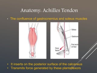
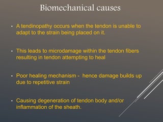
Overview of Achilles tendinopathy and its biomechanical causes. graph: load-deformation graph for Achilles tendon. Prepared by Charles Nyasa (physiotherapist)- university of malawi
1. ACHILLES TENDINOPATHY
Overview and Biomechanical causes
By:
Charles Nyasa Elizabeth Mchakama Sangwani Ngwira Hannah Mphaka
Grace Mwale Burnet Makaika & Mathews Rodgers Phiri
2. Anatomy: Achilles Tendon
• The confluence of gastrocnemius and soleus muscles
• It inserts on the posterior surface of the calcaneus
• Transmits force generated by these plantaflexors
3. Biomechanics of Achilles tendon
• A tendon is tough band of fibrous connective tissue
• Achilles tendon is not enclosed in a true synovial sheath
- But rather a paratendon – richly vascularized
• however, towards its distal end the tendon begins to
rotate
- More marked in the distal most 5-6 cm
• Angiography reveals a zone of hypovascularity 2-7cm
proximal to tendon insertion.
4. Achilles Tendinopathy
• A condition that causes pain, swelling, stiffness and
weakness of the Achilles tendon
- Caused by tiny injuries to the Achilles tendon
• Originally labelled Achilles tendinitis
- Overuse degeneration rather than inflammation revealed
5. Biomechanical causes
• A tendinopathy occurs when the tendon is unable to
adapt to the strain being placed on it.
• This leads to microdamage within the tendon fibers
resulting in tendon attempting to heal
• Poor healing mechanism - hence damage builds up
due to repetitive strain
• Causing degeneration of tendon body and/or
inflammation of the sheath.
6. Load – Deformation Curve
Causes explained:
• Repetitive overload beyond physiological threshold e.g. Vigorous P.A
• Frequent microtrauma within physiological limits e.g. Overweight
• Non-uniform stress within tendon due to abnormal load conc. and
friction btwn fibrils, e.g. weak/tight calf muscle, foot joints stiffness
But rather a paratendon – composed of a single cell layer
Tendons transmit force generated by muscle to bone. Additionally, they act as a buffer by absorbing external forces to limit muscle damage—a function that demands mechanical strength, flexibility and elasticity.5 As collagen fibres deform, they respond linearly to increasing tendon loads.8 The configuration is initially lost when the stretch exceeds 2% but is regained if the strain placed on the tendon remains at less than 4%; if strain exceeds 8% macroscopic rupture will occur.9,10 ………….foot joint stiffness and poor core stability around the hip/knee.