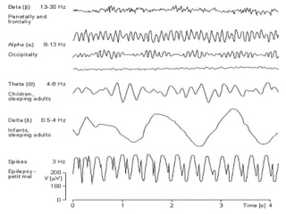
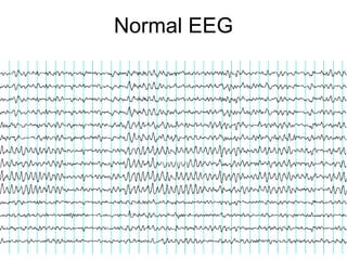
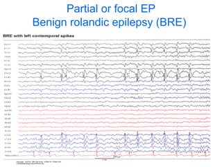
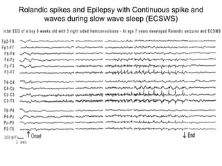
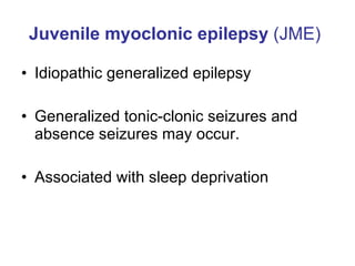
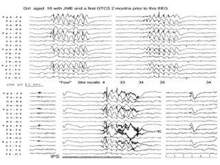
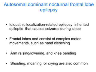
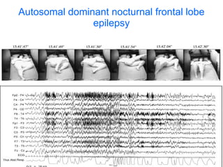
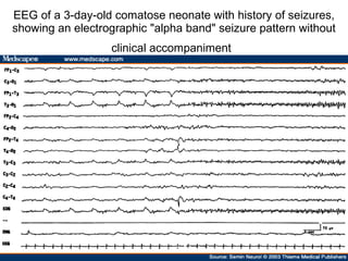
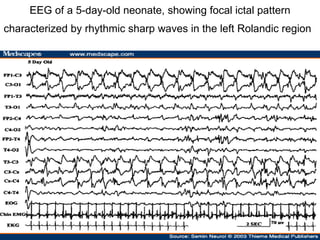
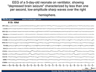
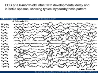
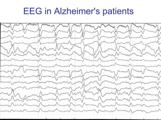
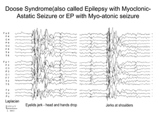
Recognizing epilepsies Dr. Francois Kouya
 
ALPHA waves Alpha waves are those between 7.5 and thirteen(13) waves per second (Hz). Alpha is usually best seen in the posterior regions of the head on each side, being higher in amplitude on the dominant side.  It is brought out by closing the eyes and by relaxation, and abolished by eye opening or alerting by any mechanism (thinking, calculating). It is the major rhythm seen in normal relaxed adults - it is present during most of life especially beyond the thirteenth year when it dominates the resting tracing .
BETA waves Beta activity is 'fast' activity. It has a frequency of 14 and greater Hz. It is usually seen on both sides in symmetrical distribution and is most evident frontally.  It is accentuated by sedative-hypnotic drugs especially the benzodiazepines and the barbiturates. It may be absent or reduced in areas of cortical damage. It is generally regarded as a normal rhythm.  It is the dominant rhythm in patients who are alert or anxious or who have their eyes open
Eyes Closed, 14 yrs. old. Very clear beta spindles Frontocentral
Eyes Open, 43 yrs. old: Clear frontocentral beta spindles, high-frequency (31-36 Hz).
THETA waves Theta activity has a frequency of 3.5 to 7.5 Hz and is classed as "slow" activity. It is abnormal in awake adults but is perfectly normal in children up to 13 years and in sleep. It can be seen as a focal disturbance in focal subcortical lesions It can be seen in generalized distribution in diffuse disorder or metabolic encephalopathy In deep midline disorders or some instances of hydrocephalus
DELTA waves Delta activity is 3 Hz or below. It tends to be the highest in amplitude and the slowest waves. It is quite normal and is the dominant rhythm in infants up to one year and in stages 3 and 4 of sleep. It may occur focally with subcortical lesions and in general distribution with diffuse lesions, metabolic encephalopathy hydrocephalus or deep midline lesions. It is usually most prominent frontally in adults (e.g. FIRDA - Frontal Intermittent Rhythmic Delta) and posteriorly in children e.g. OIRDA - Occipital Intermittent Rhythmic Delta).
Normal EEG
Benign centrotemporal lobe epilepsy of childhood:Benign Rolandic epilepsy   Idiopathic localization-related epilepsy that occurs in children between the ages of 3 and 13. Seizures are typically nocturnal and confined to sleep The patients are otherwise normal apart from the seizure
Partial or focal EP Benign rolandic epilepsy (BRE)
Rolandic spikes and Epilepsy with Continuous spike and waves during slow wave sleep (ECSWS)
Juvenile myoclonic epilepsy  (JME)   Idiopathic generalized epilepsy  Generalized tonic-clonic seizures and absence seizures may occur. Associated with sleep deprivation
Polyspike and wave discharges seen in juvenile myoclonic epilepsy
 
Autosomal dominant nocturnal frontal lobe epilepsy Idiopathic localization-related epilepsy  inherited epileptic  that causes seizures during sleep Frontal lobes and consist of complex motor movements, such as hand clenching Arm raising/lowering, and knee bending Shouting, moaning, or crying are also common
Frontal lobe EP
Autosomal dominant nocturnal frontal lobe epilepsy
Juvenile absence epilepsy   Idiopathic generalized epilepsy Typically in pre-pubertal adolescence Generalized tonic-colonic seizures can occur
 
EEG of a 53-year-old man with one day history of acute confusion and slowness of motor responses, showing almost continuous generalized spike wave activity.(status epilepticus)
Partial or focal EP  Benign Rolandic epilepsy (BRE)
EEG of an 11-year-old patient, showing rhythmic midtemporal discharges during drowsiness
EEG of a 26-year-old patient, showing 6 Hz spike wave paroxysms (phantom spike wave).
EEG of a 9-year-old child with benign Rolandic epilepsy, showing a focus of right centrotemporal spike discharges. The right half of the figure shows a spike discharge  with horizontal dipole distribution.
EEG of a 68-year-old patient with a long history of complex partial seizures, showing a focus of sharp waves and low-amplitude slow activity over the right anterior temporal region
Lennox-Gastaut syndrome (LGS) is a generalized epilepsy that consists of a triad of developmental delay or childhood dementia. Mixed generalized seizures Onset occurs between 2-18 years
EEG of an 11-year-old patient with Lennox-Gastaut syndrome, showing generalized paroxysmal fast activity (B)
EEG of a 16-year-old child with mental retardation and tonic seizures, showing slow spike wave activity superimposed on a slow background .
Epilepsies in babies
EEG of a 3-day-old comatose neonate with history of seizures, showing an electrographic "alpha band" seizure pattern without clinical accompaniment
EEG of a 5-day-old neonate, showing focal ictal pattern characterized by rhythmic sharp waves in the left Rolandic region
EEG of a 5-day-old neonate on ventilator, showing "depressed brain seizure" characterized by less than one per second, low-amplitude sharp waves over the right hemisphere.
EEG of a 6-month-old infant with developmental delay and infantile spasms, showing typical hypsarrhythmic pattern
 
Childhood absence epilepsy Idiopathic generalized epilepsy that affects children between the ages of 4 and 12 years of age  Recurrent absence seizures
Typical 3-Hz spike and wave discharges seen in absence epilepsy
EEG in Creutzfeldt-Jakob disease
Alzheimer's disease (AD) Progressive degenerative brain disease.
EEG in Alzheimer's patients
Doose Syndrome(also called Epilepsy with Myoclonic-Astatic Seizure or EP with Myo-atonic seizure

Epilepsy

  • 1.
  • 2.
  • 3.
    ALPHA waves Alphawaves are those between 7.5 and thirteen(13) waves per second (Hz). Alpha is usually best seen in the posterior regions of the head on each side, being higher in amplitude on the dominant side. It is brought out by closing the eyes and by relaxation, and abolished by eye opening or alerting by any mechanism (thinking, calculating). It is the major rhythm seen in normal relaxed adults - it is present during most of life especially beyond the thirteenth year when it dominates the resting tracing .
  • 4.
    BETA waves Betaactivity is 'fast' activity. It has a frequency of 14 and greater Hz. It is usually seen on both sides in symmetrical distribution and is most evident frontally. It is accentuated by sedative-hypnotic drugs especially the benzodiazepines and the barbiturates. It may be absent or reduced in areas of cortical damage. It is generally regarded as a normal rhythm. It is the dominant rhythm in patients who are alert or anxious or who have their eyes open
  • 5.
    Eyes Closed, 14yrs. old. Very clear beta spindles Frontocentral
  • 6.
    Eyes Open, 43yrs. old: Clear frontocentral beta spindles, high-frequency (31-36 Hz).
  • 7.
    THETA waves Thetaactivity has a frequency of 3.5 to 7.5 Hz and is classed as "slow" activity. It is abnormal in awake adults but is perfectly normal in children up to 13 years and in sleep. It can be seen as a focal disturbance in focal subcortical lesions It can be seen in generalized distribution in diffuse disorder or metabolic encephalopathy In deep midline disorders or some instances of hydrocephalus
  • 8.
    DELTA waves Deltaactivity is 3 Hz or below. It tends to be the highest in amplitude and the slowest waves. It is quite normal and is the dominant rhythm in infants up to one year and in stages 3 and 4 of sleep. It may occur focally with subcortical lesions and in general distribution with diffuse lesions, metabolic encephalopathy hydrocephalus or deep midline lesions. It is usually most prominent frontally in adults (e.g. FIRDA - Frontal Intermittent Rhythmic Delta) and posteriorly in children e.g. OIRDA - Occipital Intermittent Rhythmic Delta).
  • 9.
  • 10.
    Benign centrotemporal lobeepilepsy of childhood:Benign Rolandic epilepsy Idiopathic localization-related epilepsy that occurs in children between the ages of 3 and 13. Seizures are typically nocturnal and confined to sleep The patients are otherwise normal apart from the seizure
  • 11.
    Partial or focalEP Benign rolandic epilepsy (BRE)
  • 12.
    Rolandic spikes andEpilepsy with Continuous spike and waves during slow wave sleep (ECSWS)
  • 13.
    Juvenile myoclonic epilepsy (JME) Idiopathic generalized epilepsy Generalized tonic-clonic seizures and absence seizures may occur. Associated with sleep deprivation
  • 14.
    Polyspike and wavedischarges seen in juvenile myoclonic epilepsy
  • 15.
  • 16.
    Autosomal dominant nocturnalfrontal lobe epilepsy Idiopathic localization-related epilepsy inherited epileptic that causes seizures during sleep Frontal lobes and consist of complex motor movements, such as hand clenching Arm raising/lowering, and knee bending Shouting, moaning, or crying are also common
  • 17.
  • 18.
    Autosomal dominant nocturnalfrontal lobe epilepsy
  • 19.
    Juvenile absence epilepsy Idiopathic generalized epilepsy Typically in pre-pubertal adolescence Generalized tonic-colonic seizures can occur
  • 20.
  • 21.
    EEG of a53-year-old man with one day history of acute confusion and slowness of motor responses, showing almost continuous generalized spike wave activity.(status epilepticus)
  • 22.
    Partial or focalEP Benign Rolandic epilepsy (BRE)
  • 23.
    EEG of an11-year-old patient, showing rhythmic midtemporal discharges during drowsiness
  • 24.
    EEG of a26-year-old patient, showing 6 Hz spike wave paroxysms (phantom spike wave).
  • 25.
    EEG of a9-year-old child with benign Rolandic epilepsy, showing a focus of right centrotemporal spike discharges. The right half of the figure shows a spike discharge with horizontal dipole distribution.
  • 26.
    EEG of a68-year-old patient with a long history of complex partial seizures, showing a focus of sharp waves and low-amplitude slow activity over the right anterior temporal region
  • 27.
    Lennox-Gastaut syndrome (LGS)is a generalized epilepsy that consists of a triad of developmental delay or childhood dementia. Mixed generalized seizures Onset occurs between 2-18 years
  • 28.
    EEG of an11-year-old patient with Lennox-Gastaut syndrome, showing generalized paroxysmal fast activity (B)
  • 29.
    EEG of a16-year-old child with mental retardation and tonic seizures, showing slow spike wave activity superimposed on a slow background .
  • 30.
  • 31.
    EEG of a3-day-old comatose neonate with history of seizures, showing an electrographic "alpha band" seizure pattern without clinical accompaniment
  • 32.
    EEG of a5-day-old neonate, showing focal ictal pattern characterized by rhythmic sharp waves in the left Rolandic region
  • 33.
    EEG of a5-day-old neonate on ventilator, showing "depressed brain seizure" characterized by less than one per second, low-amplitude sharp waves over the right hemisphere.
  • 34.
    EEG of a6-month-old infant with developmental delay and infantile spasms, showing typical hypsarrhythmic pattern
  • 35.
  • 36.
    Childhood absence epilepsyIdiopathic generalized epilepsy that affects children between the ages of 4 and 12 years of age Recurrent absence seizures
  • 37.
    Typical 3-Hz spikeand wave discharges seen in absence epilepsy
  • 38.
  • 39.
    Alzheimer's disease (AD)Progressive degenerative brain disease.
  • 40.
  • 41.
    Doose Syndrome(also calledEpilepsy with Myoclonic-Astatic Seizure or EP with Myo-atonic seizure