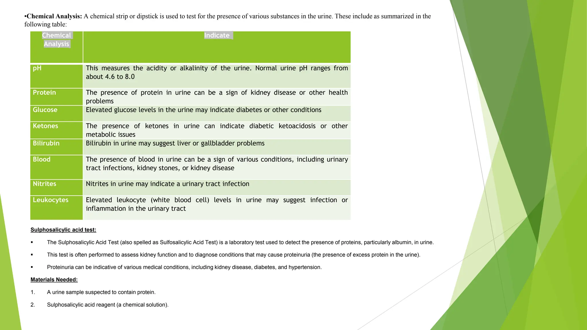
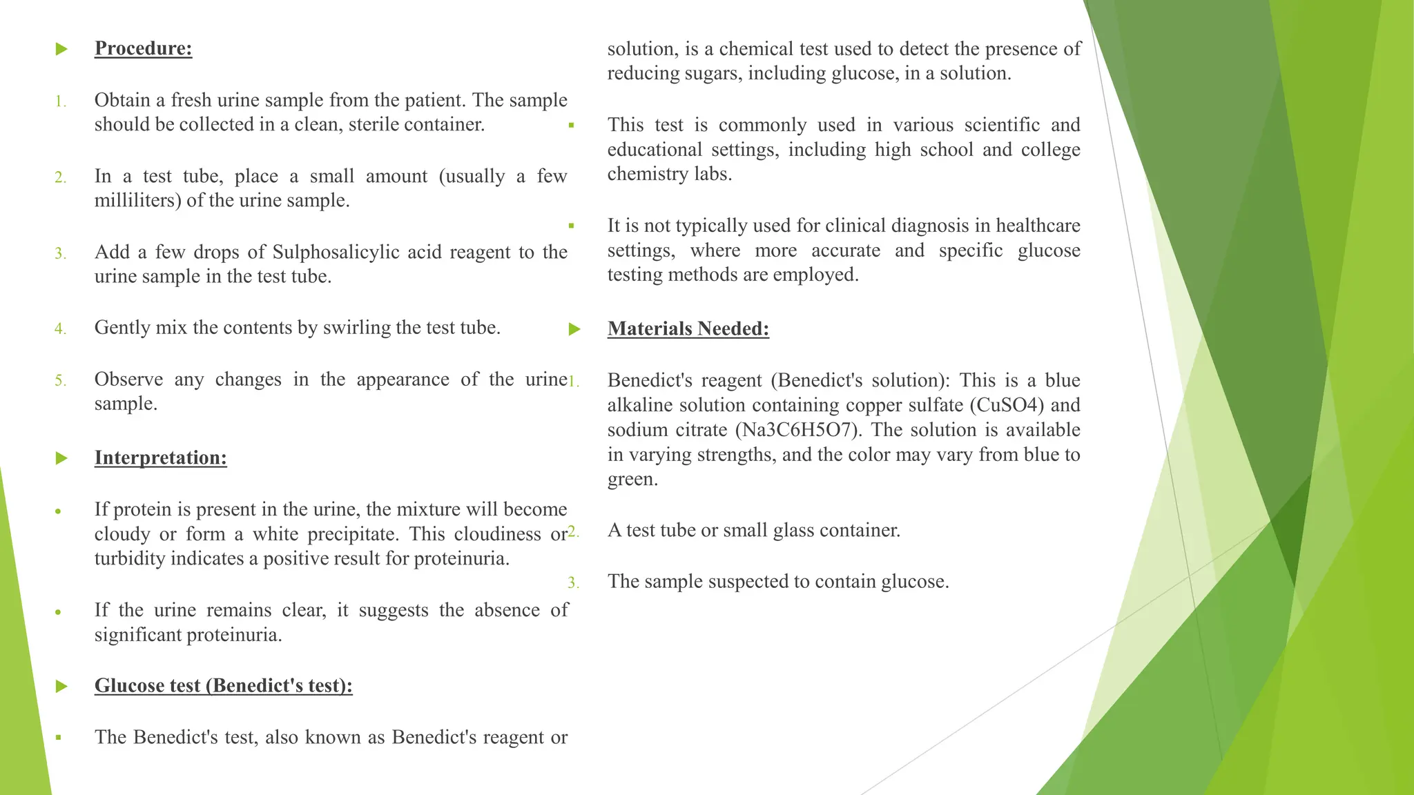
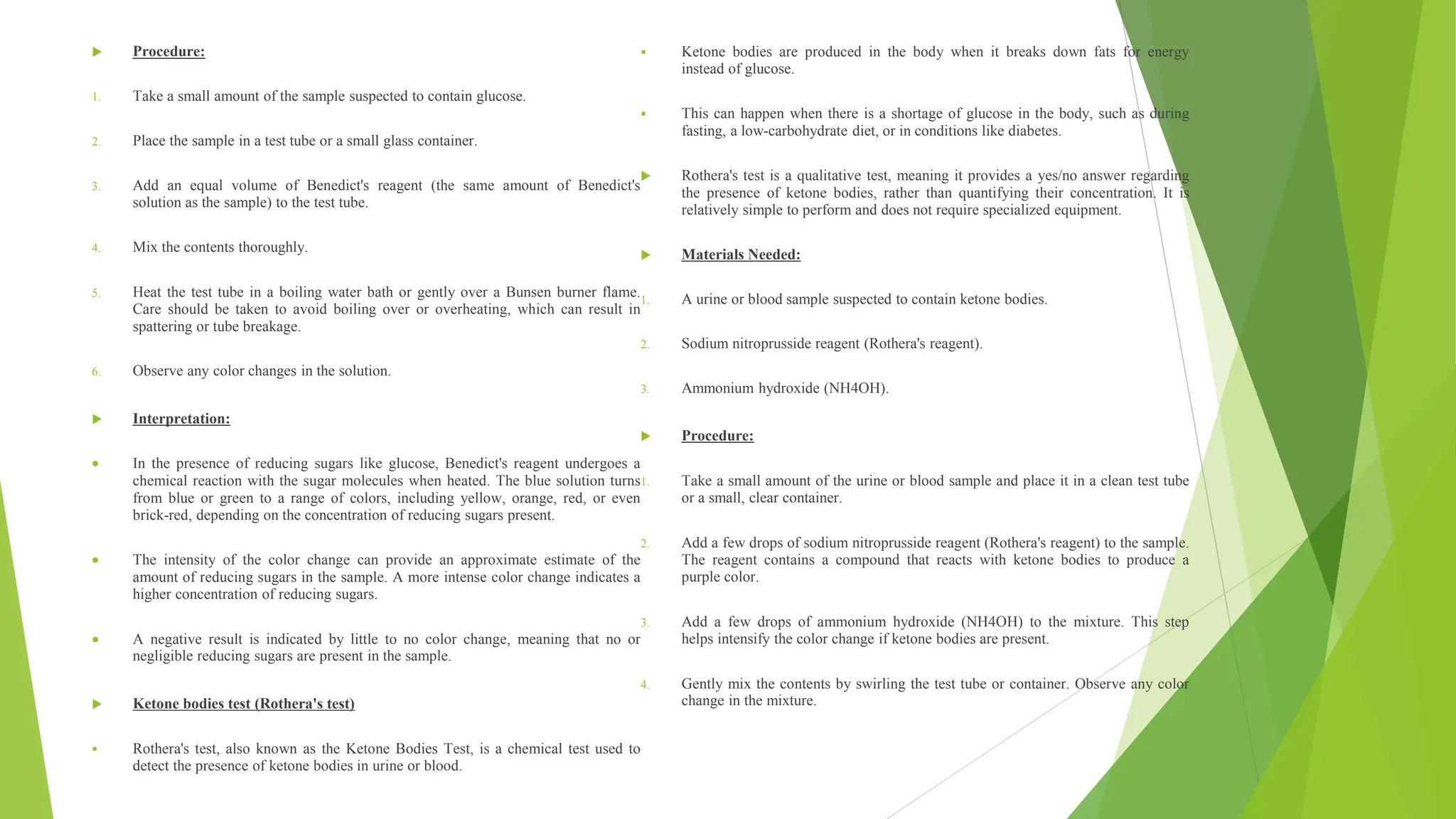
‫الطلبة‬ ‫اعداد‬
‫حسين‬ ‫راضي‬ ‫سجى‬
‫كاظم‬ ‫غفران‬
‫جاسم‬ ‫وليد‬ ‫زهراء‬
‫فليح‬ ‫حيدر‬ ‫كرار‬
‫فليح‬ ‫عدنان‬ ‫علي‬
‫والي‬ ‫العالي‬ ‫عبد‬ ‫زينب‬
‫سعيد‬ ‫ماجد‬ ‫مريم‬
‫محمد‬ ‫مصطفى‬
‫اشراف‬
‫د‬
.
‫بليغ‬
General Urine Examination (GUE) or Urinalysis
General urine examination (GUE) and urinalysis are terms often used interchangeably to describe a common medical test that involves the analysis of a
person's urine.
This test provides valuable information about a person's overall health and can help diagnose various medical conditions.
There are typically included in a general urine examination or urinalysis:
Physical Examination: This involves an assessment of the urine's color, clarity, and odor. Normal urine is typically pale to deep yellow and clear. Unusual
colors, cloudiness, or foul odors may indicate underlying health issues.
Urine color:
Colorless/pale yellow - recent fluid consumption, polyuria, DM.
Yellow - intestinal worms
,
Giardiasis.
Dark Yellow - Concentrated specimen; Streneous exercise
,
First morning specimen
Bright yellow- Riboflavin/ Multivitamins
Amber- Dehydration; fever, burns
Orange - Bilirubin (tea-colored urine)
Blue-green, Pseudomonas Infection, Methylene Blue, Phenol.
Pink/Red RBCS (Cloudy/Smoky red) Hematuria, Hemoglobin (Clear Red); Intravascular hemolysis, Myoglobin (Clear red or reddish-brown); muscle damage
Smell or Oder:
Normal - faint aromatic due to volatile acids; becomes ammoniacal as the specimen stands
Variations
Ammoniacal (freshly voided) - UTI (ex. Proteus vulgaris)
Mousy – Phenylketone urea (PKU)
Fruity/ sweet Diabetes Ketoacidosis (Ketones), Starvation
,
vomiting
Rotting fish/ Aminuria
Sulfur odor - Cystine Disorders
Chemical
Analysis
Indicate
pH This measures the acidity or alkalinity of the urine. Normal urine pH ranges from
about 4.6 to 8.0
Protein The presence of protein in urine can be a sign of kidney disease or other health
problems
Glucose Elevated glucose levels in the urine may indicate diabetes or other conditions
Ketones The presence of ketones in urine can indicate diabetic ketoacidosis or other
metabolic issues
Bilirubin Bilirubin in urine may suggest liver or gallbladder problems
Blood The presence of blood in urine can be a sign of various conditions, including urinary
tract infections, kidney stones, or kidney disease
Nitrites Nitrites in urine may indicate a urinary tract infection
Leukocytes Elevated leukocyte (white blood cell) levels in urine may suggest infection or
inflammation in the urinary tract
•Chemical Analysis: A chemical strip or dipstick is used to test for the presence of various substances in the urine. These include as summarized in the
following table:
Sulphosalicylic acid test:
 The Sulphosalicylic Acid Test (also spelled as Sulfosalicylic Acid Test) is a laboratory test used to detect the presence of proteins, particularly albumin, in urine.
 This test is often performed to assess kidney function and to diagnose conditions that may cause proteinuria (the presence of excess protein in the urine).
 Proteinuria can be indicative of various medical conditions, including kidney disease, diabetes, and hypertension.
Materials Needed:
1. A urine sample suspected to contain protein.
2. Sulphosalicylic acid reagent (a chemical solution).
 Procedure:
1. Obtain a fresh urine sample from the patient. The sample
should be collected in a clean, sterile container.
2. In a test tube, place a small amount (usually a few
milliliters) of the urine sample.
3. Add a few drops of Sulphosalicylic acid reagent to the
urine sample in the test tube.
4. Gently mix the contents by swirling the test tube.
5. Observe any changes in the appearance of the urine
sample.
 Interpretation:
 If protein is present in the urine, the mixture will become
cloudy or form a white precipitate. This cloudiness or
turbidity indicates a positive result for proteinuria.
 If the urine remains clear, it suggests the absence of
significant proteinuria.
 Glucose test (Benedict's test):
 The Benedict's test, also known as Benedict's reagent or
solution, is a chemical test used to detect the presence of
reducing sugars, including glucose, in a solution.
 This test is commonly used in various scientific and
educational settings, including high school and college
chemistry labs.
 It is not typically used for clinical diagnosis in healthcare
settings, where more accurate and specific glucose
testing methods are employed.
 Materials Needed:
1. Benedict's reagent (Benedict's solution): This is a blue
alkaline solution containing copper sulfate (CuSO4) and
sodium citrate (Na3C6H5O7). The solution is available
in varying strengths, and the color may vary from blue to
green.
2. A test tube or small glass container.
3. The sample suspected to contain glucose.
 Procedure:
1. Take a small amount of the sample suspected to contain glucose.
2. Place the sample in a test tube or a small glass container.
3. Add an equal volume of Benedict's reagent (the same amount of Benedict's
solution as the sample) to the test tube.
4. Mix the contents thoroughly.
5. Heat the test tube in a boiling water bath or gently over a Bunsen burner flame.
Care should be taken to avoid boiling over or overheating, which can result in
spattering or tube breakage.
6. Observe any color changes in the solution.
 Interpretation:
 In the presence of reducing sugars like glucose, Benedict's reagent undergoes a
chemical reaction with the sugar molecules when heated. The blue solution turns
from blue or green to a range of colors, including yellow, orange, red, or even
brick-red, depending on the concentration of reducing sugars present.
 The intensity of the color change can provide an approximate estimate of the
amount of reducing sugars in the sample. A more intense color change indicates a
higher concentration of reducing sugars.
 A negative result is indicated by little to no color change, meaning that no or
negligible reducing sugars are present in the sample.
 Ketone bodies test (Rothera's test)
 Rothera's test, also known as the Ketone Bodies Test, is a chemical test used to
detect the presence of ketone bodies in urine or blood.
 Ketone bodies are produced in the body when it breaks down fats for energy
instead of glucose.
 This can happen when there is a shortage of glucose in the body, such as during
fasting, a low-carbohydrate diet, or in conditions like diabetes.
 Rothera's test is a qualitative test, meaning it provides a yes/no answer regarding
the presence of ketone bodies, rather than quantifying their concentration. It is
relatively simple to perform and does not require specialized equipment.
 Materials Needed:
1. A urine or blood sample suspected to contain ketone bodies.
2. Sodium nitroprusside reagent (Rothera's reagent).
3. Ammonium hydroxide (NH4OH).
 Procedure:
1. Take a small amount of the urine or blood sample and place it in a clean test tube
or a small, clear container.
2. Add a few drops of sodium nitroprusside reagent (Rothera's reagent) to the sample.
The reagent contains a compound that reacts with ketone bodies to produce a
purple color.
3. Add a few drops of ammonium hydroxide (NH4OH) to the mixture. This step
helps intensify the color change if ketone bodies are present.
4. Gently mix the contents by swirling the test tube or container. Observe any color
change in the mixture.
 Interpretation:
 If the solution turns purple, it indicates the
presence of ketone bodies in the urine or blood
sample.
 If there is no color change, it suggests the
absence of significant levels of ketone bodies.
 Bile pigment test (Harrison test):
 The Harrison test, also known as the Bile
Pigment Test, is a laboratory test used to detect
the presence of bile pigments in urine.
 Bile pigments are produced by the liver and
play a crucial role in the digestion and
elimination of waste products from the body.
 Normally, bile pigments are metabolized by the
liver and excreted through the bile into the
digestive system.
 However, under certain conditions, such as liver
disease or hemolytic anemia, excessive amounts
of bile pigments may be present in the
bloodstream and subsequently appear in the
urine.
 Materials Needed:
1. A urine sample suspected to contain bile
pigments.
2. A small, clear test tube or a small glass
container.
3. A few drops of concentrated nitric acid.
 Procedure:
1. Collect a fresh urine sample from the patient,
ensuring that it is clean and free from
contaminants.
2. Place a small amount of the urine sample in a
test tube or glass container.
3. Add a few drops of concentrated nitric acid to
the urine sample. Be cautious when handling
concentrated acids, as they can be corrosive and
should be used in a well-ventilated area while
wearing appropriate protective gear.
4. Gently mix the contents by swirling the test tube
or container.
5. Observe any color changes in the urine sample
after the addition of nitric acid.
 Interpretation:
 If bile pigments are present in the urine, the
mixture may turn yellow, green, or brownish.
The appearance of these colors indicates a
positive result for the presence of bile pigments.
 If there is no color change or only a slight
change, it suggests the absence of significant
bile pigments in the urine.
 The Harrison test is a relatively simple and
qualitative way to detect bile pigments in urine.
If bile pigments are detected, it may indicate a
potential issue with liver function or hemolysis
(destruction of red blood cells), and further
diagnostic tests are typically required to identify
the underlying cause.
 Urobilinogen test (Ehrlich's test):
 The Urobilinogen Test, also known as Ehrlich's Test for
urobilinogen, is a laboratory test used to measure the
presence and quantity of urobilinogen in urine.
 Urobilinogen is a breakdown product of bilirubin, which is
formed during the normal breakdown of red blood cells in
the body.
 Bilirubin is processed by the liver and eliminated from the
body in the bile. Some of it gets converted to urobilinogen
and is excreted in the urine.
 The Urobilinogen Test is used to assess liver function and
the breakdown of red blood cells. A
 bnormal levels of urobilinogen in urine can indicate various
medical conditions, including liver disease, hemolysis (the
destruction of red blood cells), or issues with the biliary
system
 Materials Needed:
1. A urine sample suspected to contain urobilinogen.
2. Ehrlich's reagent (a chemical reagent containing p-
dimethylaminobenzaldehyde).
 Procedure:
1. Collect a fresh urine sample from the patient, ensuring that
it is clean and free from contaminants.
2. Place a small amount of the urine sample in a clean test
tube or glass container.
3. Add a few drops of Ehrlich's reagent (p-
dimethylaminobenzaldehyde) to the urine sample.
4. Gently mix the contents by swirling the test tube or
container.
5. Allow the mixture to stand for a few minutes and observe
any color changes.
 Interpretation:
 If urobilinogen is present in the urine, the mixture may turn
pink, red, or purple. The intensity of the color change can
provide an estimate of the urobilinogen concentration in the
urine.
 If there is no color change or only a slight change, it
suggests the absence of significant urobilinogen in the
urine.
1. Microscopic Examination: A sample of urine is
examined under a microscope to detect the presence
of red blood cells, white blood cells, crystals,
bacteria, and other cellular elements. This can help
diagnose conditions like urinary tract infections,
kidney stones, or kidney disease.
 Red Blood Cells
 Hematuria: is the presence of abnormal numbers of
red cells in urine due to: glomerular damage, tumors
which erode the urinary tract anywhere along its
length, kidney trauma, urinary tract stones, renal
infarcts, acute tubular necrosis, upper and lower
urinary tract infections, nephrotoxins, and physical
stress. Red cells may also contaminate the urine
from the vagina in menstruating women or from
trauma produced by bladder catherization.
Theoretically, no red cells should be found, but
some find their way into the urine even in very
healthy individuals.
 White Blood Cells
 Pyuria refers to the presence of abnormal numbers
of leukocytes that may appear with infection in
either the upper or lower urinary tract or with acute
glomerulonephritis. Usually, the WBC's are
granulocytes. White cells from the vagina, especially
in the presence of vaginal and cervical infections, or
the external urethral meatus in men and women may
contaminate the urine.
 Epithelial Cells
 Epithelial cells, usually larger than granulocytes,
contain a large round or oval nucleus and normally
slough into the urine in small numbers
 Casts
 Urinary casts are cylindrical structures that are
formed from coagulated protein secreted by tubular
cells.
 Type of Cast: Hyaline casts R.B.C casts
W.B.C Casts Granular casts
 Bacteria:
 Bacteria are common in urine specimens because of the abundant normal microbial flora of the vagina or
external urethral meatus and because of their ability to rapidly multiply in urine standing at room temperature.
Therefore, microbial organisms found in all but the most scrupulously collected urines should be interpreted in
view of clinical symptoms .
 Yeast:
 Yeast cells may be contaminants or represent a true yeast infection. They are often difficult to distinguish from
red cells and amorphous crystals but are distinguished by their tendency to bud. Most often they are Candida,
which may colonize bladder, urethra, or vagina.
 Crystals:
 Common crystals seen even in healthy patients include calcium oxalate, triple phosphate crystals and amorphous
phosphates.
1. Specific Gravity: This measures the concentration of solutes in the
urine and can provide insights into the hydration status of the
individual.
 Density of solution compared with density of similar volume of
distilled water at a
1. Cultures and Sensitivity Testing: If a urinary tract infection is
suspected, a portion of the urine sample may be cultured to identify
the specific bacteria causing the infection and determine which
antibiotics are effective against it.
- Urine culture and sensitivity testing is a laboratory procedure used
to identify and determine the susceptibility of bacteria or other
microorganisms present in a urine sample.
- This test is typically performed when a urinary tract infection (UTI)
is suspected, or in cases of recurrent or persistent UTIs. It helps
healthcare providers identify the specific bacteria causing the
infection and select an appropriate antibiotic for treatment .
 The urine culture and sensitivity testing process typically works:
 Sample Collection: A clean-catch urine sample is collected from
the patient. This involves cleaning the genital area and collecting a
midstream urine sample to minimize contamination from the skin or
genital tract.
 Sample Processing: The urine sample is transported to a clinical
laboratory, where it undergoes several steps to prepare it for testing.
This includes spreading a small amount of urine onto a special agar
(a growth medium) in a Petri dish.
 Incubation: The Petri dish with the urine sample is placed in an
incubator at a specific temperature. This allows any bacteria or
microorganisms present in the urine to grow and form colonies on
the agar over a period of 24 to 48 hours.
 Identification: After incubation, laboratory technicians examine the
Petri dish to identify the type of bacteria or microorganism that has
grown. They do this by assessing the color, size, shape, and other
characteristics of the colonies.
 Sensitivity Testing: Once the bacteria are identified, they are
subjected to sensitivity testing. This involves placing paper disks
containing different antibiotics on the agar around the bacterial
colonies. The antibiotics diffuse into the agar, creating zones of
inhibition around the disks.
 Interpretation: The zones of inhibition indicate how sensitive or
resistant the bacteria are to the various antibiotics. Larger zones
suggest greater sensitivity, while smaller or absent zones indicate
resistance.
 Reporting: The results of the culture and sensitivity testing are
reported to the healthcare provider. The report includes the name of
the bacteria identified, along with a list of antibiotics and their
respective effectiveness against the specific bacterial strain. The
healthcare provider uses this information to select an appropriate
antibiotic for treatment.

General Urine Examination (GUE) or Urinalysis

  • 1.
    ‫الطلبة‬ ‫اعداد‬ ‫حسين‬ ‫راضي‬‫سجى‬ ‫كاظم‬ ‫غفران‬ ‫جاسم‬ ‫وليد‬ ‫زهراء‬ ‫فليح‬ ‫حيدر‬ ‫كرار‬ ‫فليح‬ ‫عدنان‬ ‫علي‬ ‫والي‬ ‫العالي‬ ‫عبد‬ ‫زينب‬ ‫سعيد‬ ‫ماجد‬ ‫مريم‬ ‫محمد‬ ‫مصطفى‬ ‫اشراف‬ ‫د‬ . ‫بليغ‬
  • 2.
    General Urine Examination(GUE) or Urinalysis General urine examination (GUE) and urinalysis are terms often used interchangeably to describe a common medical test that involves the analysis of a person's urine. This test provides valuable information about a person's overall health and can help diagnose various medical conditions. There are typically included in a general urine examination or urinalysis: Physical Examination: This involves an assessment of the urine's color, clarity, and odor. Normal urine is typically pale to deep yellow and clear. Unusual colors, cloudiness, or foul odors may indicate underlying health issues. Urine color: Colorless/pale yellow - recent fluid consumption, polyuria, DM. Yellow - intestinal worms , Giardiasis. Dark Yellow - Concentrated specimen; Streneous exercise , First morning specimen Bright yellow- Riboflavin/ Multivitamins Amber- Dehydration; fever, burns Orange - Bilirubin (tea-colored urine) Blue-green, Pseudomonas Infection, Methylene Blue, Phenol. Pink/Red RBCS (Cloudy/Smoky red) Hematuria, Hemoglobin (Clear Red); Intravascular hemolysis, Myoglobin (Clear red or reddish-brown); muscle damage Smell or Oder: Normal - faint aromatic due to volatile acids; becomes ammoniacal as the specimen stands Variations Ammoniacal (freshly voided) - UTI (ex. Proteus vulgaris) Mousy – Phenylketone urea (PKU) Fruity/ sweet Diabetes Ketoacidosis (Ketones), Starvation , vomiting Rotting fish/ Aminuria Sulfur odor - Cystine Disorders
  • 3.
    Chemical Analysis Indicate pH This measuresthe acidity or alkalinity of the urine. Normal urine pH ranges from about 4.6 to 8.0 Protein The presence of protein in urine can be a sign of kidney disease or other health problems Glucose Elevated glucose levels in the urine may indicate diabetes or other conditions Ketones The presence of ketones in urine can indicate diabetic ketoacidosis or other metabolic issues Bilirubin Bilirubin in urine may suggest liver or gallbladder problems Blood The presence of blood in urine can be a sign of various conditions, including urinary tract infections, kidney stones, or kidney disease Nitrites Nitrites in urine may indicate a urinary tract infection Leukocytes Elevated leukocyte (white blood cell) levels in urine may suggest infection or inflammation in the urinary tract •Chemical Analysis: A chemical strip or dipstick is used to test for the presence of various substances in the urine. These include as summarized in the following table: Sulphosalicylic acid test:  The Sulphosalicylic Acid Test (also spelled as Sulfosalicylic Acid Test) is a laboratory test used to detect the presence of proteins, particularly albumin, in urine.  This test is often performed to assess kidney function and to diagnose conditions that may cause proteinuria (the presence of excess protein in the urine).  Proteinuria can be indicative of various medical conditions, including kidney disease, diabetes, and hypertension. Materials Needed: 1. A urine sample suspected to contain protein. 2. Sulphosalicylic acid reagent (a chemical solution).
  • 4.
     Procedure: 1. Obtaina fresh urine sample from the patient. The sample should be collected in a clean, sterile container. 2. In a test tube, place a small amount (usually a few milliliters) of the urine sample. 3. Add a few drops of Sulphosalicylic acid reagent to the urine sample in the test tube. 4. Gently mix the contents by swirling the test tube. 5. Observe any changes in the appearance of the urine sample.  Interpretation:  If protein is present in the urine, the mixture will become cloudy or form a white precipitate. This cloudiness or turbidity indicates a positive result for proteinuria.  If the urine remains clear, it suggests the absence of significant proteinuria.  Glucose test (Benedict's test):  The Benedict's test, also known as Benedict's reagent or solution, is a chemical test used to detect the presence of reducing sugars, including glucose, in a solution.  This test is commonly used in various scientific and educational settings, including high school and college chemistry labs.  It is not typically used for clinical diagnosis in healthcare settings, where more accurate and specific glucose testing methods are employed.  Materials Needed: 1. Benedict's reagent (Benedict's solution): This is a blue alkaline solution containing copper sulfate (CuSO4) and sodium citrate (Na3C6H5O7). The solution is available in varying strengths, and the color may vary from blue to green. 2. A test tube or small glass container. 3. The sample suspected to contain glucose.
  • 5.
     Procedure: 1. Takea small amount of the sample suspected to contain glucose. 2. Place the sample in a test tube or a small glass container. 3. Add an equal volume of Benedict's reagent (the same amount of Benedict's solution as the sample) to the test tube. 4. Mix the contents thoroughly. 5. Heat the test tube in a boiling water bath or gently over a Bunsen burner flame. Care should be taken to avoid boiling over or overheating, which can result in spattering or tube breakage. 6. Observe any color changes in the solution.  Interpretation:  In the presence of reducing sugars like glucose, Benedict's reagent undergoes a chemical reaction with the sugar molecules when heated. The blue solution turns from blue or green to a range of colors, including yellow, orange, red, or even brick-red, depending on the concentration of reducing sugars present.  The intensity of the color change can provide an approximate estimate of the amount of reducing sugars in the sample. A more intense color change indicates a higher concentration of reducing sugars.  A negative result is indicated by little to no color change, meaning that no or negligible reducing sugars are present in the sample.  Ketone bodies test (Rothera's test)  Rothera's test, also known as the Ketone Bodies Test, is a chemical test used to detect the presence of ketone bodies in urine or blood.  Ketone bodies are produced in the body when it breaks down fats for energy instead of glucose.  This can happen when there is a shortage of glucose in the body, such as during fasting, a low-carbohydrate diet, or in conditions like diabetes.  Rothera's test is a qualitative test, meaning it provides a yes/no answer regarding the presence of ketone bodies, rather than quantifying their concentration. It is relatively simple to perform and does not require specialized equipment.  Materials Needed: 1. A urine or blood sample suspected to contain ketone bodies. 2. Sodium nitroprusside reagent (Rothera's reagent). 3. Ammonium hydroxide (NH4OH).  Procedure: 1. Take a small amount of the urine or blood sample and place it in a clean test tube or a small, clear container. 2. Add a few drops of sodium nitroprusside reagent (Rothera's reagent) to the sample. The reagent contains a compound that reacts with ketone bodies to produce a purple color. 3. Add a few drops of ammonium hydroxide (NH4OH) to the mixture. This step helps intensify the color change if ketone bodies are present. 4. Gently mix the contents by swirling the test tube or container. Observe any color change in the mixture.
  • 6.
     Interpretation:  Ifthe solution turns purple, it indicates the presence of ketone bodies in the urine or blood sample.  If there is no color change, it suggests the absence of significant levels of ketone bodies.  Bile pigment test (Harrison test):  The Harrison test, also known as the Bile Pigment Test, is a laboratory test used to detect the presence of bile pigments in urine.  Bile pigments are produced by the liver and play a crucial role in the digestion and elimination of waste products from the body.  Normally, bile pigments are metabolized by the liver and excreted through the bile into the digestive system.  However, under certain conditions, such as liver disease or hemolytic anemia, excessive amounts of bile pigments may be present in the bloodstream and subsequently appear in the urine.  Materials Needed: 1. A urine sample suspected to contain bile pigments. 2. A small, clear test tube or a small glass container. 3. A few drops of concentrated nitric acid.  Procedure: 1. Collect a fresh urine sample from the patient, ensuring that it is clean and free from contaminants. 2. Place a small amount of the urine sample in a test tube or glass container. 3. Add a few drops of concentrated nitric acid to the urine sample. Be cautious when handling concentrated acids, as they can be corrosive and should be used in a well-ventilated area while wearing appropriate protective gear. 4. Gently mix the contents by swirling the test tube or container. 5. Observe any color changes in the urine sample after the addition of nitric acid.  Interpretation:  If bile pigments are present in the urine, the mixture may turn yellow, green, or brownish. The appearance of these colors indicates a positive result for the presence of bile pigments.  If there is no color change or only a slight change, it suggests the absence of significant bile pigments in the urine.  The Harrison test is a relatively simple and qualitative way to detect bile pigments in urine. If bile pigments are detected, it may indicate a potential issue with liver function or hemolysis (destruction of red blood cells), and further diagnostic tests are typically required to identify the underlying cause.
  • 7.
     Urobilinogen test(Ehrlich's test):  The Urobilinogen Test, also known as Ehrlich's Test for urobilinogen, is a laboratory test used to measure the presence and quantity of urobilinogen in urine.  Urobilinogen is a breakdown product of bilirubin, which is formed during the normal breakdown of red blood cells in the body.  Bilirubin is processed by the liver and eliminated from the body in the bile. Some of it gets converted to urobilinogen and is excreted in the urine.  The Urobilinogen Test is used to assess liver function and the breakdown of red blood cells. A  bnormal levels of urobilinogen in urine can indicate various medical conditions, including liver disease, hemolysis (the destruction of red blood cells), or issues with the biliary system  Materials Needed: 1. A urine sample suspected to contain urobilinogen. 2. Ehrlich's reagent (a chemical reagent containing p- dimethylaminobenzaldehyde).  Procedure: 1. Collect a fresh urine sample from the patient, ensuring that it is clean and free from contaminants. 2. Place a small amount of the urine sample in a clean test tube or glass container. 3. Add a few drops of Ehrlich's reagent (p- dimethylaminobenzaldehyde) to the urine sample. 4. Gently mix the contents by swirling the test tube or container. 5. Allow the mixture to stand for a few minutes and observe any color changes.  Interpretation:  If urobilinogen is present in the urine, the mixture may turn pink, red, or purple. The intensity of the color change can provide an estimate of the urobilinogen concentration in the urine.  If there is no color change or only a slight change, it suggests the absence of significant urobilinogen in the urine.
  • 8.
    1. Microscopic Examination:A sample of urine is examined under a microscope to detect the presence of red blood cells, white blood cells, crystals, bacteria, and other cellular elements. This can help diagnose conditions like urinary tract infections, kidney stones, or kidney disease.  Red Blood Cells  Hematuria: is the presence of abnormal numbers of red cells in urine due to: glomerular damage, tumors which erode the urinary tract anywhere along its length, kidney trauma, urinary tract stones, renal infarcts, acute tubular necrosis, upper and lower urinary tract infections, nephrotoxins, and physical stress. Red cells may also contaminate the urine from the vagina in menstruating women or from trauma produced by bladder catherization. Theoretically, no red cells should be found, but some find their way into the urine even in very healthy individuals.  White Blood Cells  Pyuria refers to the presence of abnormal numbers of leukocytes that may appear with infection in either the upper or lower urinary tract or with acute glomerulonephritis. Usually, the WBC's are granulocytes. White cells from the vagina, especially in the presence of vaginal and cervical infections, or the external urethral meatus in men and women may contaminate the urine.  Epithelial Cells  Epithelial cells, usually larger than granulocytes, contain a large round or oval nucleus and normally slough into the urine in small numbers  Casts  Urinary casts are cylindrical structures that are formed from coagulated protein secreted by tubular cells.  Type of Cast: Hyaline casts R.B.C casts W.B.C Casts Granular casts
  • 9.
     Bacteria:  Bacteriaare common in urine specimens because of the abundant normal microbial flora of the vagina or external urethral meatus and because of their ability to rapidly multiply in urine standing at room temperature. Therefore, microbial organisms found in all but the most scrupulously collected urines should be interpreted in view of clinical symptoms .  Yeast:  Yeast cells may be contaminants or represent a true yeast infection. They are often difficult to distinguish from red cells and amorphous crystals but are distinguished by their tendency to bud. Most often they are Candida, which may colonize bladder, urethra, or vagina.  Crystals:  Common crystals seen even in healthy patients include calcium oxalate, triple phosphate crystals and amorphous phosphates.
  • 10.
    1. Specific Gravity:This measures the concentration of solutes in the urine and can provide insights into the hydration status of the individual.  Density of solution compared with density of similar volume of distilled water at a 1. Cultures and Sensitivity Testing: If a urinary tract infection is suspected, a portion of the urine sample may be cultured to identify the specific bacteria causing the infection and determine which antibiotics are effective against it. - Urine culture and sensitivity testing is a laboratory procedure used to identify and determine the susceptibility of bacteria or other microorganisms present in a urine sample. - This test is typically performed when a urinary tract infection (UTI) is suspected, or in cases of recurrent or persistent UTIs. It helps healthcare providers identify the specific bacteria causing the infection and select an appropriate antibiotic for treatment .  The urine culture and sensitivity testing process typically works:  Sample Collection: A clean-catch urine sample is collected from the patient. This involves cleaning the genital area and collecting a midstream urine sample to minimize contamination from the skin or genital tract.  Sample Processing: The urine sample is transported to a clinical laboratory, where it undergoes several steps to prepare it for testing. This includes spreading a small amount of urine onto a special agar (a growth medium) in a Petri dish.  Incubation: The Petri dish with the urine sample is placed in an incubator at a specific temperature. This allows any bacteria or microorganisms present in the urine to grow and form colonies on the agar over a period of 24 to 48 hours.  Identification: After incubation, laboratory technicians examine the Petri dish to identify the type of bacteria or microorganism that has grown. They do this by assessing the color, size, shape, and other characteristics of the colonies.  Sensitivity Testing: Once the bacteria are identified, they are subjected to sensitivity testing. This involves placing paper disks containing different antibiotics on the agar around the bacterial colonies. The antibiotics diffuse into the agar, creating zones of inhibition around the disks.  Interpretation: The zones of inhibition indicate how sensitive or resistant the bacteria are to the various antibiotics. Larger zones suggest greater sensitivity, while smaller or absent zones indicate resistance.  Reporting: The results of the culture and sensitivity testing are reported to the healthcare provider. The report includes the name of the bacteria identified, along with a list of antibiotics and their respective effectiveness against the specific bacterial strain. The healthcare provider uses this information to select an appropriate antibiotic for treatment.