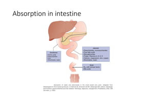
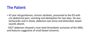
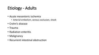
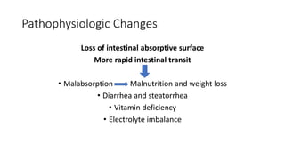
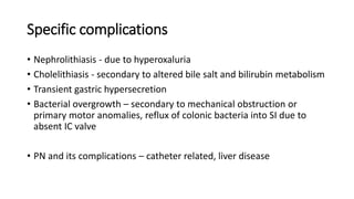
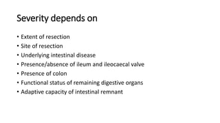
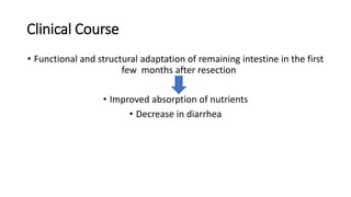
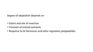
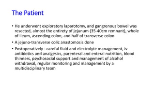
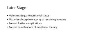
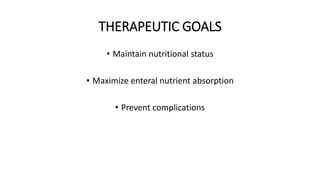
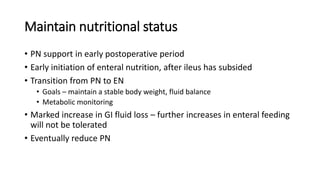
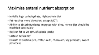
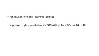
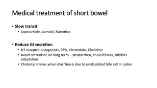
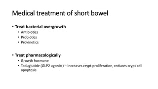
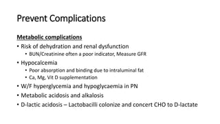
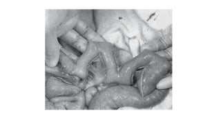
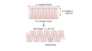
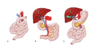
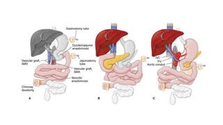
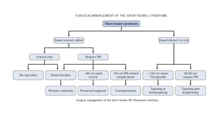
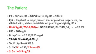
Absorption in intestine
The Patient
• 47 year old gentleman, chronic alcoholic, presented to the ED with
c/o abdominal pain, vomiting and obstipation for two days. He was
tachycardic and in shock, abdomen was tense and distended, bowel
sounds absent.
CECT abdomen showed a near total thrombotic occlusion of the SMA,
and features suggestive of small bowel ischemia.
SHORT BOWEL SYNDROME
Presented by: Anjaly Mohan
Moderator: Dr. Bharati Pandya
Dept of General Surgery
AIIMS Bhopal
Short Bowel Syndrome
• Malabsorption and malnutrition, which generally occur when less
than 180cm of functional intestine remains in adults
• Severity and clinical features dependent on several factors
• 75% cases – massive intestinal resection
• 25% cases – multiple sequential resections of SI
• Approx 2/3rd of patients survive that hospitalization
• Similar percentage are alive one year later
Etiology - Adults
• Acute mesenteric ischemia
• Arterial embolism, venous occlusion, shock
• Crohn’s disease
• Trauma
• Radiation enteritis
• Malignancy
• Recurrent intestinal obstruction
Etiology - Pediatric
• Necrotizing enterocolitis
• Intestinal atresia
• Gastroschisis
• Midgut volvulus
• Hirschsprung’s disease – Neuromuscular diseases of gut
• Congenital diseases of intestinal epithelium
Anatomic/surgical reductions of bowel
Pathophysiologic Changes
Loss of intestinal absorptive surface
More rapid intestinal transit
• Malabsorption Malnutrition and weight loss
• Diarrhea and steatorrhea
• Vitamin deficiency
• Electrolyte imbalance
Specific complications
• Nephrolithiasis - due to hyperoxaluria
• Cholelithiasis - secondary to altered bile salt and bilirubin metabolism
• Transient gastric hypersecretion
• Bacterial overgrowth – secondary to mechanical obstruction or
primary motor anomalies, reflux of colonic bacteria into SI due to
absent IC valve
• PN and its complications – catheter related, liver disease
Severity depends on
• Extent of resection
• Site of resection
• Underlying intestinal disease
• Presence/absence of ileum and ileocaecal valve
• Presence of colon
• Functional status of remaining digestive organs
• Adaptive capacity of intestinal remnant
Clinical Course
• Functional and structural adaptation of remaining intestine in the first
few months after resection
• Improved absorption of nutrients
• Decrease in diarrhea
Degree of adaptation depends on
• Extent and site of resection
• Provision of enteral nutrients
• Response to GI hormones and other regulatory polypeptides
The Patient
• He underwent exploratory laparotomy, and gangrenous bowel was
resected, almost the entirety of jejunum (35-40cm remnant), whole
of ileum, ascending colon, and half of transverse colon
• A jejuno-transverse colic anastomosis done
• Postoperatively - careful fluid and electrolyte management, iv
antibiotics and analgesics, parenteral and enteral nutrition, blood
thinners, psychosocial support and management of alcohol
withdrawal, regular monitoring and management by a
multidisciplinary team
The Patient
• He was discharged healthy ad stable a month later. Patient continued
taking TPN once a week, after the discharge, for about 2 months,
during which he encountered catheter related thrombophlebitis, but
without infection or sepsis
• He stopped coming due to inability to afford TPN and reported 4
months later.
• 6 months after the surgery, the patient returned with complaints of
abdominal pain, generalized weakness and body pain, swelling of
both legs, and was admitted
Clinical Assessment
Assessment of SBS patient
• Past medical record, surgical and pathological documentation
• Cause of SBS, anatomy and length of intestine, details of prior
surgical procedure
• Number of central lines and reasons they were changed
• Causal microorganisms for central line infections
• Nutritional assessment
• Parenteral and enteral intake, daily caloric requirements, macro &
micronutrient components of PN
Assessment of SBS patient
• S. electrolytes, LFT, GFR, S. Albumin, Prothrombin time, Vit B12, Fat
soluble vitamins, S. Ca, S. Citrulline
• Vaccination status
• Complete physical examination
• Focus on hydration status, nutritional status (ht, wt, BMI), type of central line,
signs of nutritional deficiencies, complication from PN like dermatitis, signs of
CLD
Management
EARLY MANAGEMENT
• Postoperative care
• Control of sepsis
• Maintain fluid and electrolyte balance
• Initiating nutritional support
Later Stage
• Maintain adequate nutritional status
• Maximise absorptive capacity of remaining intestine
• Prevent further complications
• Prevent complications of nutritional therapy
THERAPEUTIC GOALS
• Maintain nutritional status
• Maximize enteral nutrient absorption
• Prevent complications
Maintain nutritional status
• PN support in early postoperative period
• Early initiation of enteral nutrition, after ileus has subsided
• Transition from PN to EN
• Goals – maintain a stable body weight, fluid balance
• Metabolic monitoring
• Marked increase in GI fluid loss – further increases in enteral feeding
will not be tolerated
• Eventually reduce PN
Maximize enteral nutrient absorption
• Initially, high carbohydrate, high protein diet
• Fat requires more digestion, except MCTs
• Ability to absorb nutrients improves with time, hence diet should be
modified continually
• Restrict fat to 20-30% of caloric intake
• Lactase deficiency
• Oxalate restriction (tea, coffee, nuts, chocolate, soy products, sweet
potatoes)
• For jejunal remnants, isotonic feeding
• Ingestion of glucose-electrolyte ORS with at least 90mmol/L of Na
Medical treatment of short bowel
• Slow transit
• Loperamide, Lomotil, Narcotics
• Reduce GI secretion
• H2 receptor antagonists, PPIs, Octreotide, Clonidine
• Avoid octreotide on long term – steatorrhea, cholelithiasis, inhibits
adaptation
• Cholestyramine, when diarrhea is due to unabsorbed bile salt in colon
Medical treatment of short bowel
• Treat bacterial overgrowth
• Antibiotics
• Probiotics
• Prokinetics
• Treat pharmacologically
• Growth hormone
• Teduglutide (GLP2 agonist) – increases crypt proliferation, reduces crypt cell
apoptosis
Prevent Complications
Metabolic complications
• Risk of dehydration and renal dysfunction
• BUN/Creatinine often a poor indicator, Measure GFR
• Hypocalcemia
• Poor absorption and binding due to intraluminal fat
• Ca, Mg, Vit D supplementation
• W/F hyperglycemia and hypoglycaemia in PN
• Metabolic acidosis and alkalosis
• D-lactic acidosis – Lactobacilli colonize and concert CHO to D-lactate
Prevent Complications
• Specific nutrient deficiencies
• Iron and vitamin deficiencies
• Micronutrients – Se, Zn, Cu
• Fatty acid deficiency
• PN related
• Fluid and electrolyte disturbances
• Silent renal insufficiency
• IFALD
• Organ dysfunction – skeletal system (osteopenia, osteoporosis), intestine
(bacterial overgrowth, translocation), neurologic(memory disturbance), GB
(cholelithiasis), liver (steatosis, cholestasis, fibrosis, cirrhosis)
Prevent Complications
Catheter Related
• Catheter related sepsis
• Attention to technique and meticulous patient education
• Catheter thrombosis
• Difficult vascular access
Prevent Complications
PN-induced liver disease
• Multifactorial process
• More frequently in children
• Steatosis, cholestasis, eventually cirrhosis
• Increase enteral nutrition, avoid overfeeding, lipid minimization,
prevent specific deficiencies
Prevent Complications
Bacterial overgrowth
• Impaired motility or stasis
• Secretory diarrhea may occur
• Suspected when a patient’s absorptive capacity and stool habits
change acutely
• If mechanical obstruction or blind loop – surgery
• Primary motor abnormality – intermittent antibiotic therapy
• Colonization of lumen with probiotics
Prevent Complications
Cholelithiasis
• 30-40% of SBS patients
• Long term PN – altered hepatic bile metabolism, GB stasis
• Ileal resection – bile acid malabsorption, lithogenic bile -> cholesterol
stones
• Provide nutrients enterally, whenever possible
• Requires surgical treatment
• May be done prophylactically, while doing laparotomy for other
reasons
Prevent Complications
Nephrolithiasis
• Normally, oxalate is bound to Ca in the intestinal lumen, hence not
absorbed
• Decreased availability of Ca – reduced intake, binding by intraluminal
fat
• Hence, free oxalate in lumen – absorbed in colon - forms ca oxalate in
urine
• Diet low in oxalate, min intraluminal fat, supplement Ca porally,
maintain high urinary volume
• Cholestyramine – binds oxalic acid in colon
Prevent Complications
Gastric hypersecretion
• Massive intestinal resection – parietal cell hyperplasia,
hypergastrinemia
• Usually transient
• Presumably involves loss of an inhibitor from resected intestine
• Hyperacidity – exacerbates malabsorption and diarrhea
• H2 receptor antagonists, PPIs
SURGICAL MANAGEMENT
Goals:
• To slow intestinal transit
• Reversing intestinal segments
• Interposing colonic segments into SI
• To improve the function of existing intestine
• Stenotic segments – strictureoplasty
• Dilated segments – tapering enteroplasty
• Intestinal lengthening procedures
• Intestinal transplantation
Cautious patient selection
• Worsening malabsorption
• Patients stable on EN, going into risk of requiring PN, or going into
malabsorption
• PN associated complications
Bianchi Procedure
Longitudinal intestinal lengthening
STEP Procedure
Serial Transverse EnteroPlasty
Intestinal Transplantaion
• Final available therapy for patients with intestinal failure or SBS, who
in general have failed PN therapy
The Patient
• PR – 96/min, BP – 80/50mm of Hg, RR – 18/min
• P/A – Scaphoid in shape, healed scar of previous surgery see, no
dilated veins, visible peristalsis, no guarding or rigidity, BS +
• Hb-6.5g/dL, TC-16,600/uL, N92L03M05, Plt-2.81L/uL, Hct – 20.9%
• FBS – 101mg%
• BUN/Creat – 22.17/0.85mg/d
• T.Bil/D.Bil – 0.63/0.09U/L
• Tot.Prot/alb – 6.5/3.9
• S. Na+/K+ – 133/2.7mmol/L
• S. Ca++ – 3.5mg/dL
References
• Maingot’s Abdominal Operations 13th Edition
• Current Surgical Therapy 12th Edition, Cameron et al
• Sabiston Textbook of Surgery 20th edition
THANK YOU
FOR YOUR TIME

SHORT_BOWEL_SYNDROME.pptx

  • 1.
  • 2.
    The Patient • 47year old gentleman, chronic alcoholic, presented to the ED with c/o abdominal pain, vomiting and obstipation for two days. He was tachycardic and in shock, abdomen was tense and distended, bowel sounds absent. CECT abdomen showed a near total thrombotic occlusion of the SMA, and features suggestive of small bowel ischemia.
  • 3.
    SHORT BOWEL SYNDROME Presentedby: Anjaly Mohan Moderator: Dr. Bharati Pandya Dept of General Surgery AIIMS Bhopal
  • 4.
    Short Bowel Syndrome •Malabsorption and malnutrition, which generally occur when less than 180cm of functional intestine remains in adults • Severity and clinical features dependent on several factors • 75% cases – massive intestinal resection • 25% cases – multiple sequential resections of SI • Approx 2/3rd of patients survive that hospitalization • Similar percentage are alive one year later
  • 5.
    Etiology - Adults •Acute mesenteric ischemia • Arterial embolism, venous occlusion, shock • Crohn’s disease • Trauma • Radiation enteritis • Malignancy • Recurrent intestinal obstruction
  • 6.
    Etiology - Pediatric •Necrotizing enterocolitis • Intestinal atresia • Gastroschisis • Midgut volvulus • Hirschsprung’s disease – Neuromuscular diseases of gut • Congenital diseases of intestinal epithelium Anatomic/surgical reductions of bowel
  • 7.
    Pathophysiologic Changes Loss ofintestinal absorptive surface More rapid intestinal transit • Malabsorption Malnutrition and weight loss • Diarrhea and steatorrhea • Vitamin deficiency • Electrolyte imbalance
  • 8.
    Specific complications • Nephrolithiasis- due to hyperoxaluria • Cholelithiasis - secondary to altered bile salt and bilirubin metabolism • Transient gastric hypersecretion • Bacterial overgrowth – secondary to mechanical obstruction or primary motor anomalies, reflux of colonic bacteria into SI due to absent IC valve • PN and its complications – catheter related, liver disease
  • 9.
    Severity depends on •Extent of resection • Site of resection • Underlying intestinal disease • Presence/absence of ileum and ileocaecal valve • Presence of colon • Functional status of remaining digestive organs • Adaptive capacity of intestinal remnant
  • 10.
    Clinical Course • Functionaland structural adaptation of remaining intestine in the first few months after resection • Improved absorption of nutrients • Decrease in diarrhea
  • 11.
    Degree of adaptationdepends on • Extent and site of resection • Provision of enteral nutrients • Response to GI hormones and other regulatory polypeptides
  • 12.
    The Patient • Heunderwent exploratory laparotomy, and gangrenous bowel was resected, almost the entirety of jejunum (35-40cm remnant), whole of ileum, ascending colon, and half of transverse colon • A jejuno-transverse colic anastomosis done • Postoperatively - careful fluid and electrolyte management, iv antibiotics and analgesics, parenteral and enteral nutrition, blood thinners, psychosocial support and management of alcohol withdrawal, regular monitoring and management by a multidisciplinary team
  • 13.
    The Patient • Hewas discharged healthy ad stable a month later. Patient continued taking TPN once a week, after the discharge, for about 2 months, during which he encountered catheter related thrombophlebitis, but without infection or sepsis • He stopped coming due to inability to afford TPN and reported 4 months later. • 6 months after the surgery, the patient returned with complaints of abdominal pain, generalized weakness and body pain, swelling of both legs, and was admitted
  • 14.
  • 15.
    Assessment of SBSpatient • Past medical record, surgical and pathological documentation • Cause of SBS, anatomy and length of intestine, details of prior surgical procedure • Number of central lines and reasons they were changed • Causal microorganisms for central line infections • Nutritional assessment • Parenteral and enteral intake, daily caloric requirements, macro & micronutrient components of PN
  • 16.
    Assessment of SBSpatient • S. electrolytes, LFT, GFR, S. Albumin, Prothrombin time, Vit B12, Fat soluble vitamins, S. Ca, S. Citrulline • Vaccination status • Complete physical examination • Focus on hydration status, nutritional status (ht, wt, BMI), type of central line, signs of nutritional deficiencies, complication from PN like dermatitis, signs of CLD
  • 17.
  • 18.
    EARLY MANAGEMENT • Postoperativecare • Control of sepsis • Maintain fluid and electrolyte balance • Initiating nutritional support
  • 19.
    Later Stage • Maintainadequate nutritional status • Maximise absorptive capacity of remaining intestine • Prevent further complications • Prevent complications of nutritional therapy
  • 20.
    THERAPEUTIC GOALS • Maintainnutritional status • Maximize enteral nutrient absorption • Prevent complications
  • 21.
    Maintain nutritional status •PN support in early postoperative period • Early initiation of enteral nutrition, after ileus has subsided • Transition from PN to EN • Goals – maintain a stable body weight, fluid balance • Metabolic monitoring • Marked increase in GI fluid loss – further increases in enteral feeding will not be tolerated • Eventually reduce PN
  • 22.
    Maximize enteral nutrientabsorption • Initially, high carbohydrate, high protein diet • Fat requires more digestion, except MCTs • Ability to absorb nutrients improves with time, hence diet should be modified continually • Restrict fat to 20-30% of caloric intake • Lactase deficiency • Oxalate restriction (tea, coffee, nuts, chocolate, soy products, sweet potatoes)
  • 23.
    • For jejunalremnants, isotonic feeding • Ingestion of glucose-electrolyte ORS with at least 90mmol/L of Na
  • 24.
    Medical treatment ofshort bowel • Slow transit • Loperamide, Lomotil, Narcotics • Reduce GI secretion • H2 receptor antagonists, PPIs, Octreotide, Clonidine • Avoid octreotide on long term – steatorrhea, cholelithiasis, inhibits adaptation • Cholestyramine, when diarrhea is due to unabsorbed bile salt in colon
  • 25.
    Medical treatment ofshort bowel • Treat bacterial overgrowth • Antibiotics • Probiotics • Prokinetics • Treat pharmacologically • Growth hormone • Teduglutide (GLP2 agonist) – increases crypt proliferation, reduces crypt cell apoptosis
  • 26.
    Prevent Complications Metabolic complications •Risk of dehydration and renal dysfunction • BUN/Creatinine often a poor indicator, Measure GFR • Hypocalcemia • Poor absorption and binding due to intraluminal fat • Ca, Mg, Vit D supplementation • W/F hyperglycemia and hypoglycaemia in PN • Metabolic acidosis and alkalosis • D-lactic acidosis – Lactobacilli colonize and concert CHO to D-lactate
  • 27.
    Prevent Complications • Specificnutrient deficiencies • Iron and vitamin deficiencies • Micronutrients – Se, Zn, Cu • Fatty acid deficiency • PN related • Fluid and electrolyte disturbances • Silent renal insufficiency • IFALD • Organ dysfunction – skeletal system (osteopenia, osteoporosis), intestine (bacterial overgrowth, translocation), neurologic(memory disturbance), GB (cholelithiasis), liver (steatosis, cholestasis, fibrosis, cirrhosis)
  • 28.
    Prevent Complications Catheter Related •Catheter related sepsis • Attention to technique and meticulous patient education • Catheter thrombosis • Difficult vascular access
  • 29.
    Prevent Complications PN-induced liverdisease • Multifactorial process • More frequently in children • Steatosis, cholestasis, eventually cirrhosis • Increase enteral nutrition, avoid overfeeding, lipid minimization, prevent specific deficiencies
  • 30.
    Prevent Complications Bacterial overgrowth •Impaired motility or stasis • Secretory diarrhea may occur • Suspected when a patient’s absorptive capacity and stool habits change acutely • If mechanical obstruction or blind loop – surgery • Primary motor abnormality – intermittent antibiotic therapy • Colonization of lumen with probiotics
  • 31.
    Prevent Complications Cholelithiasis • 30-40%of SBS patients • Long term PN – altered hepatic bile metabolism, GB stasis • Ileal resection – bile acid malabsorption, lithogenic bile -> cholesterol stones • Provide nutrients enterally, whenever possible • Requires surgical treatment • May be done prophylactically, while doing laparotomy for other reasons
  • 32.
    Prevent Complications Nephrolithiasis • Normally,oxalate is bound to Ca in the intestinal lumen, hence not absorbed • Decreased availability of Ca – reduced intake, binding by intraluminal fat • Hence, free oxalate in lumen – absorbed in colon - forms ca oxalate in urine • Diet low in oxalate, min intraluminal fat, supplement Ca porally, maintain high urinary volume • Cholestyramine – binds oxalic acid in colon
  • 33.
    Prevent Complications Gastric hypersecretion •Massive intestinal resection – parietal cell hyperplasia, hypergastrinemia • Usually transient • Presumably involves loss of an inhibitor from resected intestine • Hyperacidity – exacerbates malabsorption and diarrhea • H2 receptor antagonists, PPIs
  • 34.
    SURGICAL MANAGEMENT Goals: • Toslow intestinal transit • Reversing intestinal segments • Interposing colonic segments into SI • To improve the function of existing intestine • Stenotic segments – strictureoplasty • Dilated segments – tapering enteroplasty • Intestinal lengthening procedures • Intestinal transplantation
  • 35.
    Cautious patient selection •Worsening malabsorption • Patients stable on EN, going into risk of requiring PN, or going into malabsorption • PN associated complications
  • 36.
  • 38.
  • 42.
  • 43.
    • Final availabletherapy for patients with intestinal failure or SBS, who in general have failed PN therapy
  • 47.
    The Patient • PR– 96/min, BP – 80/50mm of Hg, RR – 18/min • P/A – Scaphoid in shape, healed scar of previous surgery see, no dilated veins, visible peristalsis, no guarding or rigidity, BS + • Hb-6.5g/dL, TC-16,600/uL, N92L03M05, Plt-2.81L/uL, Hct – 20.9% • FBS – 101mg% • BUN/Creat – 22.17/0.85mg/d • T.Bil/D.Bil – 0.63/0.09U/L • Tot.Prot/alb – 6.5/3.9 • S. Na+/K+ – 133/2.7mmol/L • S. Ca++ – 3.5mg/dL
  • 48.
    References • Maingot’s AbdominalOperations 13th Edition • Current Surgical Therapy 12th Edition, Cameron et al • Sabiston Textbook of Surgery 20th edition
  • 49.

Editor's Notes

  • #2 normal length of the small intestine is estimated at 600cm , and the colon is about 150cm. Individuals who have only 150-200cm of the small intestine and lack a colon may have significant fluid and nutrient losses that often require the use of intravenous fluid and/or nutrients. duodenum (about 30cm) and is connected to the stomach. The duodenum continues the breakdown of food particles from the stomach which prepares particles to be absorbed by the intestinal lining. The duodenum also absorbs iron, calcium and magnesium. The jejunum is where most nutrient absorption occurs. It is about 200cm in length. Carbohydrates, fats, proteins, and vitamins are all absorbed in the jejunum. The ileum is the last portion of the small intestine and is connected to the large intestine. Some products of digestion that are not absorbed by the jejunum may also be absorbed by the ileum as this area can adapt to compensate for many of the jejunal functions. The reverse is not true. As there are special binding sites for the absorption of bile acids and vitamin B12 found only in the final portion of the ileum, referred to as the terminal ileum
  • #5 <25cm in presence of ileocaecal valve and <40cm without ileocaecal valve in pediatric patients
  • #17 Citrulline – a nonprotein aminoacid produced by intestinal mucosa; its serum levels in patients with SBS may differentiate transient from permanent intestinal failure
  • #25 Loperamide – peripherally acting narcotic Lomotil – diphenoxylate and atropine Narcotics such as codeine
  • #35 Improve function
  • #38 The small bowel is divided at either end of a dilated loop. The mesentery is dissected to create a plane along the axis of the intestine between branches of mesenteric blood vessels. (B) The mesentery has two leaves. Arterial and venous branches of mesenteric vessels alternate from one leaf of the mesentery to the other. (C)A gastrointestinal stapling device can be passed between the leaves of the mesentery. (D) When the stapler is fired, the single loop of dilated intestine is divided into two parallel loops. (E) The parallel loops can then be turned in a “lazy S” fashion to approximate the distal end of one loop to the proximal end of the second loop. In this way, the parallel loops are anastomosed end to-end to reestablish continuity and double the length of the small bowel. In addition, the lengthened segment is then reanastomosed to the normal small bowel or colon proximally and distally (not shown).
  • #40 the dilated segment of small bowel is narrowed by alternate firings of the GI stapling device from the mesenteric and antimesenteric borders of the bowel. This would result in luminal diameters between 1 cm and 2.5 cm and a resultant increase in bowel length
  • #45 A – jejunoileal graft, procured with vascular pedicle SMA and SMV B – Liver intestine graft C – Multivisceral allograft