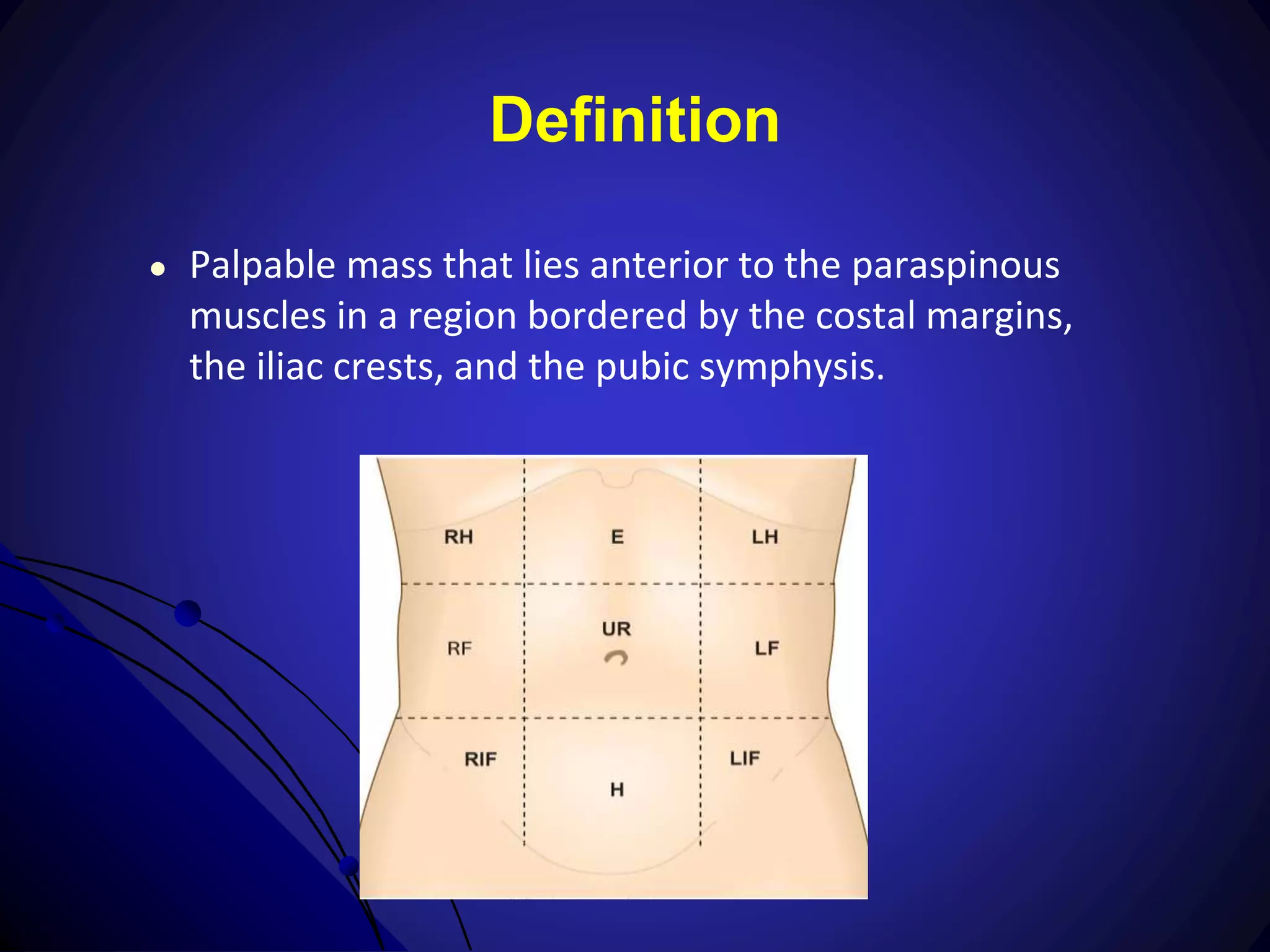
Clinical Assessment of
Abdominal mass
Ketrampilan Klinik
Pra Bedah Dasar
PPDS I Bedah FK Unpad/RSHS Bandung
Objectives :
● To explain and demonstrate the history
taking for patients with abdominal mass
● To demonstrate the physical diagnosis in
patients with abdominal mass
● To explain investigative studies for abdominal
mass
● To discuss the decision making for the
management of abdominal mass
Introduction
● Why is it important?
● Abdominal masses
● are commonly addressed by surgeons,
● cover a broad spectrum: no apparent
consequences, others significantly impair quality
of life, others represent severe conditions
● For each patient, therefore, it is essential to formulate a
management approach that is tailored to the particular
clinical situation.
Introduction
● Commonly, an abnormal enlargement or mass in
the abdomen comes to the clinician's attention
in one of three ways:
● detected and reported by the patient,
● discovered by the clinician on physical
examination,
● noticed as an unrelated incidental finding on
a radiographic study.
Introduction
● Effective decision-making involves:
● establishing the correct diagnosis,
● introducing an effective treatment plan,
● eliminating risks and complicating factors,
● initiating preventive measures,
● determining the prognosis.
Definition
● Palpable mass that lies anterior to the paraspinous
muscles in a region bordered by the costal margins,
the iliac crests, and the pubic symphysis.
History taking
● Establishing a solid surgeon-patient relationship is
vital for building patient trust and confidence,
● philosophy in dealing with an abdominal mass is:
● to evaluate the patient
● consider radiographic and laboratory studies if
the initial assessment does not yield a diagnosis.
History Taking
Introduction
• Greet the patient, and develop a warm and
helpful environment
• Introduce yourself to the patient
Patient Identity
» Ask the patient politely concerning his/her:
name age
» Record the gender:
– Male
– Female
» Ask the marital status of the patient (especially for
female)
Current history of illness
● A careful and methodical clinical history
should be taken that includes all factors
pertaining to the lesion.
● Ask concerning the mass (non directive):
● mode of onset, duration, character,
chronology, and location.
● Presence or absence of associated
symptoms.
History
● Associated GI symptoms with the mass:
● concerned with nausea
● vomiting,
● diarrhea,
● constipation
● Non specific GI symptoms
● jaundice, melena, hematochezia, hematemesis,
hematuria, or changes in stool caliber
History
● Non-GI symptoms
● urologic, gynecologic or obstetric,
vascular, and endocrinologic symptoms
● A history of surgery, trauma, or neoadjuvant
or adjuvant cancer therapy.
Develop Differential Diagnosis
Organ Specific Causes
Physical Diagnosis
● Practice in examining patients with an
abdominal mass is to follow an
organized, systematic approach
consisting of:
● inspection,
● auscultation,
● percussion, and
● palpation,
Physical Diagnosis
● Three main objectives:
● evaluate the patient's condition as it directly or
indirectly relates to the mass (e.g., by noting
associated systemic illness, pain, malaise, or cachexia)
● assess the acuteness of the patient's condition (e.g.,
by determining whether a left upper quadrant mass is
likely to be a ruptured spleen or simply a long-
standing mass in the abdominal wall)
● must carefully examine each abdominal quadrant,
assessing both normal and abnormal anatomic
relations as possible sources of the presumed mass.
Physical Diagnosis
● General examination should seek systemic signs
of disease (e.g. cachexia, anaemia and jaundice)
or signs of malignant dissemination (e.g.
supraclavicular lymphadenopathy in suspected
stomach cancer).
● Abdominal and pelvic examination must be
thorough and, if appropriate, proctoscopy and
sigmoidoscopy should be performed.
Physical Diagnosis
● Carnett test: to determine the mass, whether on
the abdominal wall or intra abdominal
Physical Diagnosis
● Palpable intra abdominal mass:
● Intra peritoneal
● Reproperitoneal
● Pre peritoneal
Physical Diagnosis
● Determine (Inspection, Palpation, Percussion)
● The location of the mass, its relations to other structures,
● Mobility
● Physical characteristics, such as size, shape, consistency
and pulsatility (information: organ of origin and the likely
pathology).
● Hernias, e.g. incisional, umbilical and sometimes interstitial
(Spigelian) hernias (see Ch. 32), may present as localised
swellings but they usually shrink or reduce completely
when the patient is supine or under anaesthesia.
● An incarcerated (irreducible but not obstructed) hernia is
more appropriately considered a true 'mass'.
Physical Diagnosis
● Palpable or discrete masses should always be localized
(abdominal land marks) , and described :
● terms of size, shape, consistency, contour, presence or
absence of tenderness, pulsatility, and fixation
● The mass's location can be vaguely outlined: fluid is present,
the abdomen is tender or tense, the patient is obese.
● Gastric neoplasms, pancreatic neoplasms, colonic neoplasms,
sarcomas, pancreatic cysts, and distended gallbladders are
palpable (adsvanced stages) and recognition can be facilitated
by repeating the abdominal examination after analgesics have
been administered or after the patient has been anesthetized in
preparation for a procedure.
Working or Presumed Diagnosis
» generate a working diagnosis.
» Consider subsequent management
» Sometimes, however, the diagnosis remains unknown
even after a comprehensive clinical history and physical
examination; in such cases, further studies are required.
» Investigative methods: A wide range of laboratory and
imaging studies are now available for establishing the
diagnosis.
» If these studies do not resolve the diagnostic uncertainty,
additional procedures, including image-guided
percutaneous biopsy, diagnostic laparoscopy, and
exploratory laparotomy, may be employed as necessary.
Investigative Studies
» Collaborative management effort and the
choice of appropriate investigative studies.
» Be familiar with every available method for
efficient and cost-effective diagnosis of an
abdominal mass.
» Be based on:
– the preferences of the patient,
– the knowledge and judgment of the
surgeon, and the capabilities of the
institution
Imaging
● Plain Abdominal Radiographs
● Conventional Gastrointestinal Imaging
● Ultrasonography
● Computed Tomography
● Magnetic Resonance Imaging
● Positron Emission Tomography
● Image-Guided Percutaneous Biopsy
● EUS-Guided Imaging and Biopsy
● Diagnostic Laparoscopy
● Limited indications for Exoploratory Laparotomy
Decision making
Abdominal mass .pptx

Abdominal mass .pptx

  • 1.
    Clinical Assessment of Abdominalmass Ketrampilan Klinik Pra Bedah Dasar PPDS I Bedah FK Unpad/RSHS Bandung
  • 2.
    Objectives : ● Toexplain and demonstrate the history taking for patients with abdominal mass ● To demonstrate the physical diagnosis in patients with abdominal mass ● To explain investigative studies for abdominal mass ● To discuss the decision making for the management of abdominal mass
  • 3.
    Introduction ● Why isit important? ● Abdominal masses ● are commonly addressed by surgeons, ● cover a broad spectrum: no apparent consequences, others significantly impair quality of life, others represent severe conditions ● For each patient, therefore, it is essential to formulate a management approach that is tailored to the particular clinical situation.
  • 4.
    Introduction ● Commonly, anabnormal enlargement or mass in the abdomen comes to the clinician's attention in one of three ways: ● detected and reported by the patient, ● discovered by the clinician on physical examination, ● noticed as an unrelated incidental finding on a radiographic study.
  • 5.
    Introduction ● Effective decision-makinginvolves: ● establishing the correct diagnosis, ● introducing an effective treatment plan, ● eliminating risks and complicating factors, ● initiating preventive measures, ● determining the prognosis.
  • 6.
    Definition ● Palpable massthat lies anterior to the paraspinous muscles in a region bordered by the costal margins, the iliac crests, and the pubic symphysis.
  • 8.
    History taking ● Establishinga solid surgeon-patient relationship is vital for building patient trust and confidence, ● philosophy in dealing with an abdominal mass is: ● to evaluate the patient ● consider radiographic and laboratory studies if the initial assessment does not yield a diagnosis.
  • 9.
    History Taking Introduction • Greetthe patient, and develop a warm and helpful environment • Introduce yourself to the patient
  • 10.
    Patient Identity » Askthe patient politely concerning his/her: name age » Record the gender: – Male – Female » Ask the marital status of the patient (especially for female)
  • 11.
    Current history ofillness ● A careful and methodical clinical history should be taken that includes all factors pertaining to the lesion. ● Ask concerning the mass (non directive): ● mode of onset, duration, character, chronology, and location. ● Presence or absence of associated symptoms.
  • 12.
    History ● Associated GIsymptoms with the mass: ● concerned with nausea ● vomiting, ● diarrhea, ● constipation ● Non specific GI symptoms ● jaundice, melena, hematochezia, hematemesis, hematuria, or changes in stool caliber
  • 13.
    History ● Non-GI symptoms ●urologic, gynecologic or obstetric, vascular, and endocrinologic symptoms ● A history of surgery, trauma, or neoadjuvant or adjuvant cancer therapy.
  • 14.
  • 15.
  • 16.
    Physical Diagnosis ● Practicein examining patients with an abdominal mass is to follow an organized, systematic approach consisting of: ● inspection, ● auscultation, ● percussion, and ● palpation,
  • 17.
    Physical Diagnosis ● Threemain objectives: ● evaluate the patient's condition as it directly or indirectly relates to the mass (e.g., by noting associated systemic illness, pain, malaise, or cachexia) ● assess the acuteness of the patient's condition (e.g., by determining whether a left upper quadrant mass is likely to be a ruptured spleen or simply a long- standing mass in the abdominal wall) ● must carefully examine each abdominal quadrant, assessing both normal and abnormal anatomic relations as possible sources of the presumed mass.
  • 18.
    Physical Diagnosis ● Generalexamination should seek systemic signs of disease (e.g. cachexia, anaemia and jaundice) or signs of malignant dissemination (e.g. supraclavicular lymphadenopathy in suspected stomach cancer). ● Abdominal and pelvic examination must be thorough and, if appropriate, proctoscopy and sigmoidoscopy should be performed.
  • 19.
    Physical Diagnosis ● Carnetttest: to determine the mass, whether on the abdominal wall or intra abdominal
  • 20.
    Physical Diagnosis ● Palpableintra abdominal mass: ● Intra peritoneal ● Reproperitoneal ● Pre peritoneal
  • 21.
    Physical Diagnosis ● Determine(Inspection, Palpation, Percussion) ● The location of the mass, its relations to other structures, ● Mobility ● Physical characteristics, such as size, shape, consistency and pulsatility (information: organ of origin and the likely pathology). ● Hernias, e.g. incisional, umbilical and sometimes interstitial (Spigelian) hernias (see Ch. 32), may present as localised swellings but they usually shrink or reduce completely when the patient is supine or under anaesthesia. ● An incarcerated (irreducible but not obstructed) hernia is more appropriately considered a true 'mass'.
  • 22.
    Physical Diagnosis ● Palpableor discrete masses should always be localized (abdominal land marks) , and described : ● terms of size, shape, consistency, contour, presence or absence of tenderness, pulsatility, and fixation ● The mass's location can be vaguely outlined: fluid is present, the abdomen is tender or tense, the patient is obese. ● Gastric neoplasms, pancreatic neoplasms, colonic neoplasms, sarcomas, pancreatic cysts, and distended gallbladders are palpable (adsvanced stages) and recognition can be facilitated by repeating the abdominal examination after analgesics have been administered or after the patient has been anesthetized in preparation for a procedure.
  • 23.
    Working or PresumedDiagnosis » generate a working diagnosis. » Consider subsequent management » Sometimes, however, the diagnosis remains unknown even after a comprehensive clinical history and physical examination; in such cases, further studies are required. » Investigative methods: A wide range of laboratory and imaging studies are now available for establishing the diagnosis. » If these studies do not resolve the diagnostic uncertainty, additional procedures, including image-guided percutaneous biopsy, diagnostic laparoscopy, and exploratory laparotomy, may be employed as necessary.
  • 24.
    Investigative Studies » Collaborativemanagement effort and the choice of appropriate investigative studies. » Be familiar with every available method for efficient and cost-effective diagnosis of an abdominal mass. » Be based on: – the preferences of the patient, – the knowledge and judgment of the surgeon, and the capabilities of the institution
  • 25.
    Imaging ● Plain AbdominalRadiographs ● Conventional Gastrointestinal Imaging ● Ultrasonography ● Computed Tomography ● Magnetic Resonance Imaging ● Positron Emission Tomography ● Image-Guided Percutaneous Biopsy ● EUS-Guided Imaging and Biopsy ● Diagnostic Laparoscopy ● Limited indications for Exoploratory Laparotomy
  • 26.