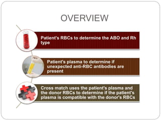
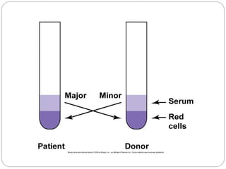
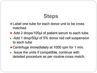
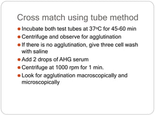
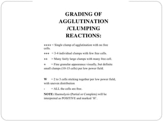
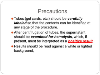
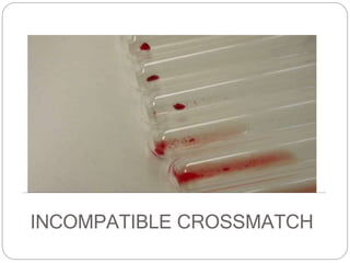
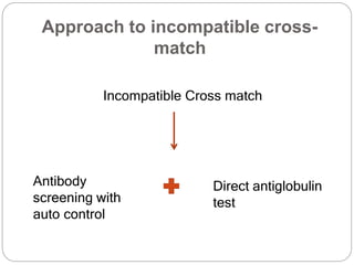
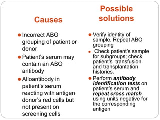
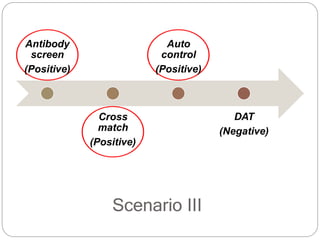
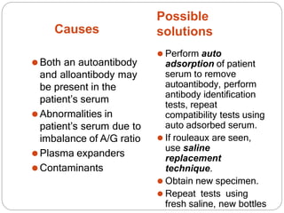
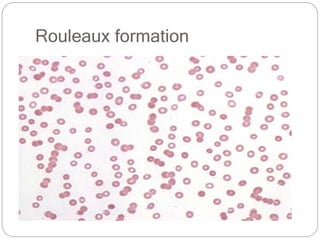
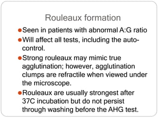
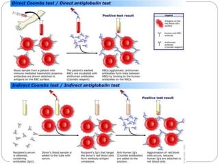
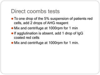
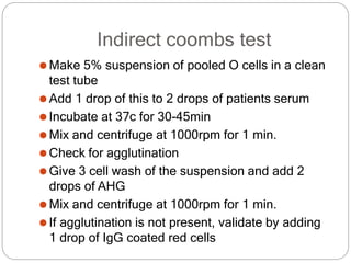
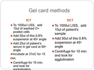
By: Sivaranjini Narayanan
Guide: Dr. Roopam Gidwani
CROSSMATCHING
OVERVIEW
Is the minor cross match
unnecessary?
⚫Donated units are tested for antibodies
⚫Most blood is transfused as packed cells, having
little antibodies
⚫The plasma volume is small, and Abs will be diluted
in recipient circulation
⚫This applies for packed cells. For whole blood and
FFP, minor cross match is significant
Types of tests
Manual (IS and AHG)
Gel Technology
Electronic (Computerized) Cross match
Solid Phase Adherence Assays (SPAA)
⚫For major cross match: 2 drops of patient’s
serum + 1 drop of 5% suspension of donor red
cells
⚫For minor cross match: 2 drops of donor’s
serum + 1 drop of 5% suspension of patient’s red
cells
Immediate spin cross matching
⚫ When no clinically significant unexpected
antibodies are
detected and there are no previous records of such
antibodies,
a serologic test to detect ABO incompatibility is
sufficient.
⚫ This is accomplished by mixing the recipient’s serum
with
donor RBCs and centrifuging immediately (i.e..,
immediate
spin).
Steps
⚫Label one tube for each donor unit to be cross
matched
⚫ Add 2 drops/100μl of patient serum to each tube.
⚫ Add 1 drop/50μl of 5% donor red cell suspension
to each tube
⚫Centrifuge immediately at 1000 rpm for 1 min.
⚫ Issue the units if compatible, continue with
detailed procedure as per routine cross match.
Cross match using tube method
⚫Incubate both test tubes at 37oC for 45-60 min
⚫Centrifuge and observe for agglutination
⚫If there is no agglutination, give three cell wash
with saline
⚫Add 2 drops of AHG serum
⚫Centrifuge at 1000 rpm for 1 min.
⚫Look for agglutination macroscopically and
microscopically
Precautions
⚫Tubes (gel cards, etc.) should be carefully
labeled so that the contents can be identified at
any stage of the procedure.
⚫After centrifugation of tubes, the supernatant
should be examined for hemolysis, which, if
present, must be interpreted as a positive result.
⚫Results should be read against a white or lighted
background,
Gel card method
⚫ Prepare 0.8 – 1% red cell suspension of donor cells
by adding 10µl of packed washed red cells to 1 ml
LISS .
⚫ Pipette 50μl of red cell suspension in micro tube of
the gel card
⚫ Add 25 µl of serum in that micro tube of gel card
⚫ Incubate for 15 min at 37 degree and then centrifuge
for 10 min at 3000rpm. Read and record the results
Grading of agglutination
reactions using gel cards
Role of LISS
LISS contains glycine in an albumin solution. In addition
to lowering the zeta potential, LISS increases the
uptake of antibody onto the
RBC during the sensitization phase. This increases the
possibility
of agglutination.
Electronic cross matching
⚫Donor blood is issued based on blood bank
information (computer).
⚫ The Electronic Cross match (Electronic Issue)
allows for donor blood to be issued to a patient
instantaneously
Criteria for electronic cross matching
1.Two Concordant blood groups of the recipient
which have been recorded electronically
2.The patients serum/plasma does not contain,
and has not been known to contain clinically
significant red cell alloantibodies reactive at 37°C
Solid phase adherence assays
⚫Solid phase testing system uses microtiter plates,
with wells coated with stroma from reagent RBCs
of known phenotype
⚫Patient plasma and LISS added to the RBC well
then incubate at 37 degree for 15min
⚫ If antibody present, will bind
to the target antigen present
on the RBCs coating the well
⚫ Plate washed to remove
unbound antibodies, and
indicator RBCs coated with
anti-IgG are added,
⚫ Spin and read:-
⚫ Positive: Uniform layer of
indicator RBCs at the bottom
of the well
⚫ Negative: Indicator RBCs
from a tight bottom at centre.
INCOMPATIBLE CROSSMATCH
Approach to incompatible cross-
match
Incompatible Cross match
Antibody
screening with
auto control
Direct antiglobulin
test
Scenario I
Antibody
screen
(Negative)
Cross
match
(Positive)
Auto
control
(Negative)
DAT
(Negative)
Possible
solutions
Causes
⚫Incorrect ABO
grouping of patient or
donor
⚫Patient’s serum may
contain an ABO
antibody
⚫Alloantibody in
patient’s serum
reacting with antigen
donor’s red cells but
not present on
screening cells
⚫ Verify identity of
sample. Repeat ABO
grouping
⚫ Check patient’s sample
for subgroups; check
patient’s transfusion
and transplantation
histories.
⚫ Perform antibody
identification tests on
patient’s serum and
repeat cross match
using units negative for
the corresponding
antigen
Scenario II
Antibody
screen
(Positive)
Cross
match
(Positive)
Auto
control
(Negative)
DAT
(Negative)
Causes
Possible
solutions
⚫Donor unit may have
a positive DAT
⚫Alloantibody in
patient’s serum
reacting with antigens
on donor’s cells and
screening cells
⚫Perform DAT on
donor unit; if
positive, do not use
the unit.
⚫Perform antibody
identification
studies on patient’s
serum and repeat
cross match using
units negative for the
corresponding
antigen.
Alloantibodies
 Prevalence - 0.3% - 38%
 Immunizing stimulus
⚫Red cell antigens
 Pregnancy
 Transfusion
 Transplantation
⚫Passively acquired antibodies
 Intra-venous immunoglobulin
 Donor plasma
Common alloantibodies
Most common- anti-E, anti-Le(a), anti-K, anti-D, and
anti-
Le(b) (variable)
Always Clinically
significant
Rarely or never
considered Clinically
significant
ABO (A, B) Lewis (Lea, Leb),
Rh (D, C, c, E, e) Lutheran (Lua)
Duffy (Fya, Fyb) P1
Kidd (Jka, Jkb) Xga
Kell (K, k)
MN
Ss (S, s)
Lutheran (Lub)
Scenario III
Antibody
screen
(Positive)
Cross
match
(Positive)
Auto
control
(Positive)
DAT
(Negative)
Causes
Possible
solutions
⚫Both an autoantibody
and alloantibody may
be present in the
patient’s serum
⚫Abnormalities in
patient’s serum due to
imbalance of A/G ratio
⚫Plasma expanders
⚫Contaminants
⚫ Perform auto
adsorption of patient
serum to remove
autoantibody, perform
antibody identification
tests, repeat
compatibility tests using
auto adsorbed serum.
⚫ If rouleaux are seen,
use saline
replacement
technique.
⚫ Obtain new specimen.
⚫ Repeat tests using
fresh saline, new bottles
Rouleaux formation
Rouleaux formation
⚫Seen in patients with abnormal A:G ratio
⚫Will affect all tests, including the auto-
control.
⚫Strong rouleaux may mimic true
agglutination; however, agglutination
clumps are refractile when viewed under
the microscope.
⚫Rouleaux are usually strongest after
37C incubation but do not persist
through washing before the AHG test.
Scenario IV
Antibody
screen
(Positive)
Cross
match
(Positive)
Auto
control
(Positive)
DAT
(Positive)
Causes
Possible
solution
⚫Passively acquired
autoantibody (e.g.,
intravenous immune
globulin) is present.
⚫Cold- or warm-
reactive autoantibody
is present
⚫ Cold-AIHA – not a
major challenge as long
as all testing and cross
matching is performed
at 37ºC
⚫ Warm AIHA:-
⚫Best matched blood
(least incompatible)
⚫Phenotypically matched
RBCs
⚫Auto/Alloadsorbed
compatible RBCs
Autoantibodies
⚫Warm reactive autoantibodies (usually IgG) :
antibodies attach to red cells with maximal reactivity
at 37c
⚫Cold reactive autoantibodies (usually IgM):
antibodies bind to RBCs only at low body
temperature (28-31c)
⚫Drug-induced autoantibodies
Coombs’ tests
⚫The direct coombs
test detects
antibodies or
complement bound to
a patient’s RBC i.e.
previously sensitized
cells
⚫The indirect coombs
test detects
antibodies against
RBCs present
unbound in the
patients serum
Direct coombs tests
⚫To one drop of the 5% suspension of patients red
cells, add 2 drops of AHG reagent
⚫Mix and centrifuge at 1000rpm for 1 min
⚫If agglutination is absent, add 1 drop of IgG
coated red cells
⚫Mix and centrifuge at 1000rpm for 1 min.
Indirect coombs test
⚫Make 5% suspension of pooled O cells in a clean
test tube
⚫Add 1 drop of this to 2 drops of patients serum
⚫Incubate at 37c for 30-45min
⚫Mix and centrifuge at 1000rpm for 1 min.
⚫Check for agglutination
⚫Give 3 cell wash of the suspension and add 2
drops of AHG
⚫Mix and centrifuge at 1000rpm for 1 min.
⚫If agglutination is not present, validate by adding
1 drop of IgG coated red cells
Gel card methods
ICT
⚫ To 1000ul LISS, add
10ul of washed O+
pooled cells
⚫ Add 50ul of this 0.8%
suspension at 45o angle
⚫ Add 25ul of patient’s
serum in gel card at 90o
angle
⚫Incubate at 37oC for 15
min.
⚫ Centrifuge for 10 min.
and look for
DCT
⚫To 1000ul LISS, add
10ul of patient's
sample
⚫Add 50ul of this 0.8%
suspension at 45o
angle
⚫Centrifuge for 10 min.
and look for
agglutination
Antibody titration
Semi quantitative method used to
determine the concentration of
antibody
Procedure
⚫Using 0.5ml volumes, prepare serial two-fold
dilutions of serum in saline.
⚫Initial tube contains undiluted serum and the
doubling dilution range should be from 1:2 to
1:2048 i.e.12 tubes in all.
⚫Add 0.1ml of 2% suspension of red cells to each
test tube and gently agitate. Incubate at 37C for 1
hour.
⚫Following 3 cell wash, decant the final
supernatant
⚫Add AHG to the red cell buttons obtained
⚫Centrifuge and observe for agglutination.
Validate with IgG coated red cells
Alternative method
Interpretation of results
The titer is reported as the reciprocal of the
highest dilution of serum at which 1+
agglutination is observed.
• Titer level studies are useful in monitoring
the obstetric patient who has an IgG
antibody that may cause HDN.
• An increase in antibody titer level during
pregnancy suggests that the fetus is antigen-
positive and therefore at risk of developing
HDN.
• An increasing titer level may indicate the
need for intrauterine exchange transfusion
References
⚫Blood bank manual
⚫Modern blood banking and transfusion practices;
Harmening
Thank you

crossmatching-200827073022.pptx

  • 1.
    By: Sivaranjini Narayanan Guide:Dr. Roopam Gidwani CROSSMATCHING
  • 2.
  • 4.
    Is the minorcross match unnecessary? ⚫Donated units are tested for antibodies ⚫Most blood is transfused as packed cells, having little antibodies ⚫The plasma volume is small, and Abs will be diluted in recipient circulation ⚫This applies for packed cells. For whole blood and FFP, minor cross match is significant
  • 5.
    Types of tests Manual(IS and AHG) Gel Technology Electronic (Computerized) Cross match Solid Phase Adherence Assays (SPAA)
  • 6.
    ⚫For major crossmatch: 2 drops of patient’s serum + 1 drop of 5% suspension of donor red cells ⚫For minor cross match: 2 drops of donor’s serum + 1 drop of 5% suspension of patient’s red cells
  • 7.
    Immediate spin crossmatching ⚫ When no clinically significant unexpected antibodies are detected and there are no previous records of such antibodies, a serologic test to detect ABO incompatibility is sufficient. ⚫ This is accomplished by mixing the recipient’s serum with donor RBCs and centrifuging immediately (i.e.., immediate spin).
  • 8.
    Steps ⚫Label one tubefor each donor unit to be cross matched ⚫ Add 2 drops/100μl of patient serum to each tube. ⚫ Add 1 drop/50μl of 5% donor red cell suspension to each tube ⚫Centrifuge immediately at 1000 rpm for 1 min. ⚫ Issue the units if compatible, continue with detailed procedure as per routine cross match.
  • 9.
    Cross match usingtube method ⚫Incubate both test tubes at 37oC for 45-60 min ⚫Centrifuge and observe for agglutination ⚫If there is no agglutination, give three cell wash with saline ⚫Add 2 drops of AHG serum ⚫Centrifuge at 1000 rpm for 1 min. ⚫Look for agglutination macroscopically and microscopically
  • 11.
    Precautions ⚫Tubes (gel cards,etc.) should be carefully labeled so that the contents can be identified at any stage of the procedure. ⚫After centrifugation of tubes, the supernatant should be examined for hemolysis, which, if present, must be interpreted as a positive result. ⚫Results should be read against a white or lighted background,
  • 12.
    Gel card method ⚫Prepare 0.8 – 1% red cell suspension of donor cells by adding 10µl of packed washed red cells to 1 ml LISS . ⚫ Pipette 50μl of red cell suspension in micro tube of the gel card ⚫ Add 25 µl of serum in that micro tube of gel card ⚫ Incubate for 15 min at 37 degree and then centrifuge for 10 min at 3000rpm. Read and record the results
  • 13.
  • 14.
    Role of LISS LISScontains glycine in an albumin solution. In addition to lowering the zeta potential, LISS increases the uptake of antibody onto the RBC during the sensitization phase. This increases the possibility of agglutination.
  • 15.
    Electronic cross matching ⚫Donorblood is issued based on blood bank information (computer). ⚫ The Electronic Cross match (Electronic Issue) allows for donor blood to be issued to a patient instantaneously
  • 16.
    Criteria for electroniccross matching 1.Two Concordant blood groups of the recipient which have been recorded electronically 2.The patients serum/plasma does not contain, and has not been known to contain clinically significant red cell alloantibodies reactive at 37°C
  • 17.
    Solid phase adherenceassays ⚫Solid phase testing system uses microtiter plates, with wells coated with stroma from reagent RBCs of known phenotype ⚫Patient plasma and LISS added to the RBC well then incubate at 37 degree for 15min
  • 19.
    ⚫ If antibodypresent, will bind to the target antigen present on the RBCs coating the well ⚫ Plate washed to remove unbound antibodies, and indicator RBCs coated with anti-IgG are added, ⚫ Spin and read:- ⚫ Positive: Uniform layer of indicator RBCs at the bottom of the well ⚫ Negative: Indicator RBCs from a tight bottom at centre.
  • 20.
  • 22.
    Approach to incompatiblecross- match Incompatible Cross match Antibody screening with auto control Direct antiglobulin test
  • 23.
  • 24.
    Possible solutions Causes ⚫Incorrect ABO grouping ofpatient or donor ⚫Patient’s serum may contain an ABO antibody ⚫Alloantibody in patient’s serum reacting with antigen donor’s red cells but not present on screening cells ⚫ Verify identity of sample. Repeat ABO grouping ⚫ Check patient’s sample for subgroups; check patient’s transfusion and transplantation histories. ⚫ Perform antibody identification tests on patient’s serum and repeat cross match using units negative for the corresponding antigen
  • 25.
  • 26.
    Causes Possible solutions ⚫Donor unit mayhave a positive DAT ⚫Alloantibody in patient’s serum reacting with antigens on donor’s cells and screening cells ⚫Perform DAT on donor unit; if positive, do not use the unit. ⚫Perform antibody identification studies on patient’s serum and repeat cross match using units negative for the corresponding antigen.
  • 27.
    Alloantibodies  Prevalence -0.3% - 38%  Immunizing stimulus ⚫Red cell antigens  Pregnancy  Transfusion  Transplantation ⚫Passively acquired antibodies  Intra-venous immunoglobulin  Donor plasma
  • 28.
    Common alloantibodies Most common-anti-E, anti-Le(a), anti-K, anti-D, and anti- Le(b) (variable) Always Clinically significant Rarely or never considered Clinically significant ABO (A, B) Lewis (Lea, Leb), Rh (D, C, c, E, e) Lutheran (Lua) Duffy (Fya, Fyb) P1 Kidd (Jka, Jkb) Xga Kell (K, k) MN Ss (S, s) Lutheran (Lub)
  • 29.
  • 30.
    Causes Possible solutions ⚫Both an autoantibody andalloantibody may be present in the patient’s serum ⚫Abnormalities in patient’s serum due to imbalance of A/G ratio ⚫Plasma expanders ⚫Contaminants ⚫ Perform auto adsorption of patient serum to remove autoantibody, perform antibody identification tests, repeat compatibility tests using auto adsorbed serum. ⚫ If rouleaux are seen, use saline replacement technique. ⚫ Obtain new specimen. ⚫ Repeat tests using fresh saline, new bottles
  • 31.
  • 32.
    Rouleaux formation ⚫Seen inpatients with abnormal A:G ratio ⚫Will affect all tests, including the auto- control. ⚫Strong rouleaux may mimic true agglutination; however, agglutination clumps are refractile when viewed under the microscope. ⚫Rouleaux are usually strongest after 37C incubation but do not persist through washing before the AHG test.
  • 33.
  • 34.
    Causes Possible solution ⚫Passively acquired autoantibody (e.g., intravenousimmune globulin) is present. ⚫Cold- or warm- reactive autoantibody is present ⚫ Cold-AIHA – not a major challenge as long as all testing and cross matching is performed at 37ºC ⚫ Warm AIHA:- ⚫Best matched blood (least incompatible) ⚫Phenotypically matched RBCs ⚫Auto/Alloadsorbed compatible RBCs
  • 35.
    Autoantibodies ⚫Warm reactive autoantibodies(usually IgG) : antibodies attach to red cells with maximal reactivity at 37c ⚫Cold reactive autoantibodies (usually IgM): antibodies bind to RBCs only at low body temperature (28-31c) ⚫Drug-induced autoantibodies
  • 36.
  • 38.
    ⚫The direct coombs testdetects antibodies or complement bound to a patient’s RBC i.e. previously sensitized cells ⚫The indirect coombs test detects antibodies against RBCs present unbound in the patients serum
  • 39.
    Direct coombs tests ⚫Toone drop of the 5% suspension of patients red cells, add 2 drops of AHG reagent ⚫Mix and centrifuge at 1000rpm for 1 min ⚫If agglutination is absent, add 1 drop of IgG coated red cells ⚫Mix and centrifuge at 1000rpm for 1 min.
  • 40.
    Indirect coombs test ⚫Make5% suspension of pooled O cells in a clean test tube ⚫Add 1 drop of this to 2 drops of patients serum ⚫Incubate at 37c for 30-45min ⚫Mix and centrifuge at 1000rpm for 1 min. ⚫Check for agglutination ⚫Give 3 cell wash of the suspension and add 2 drops of AHG ⚫Mix and centrifuge at 1000rpm for 1 min. ⚫If agglutination is not present, validate by adding 1 drop of IgG coated red cells
  • 41.
    Gel card methods ICT ⚫To 1000ul LISS, add 10ul of washed O+ pooled cells ⚫ Add 50ul of this 0.8% suspension at 45o angle ⚫ Add 25ul of patient’s serum in gel card at 90o angle ⚫Incubate at 37oC for 15 min. ⚫ Centrifuge for 10 min. and look for DCT ⚫To 1000ul LISS, add 10ul of patient's sample ⚫Add 50ul of this 0.8% suspension at 45o angle ⚫Centrifuge for 10 min. and look for agglutination
  • 42.
    Antibody titration Semi quantitativemethod used to determine the concentration of antibody
  • 43.
    Procedure ⚫Using 0.5ml volumes,prepare serial two-fold dilutions of serum in saline. ⚫Initial tube contains undiluted serum and the doubling dilution range should be from 1:2 to 1:2048 i.e.12 tubes in all. ⚫Add 0.1ml of 2% suspension of red cells to each test tube and gently agitate. Incubate at 37C for 1 hour. ⚫Following 3 cell wash, decant the final supernatant ⚫Add AHG to the red cell buttons obtained ⚫Centrifuge and observe for agglutination. Validate with IgG coated red cells
  • 44.
  • 45.
    Interpretation of results Thetiter is reported as the reciprocal of the highest dilution of serum at which 1+ agglutination is observed.
  • 46.
    • Titer levelstudies are useful in monitoring the obstetric patient who has an IgG antibody that may cause HDN. • An increase in antibody titer level during pregnancy suggests that the fetus is antigen- positive and therefore at risk of developing HDN. • An increasing titer level may indicate the need for intrauterine exchange transfusion
  • 47.
    References ⚫Blood bank manual ⚫Modernblood banking and transfusion practices; Harmening
  • 48.