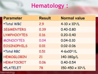
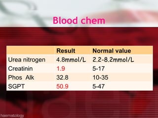
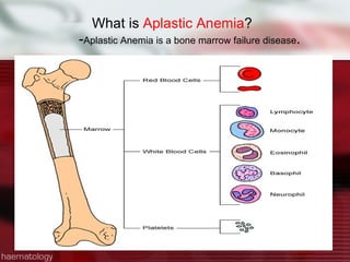
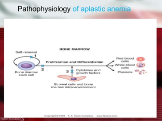
CASE  MANAGEMENT  Group 1
GENERAL DATA J.G., 34 y/o, male Filipino, from Dagupan City  admitted for the first time at PMC on November 2, 2009.
INFORMANT:  Patient RELIABILITY:  80 % CHIEF COMPLAINT:  Dizziness
HISTORY OF PRESENT ILLNESS 5 days  prior to admission dizziness associated with loss of appetite  Vomiting  non-billous non-projectile amounting to 5-10 ml per bout.  generalized body weakness
HPI..cont Few hours PTA  Dizziness---px can’t walk and stand alone Consultation --admission
Past Medical History  Year 2000 Dizziness Philippine General Hospital due ---- diagnosed to have  “ Paroxysmal Nocturnal Hemoglubinuria” Medication: Folic acid  OD  and  Vit B6 & B1 OD September 2009 Skin discoloration At Philippine General Hospital  Px  already had 14x blood transfusion(4-5bags/time)
Family History Mother has  breast cancer Father has  hypertension.  3 siblings have  anemia.
Social, Personal, and Environmental History   P atient is a farmer Lives with his wife and 3 children Alcoholic drinker Smoker
Review of system +Dizziness +loss of appetite  +Vomiting  +generalized body weakness
PHYSICAL EXAMINATION   SKIN:  (+) jaundice HEENT:  Pale palpebral conjunctivae ABDOMEN:  Globular , liver span - 13cm
Vital signs BP - 110/80 mmHg HR - 78 bpm RR - 16 cpm TEM - 36.8 C( upon admission  37.8)
Laboratory test
Hematology : Parameter  Result  Normal value  Total WBC 2.9 4-10 x 10 9 /L SEGMENTERS 0.39 0.40-0.80 LYMPHOCYTES 0.16 0.20-0.40 MONOCYTES 0.04 0.04-0.10 EOSINOPHILS 0.01 0.02-0.06 Total RBC 0.51 4-6x10 12 /L HEMOGLOBIN 21 140-180g/L HEMATOCRIT 0.06 0.40-0.54 PLATELET 78 150-450 x 10 9 /L
Blood chem Result  Normal value  Urea nitrogen 4.8 mmol/L 2.2-8.2mmol/L Creatinin 1.9 5-17 Phos  Alk 32.8 10-35 SGPT  50.9 5-47
HBsAg screening  Nonreaction
What are you impression?
APLASTIC ANEMIA
DIFFERENTIAL  DIAGNOSIS Role out  Leukemia--- WBC  increased in BM Multiple Myeloma--- plasma cell  increased in BM Lymphoma(Hodgkins)--- Lymphocyte  increased in BM
APLASTIC  ANEMIA What happens when the bone marrow shuts down?
What is  Aplastic Anemia ? - Aplastic Anemia is a bone marrow failure disease .
APLASTIC ANEMIA Aplastic anemia is a severe, life threatening syndrome in which production of erythrocytes, WBCs, and platlets has failed. The disease is characterized by  peripheral pancytopenia  and accompanied by a  hypocellular bone marrow.
Peripheral blood  Smear  Peripheral  blood  Smear
HYPOCELLULAR  BONE MARROW IN APLASTIC ANEMIA
Aplastic Anemia  patients Aplastic Anemia  patients have  decreased  amounts of: -  Red Blood Cells -  White Blood Cells -  Platelets
Functions of Blood Cells Red Blood Cells Carry oxygen to all body organs White Blood Cells Fight infection and keep you healthy Platelets Help control bleeding
Clinical Manifestations  Symptoms caused by  suppression of any or all bone marrow elements Low Red Blood Cell Fatigue, Headache, Inability to Concentrate,  Dyspnea ,Pale skin Low White Blood Cell  Frequent or prolonged infections Viral Infections, Bacterial Infections Low Platelets Easy Bruising, Petichiae,  Nosebleed and bleeding gums,Prolonged bleeding
PATHOGENESIS Stem cell failure resulting from: 1-An acquired intrinsic stem cell defect 2-An environmental cause Immune mechanisms Growth factor deficiency  Defects in the microenvironment
Pathophysiology  of aplastic anemia
Epidemiology Incidence:  5-10 persons:10 6  per year Aplastic anemia may occur in all age groups and both genders
Etiology  : Hereditary 1-Schwacman – Diamond Syndrome  2-Fanconi’s anemia syndrome Autosomal recessive disorder congenital developmental anomalies progressive pancytopenia increase risk of malignancy 3-Dyskeratosis congenita characterized by mucous membrane leukoplasia dystrophic nails, reticular hyperpigmentation development of aplastic anemia during childhood
Etiology:  Acquired 1-Idiopathic 2- Drugs: dose related idiosyncratic Nonsteroidal analgesics Sulfonamides Thyrostatic drugs Some psychotropics Penicillamine Allopurinol Chlorampenicol
Etiology:  Acquired  3-Radiation- Damages DNA 4-Chemicals-  BENZENE 5-Viruses-  Hepatitis – most common  6-Pregnancy  7-PNH ( Paroxysmal Nocturnal Hemoglobinuria ) 8-Disorders of immune system-  SLE.
What causes Paroxysmal Nocturnal Hemoglobinuria  Defect in PIG-A gene  which is required for the biosynthesis of cellular anchors,  so partial or complete inability to construct Glycosylphosphatidylinositol(GPI) anchor for the attachment of CD55, CD59 Diagnosis:  Ham’s test ,  sucrose hemolytic test , Flow cytometry  using antibodies against cell surface antigens CD55, CD59 which are lacking in disease
Clinical manifestations of  PNH Thrombosis Hepatic vein most common common cause of fatality Cerebral vein thrombosis sagittal sinus in particular Abdominal veins Dermal veins Pulmonary embolism unusual
Clinical manifestations of  PNH Relative/absolute bone marrow failure present to some degree in all patients relative granulocytopenia/thrombocytopenia decreased capacity to form myeloid colonies Two stage model somatic mutation in PIG-A gene some cause for bone marrow failure
Diagnosis  of AA:  Lab findings Peripheral blood Smear  Bone marrow biopsy Pancytopenia Normocytic-normochromic anemia (may be slightly macrocytic) Low reticulocyte index Empty fatty spaces Few hematopoietic cells Lymphocytes and plasma cells Hypocellular bone marrow
CLASSIFICATION Designation Criteria PBS BM biopsy Severe aplastic anemia -2 / 3 values- Neutro < 500/  L -Platelets < 20,000/ ul- -Reticulocyte index < 1% -Marked hypocellular < 25% cellularity -Moderate hypocellular <25-50%  -normal cellularity with <30% of remaining cell hematopoietic  Very severe aplastic anemia As above but neutrophils < 200/  L Infection present
Presentation of Anemia, Neutropenia and Thrombocytopenia Hemorrhagic lesion of the gums in a patient with aplastic anemia caused by infection with  Capnocytophaga ochraceus;  such lesions are easily confused with those of herpes simplex.
26-year-old woman with acute aplastic anemia and 1 day of facial pain/swelling. Mouth open involuntarily due to perioral edema. Needle aspirate of small purplish area near right alar revealed  P. aeruginosa.
Aplastic anemia. Oral leukoplakia in dyskeratosis congenita.
Treatment Options Bone Marrow  Transplant Growth Hormones Immune Suppressive Therapy Supportive Care
TREATMENT 1-Withdrawal of the etiologic agent 2-Supportive treatment transfusion of the blood products,  CMV seronegative should be given transfusion from the family members should be avoided to prevent sensitization. pooled donor platelets but leads to sensitization in refractory cases need HLA matched transfusion packed cells filtrated to remove leukocyte and platelets iron overload : give chelating therapy deferoxamine CMV prophylaxis Staph. Aeureus *  hospitalization * menses
3.Allogeneic BMT This is the best therapy for the young patient with a fully histocompatible sibling donor  usually indicated for most patients with severe disease.  -Preferably from sibling -Curative in 60-90% of patients -Applicable only for a third of patients
4.SPLENECTOMY Removal of the spleen  does not increase hematopoiesis but may increase neutrophil  increase platelet counts two- to threefold  improve survival of transfused red cells  platelets in highly sensitized individuals
5.Immunosuppression Antithymocyte globulin(ATG) induces hematologic recovery (independence from transfusion and a leukocyte count adequate to prevent infection. Cyclosporin + ATG Corticosteroids High dose cyclophosphamide  6. G-CSF/ GM-CSF/ EPO  **Response rate 50-70%  Occurs 2-3 months  post Rx.
Newer *Mycophenolate mofetil (MMF) - cytotoxic to T cells *Monoclonal Ab against IL-2 receptor which is present on activated lymphocytes
Outcomes Age,  Younger is  better BMT < 20 yr with a sib… 75% 20 - 40 yr with a sib…60% < 20 yr unrelated BMT… 40% 20 - 40 yr unrelated BMT…35% Immunosuppression - 60 - 80%
 
Thank you ?

Aa

  • 1.
  • 2.
    GENERAL DATA J.G.,34 y/o, male Filipino, from Dagupan City admitted for the first time at PMC on November 2, 2009.
  • 3.
    INFORMANT: PatientRELIABILITY: 80 % CHIEF COMPLAINT: Dizziness
  • 4.
    HISTORY OF PRESENTILLNESS 5 days prior to admission dizziness associated with loss of appetite Vomiting non-billous non-projectile amounting to 5-10 ml per bout. generalized body weakness
  • 5.
    HPI..cont Few hoursPTA Dizziness---px can’t walk and stand alone Consultation --admission
  • 6.
    Past Medical History Year 2000 Dizziness Philippine General Hospital due ---- diagnosed to have “ Paroxysmal Nocturnal Hemoglubinuria” Medication: Folic acid OD and Vit B6 & B1 OD September 2009 Skin discoloration At Philippine General Hospital Px already had 14x blood transfusion(4-5bags/time)
  • 7.
    Family History Motherhas breast cancer Father has hypertension. 3 siblings have anemia.
  • 8.
    Social, Personal, andEnvironmental History P atient is a farmer Lives with his wife and 3 children Alcoholic drinker Smoker
  • 9.
    Review of system+Dizziness +loss of appetite +Vomiting +generalized body weakness
  • 10.
    PHYSICAL EXAMINATION SKIN: (+) jaundice HEENT: Pale palpebral conjunctivae ABDOMEN: Globular , liver span - 13cm
  • 11.
    Vital signs BP- 110/80 mmHg HR - 78 bpm RR - 16 cpm TEM - 36.8 C( upon admission 37.8)
  • 12.
  • 13.
    Hematology : Parameter Result Normal value Total WBC 2.9 4-10 x 10 9 /L SEGMENTERS 0.39 0.40-0.80 LYMPHOCYTES 0.16 0.20-0.40 MONOCYTES 0.04 0.04-0.10 EOSINOPHILS 0.01 0.02-0.06 Total RBC 0.51 4-6x10 12 /L HEMOGLOBIN 21 140-180g/L HEMATOCRIT 0.06 0.40-0.54 PLATELET 78 150-450 x 10 9 /L
  • 14.
    Blood chem Result Normal value Urea nitrogen 4.8 mmol/L 2.2-8.2mmol/L Creatinin 1.9 5-17 Phos Alk 32.8 10-35 SGPT 50.9 5-47
  • 15.
    HBsAg screening Nonreaction
  • 16.
    What are youimpression?
  • 17.
  • 18.
    DIFFERENTIAL DIAGNOSISRole out Leukemia--- WBC increased in BM Multiple Myeloma--- plasma cell increased in BM Lymphoma(Hodgkins)--- Lymphocyte increased in BM
  • 19.
    APLASTIC ANEMIAWhat happens when the bone marrow shuts down?
  • 20.
    What is Aplastic Anemia ? - Aplastic Anemia is a bone marrow failure disease .
  • 21.
    APLASTIC ANEMIA Aplasticanemia is a severe, life threatening syndrome in which production of erythrocytes, WBCs, and platlets has failed. The disease is characterized by peripheral pancytopenia and accompanied by a hypocellular bone marrow.
  • 22.
    Peripheral blood Smear Peripheral blood Smear
  • 23.
    HYPOCELLULAR BONEMARROW IN APLASTIC ANEMIA
  • 24.
    Aplastic Anemia patients Aplastic Anemia patients have decreased amounts of: - Red Blood Cells - White Blood Cells - Platelets
  • 25.
    Functions of BloodCells Red Blood Cells Carry oxygen to all body organs White Blood Cells Fight infection and keep you healthy Platelets Help control bleeding
  • 26.
    Clinical Manifestations Symptoms caused by suppression of any or all bone marrow elements Low Red Blood Cell Fatigue, Headache, Inability to Concentrate, Dyspnea ,Pale skin Low White Blood Cell Frequent or prolonged infections Viral Infections, Bacterial Infections Low Platelets Easy Bruising, Petichiae, Nosebleed and bleeding gums,Prolonged bleeding
  • 27.
    PATHOGENESIS Stem cellfailure resulting from: 1-An acquired intrinsic stem cell defect 2-An environmental cause Immune mechanisms Growth factor deficiency Defects in the microenvironment
  • 28.
    Pathophysiology ofaplastic anemia
  • 29.
    Epidemiology Incidence: 5-10 persons:10 6 per year Aplastic anemia may occur in all age groups and both genders
  • 30.
    Etiology :Hereditary 1-Schwacman – Diamond Syndrome 2-Fanconi’s anemia syndrome Autosomal recessive disorder congenital developmental anomalies progressive pancytopenia increase risk of malignancy 3-Dyskeratosis congenita characterized by mucous membrane leukoplasia dystrophic nails, reticular hyperpigmentation development of aplastic anemia during childhood
  • 31.
    Etiology: Acquired1-Idiopathic 2- Drugs: dose related idiosyncratic Nonsteroidal analgesics Sulfonamides Thyrostatic drugs Some psychotropics Penicillamine Allopurinol Chlorampenicol
  • 32.
    Etiology: Acquired 3-Radiation- Damages DNA 4-Chemicals- BENZENE 5-Viruses- Hepatitis – most common 6-Pregnancy 7-PNH ( Paroxysmal Nocturnal Hemoglobinuria ) 8-Disorders of immune system- SLE.
  • 33.
    What causes ParoxysmalNocturnal Hemoglobinuria Defect in PIG-A gene which is required for the biosynthesis of cellular anchors, so partial or complete inability to construct Glycosylphosphatidylinositol(GPI) anchor for the attachment of CD55, CD59 Diagnosis: Ham’s test , sucrose hemolytic test , Flow cytometry using antibodies against cell surface antigens CD55, CD59 which are lacking in disease
  • 34.
    Clinical manifestations of PNH Thrombosis Hepatic vein most common common cause of fatality Cerebral vein thrombosis sagittal sinus in particular Abdominal veins Dermal veins Pulmonary embolism unusual
  • 35.
    Clinical manifestations of PNH Relative/absolute bone marrow failure present to some degree in all patients relative granulocytopenia/thrombocytopenia decreased capacity to form myeloid colonies Two stage model somatic mutation in PIG-A gene some cause for bone marrow failure
  • 36.
    Diagnosis ofAA: Lab findings Peripheral blood Smear Bone marrow biopsy Pancytopenia Normocytic-normochromic anemia (may be slightly macrocytic) Low reticulocyte index Empty fatty spaces Few hematopoietic cells Lymphocytes and plasma cells Hypocellular bone marrow
  • 37.
    CLASSIFICATION Designation CriteriaPBS BM biopsy Severe aplastic anemia -2 / 3 values- Neutro < 500/  L -Platelets < 20,000/ ul- -Reticulocyte index < 1% -Marked hypocellular < 25% cellularity -Moderate hypocellular <25-50% -normal cellularity with <30% of remaining cell hematopoietic Very severe aplastic anemia As above but neutrophils < 200/  L Infection present
  • 38.
    Presentation of Anemia,Neutropenia and Thrombocytopenia Hemorrhagic lesion of the gums in a patient with aplastic anemia caused by infection with Capnocytophaga ochraceus; such lesions are easily confused with those of herpes simplex.
  • 39.
    26-year-old woman withacute aplastic anemia and 1 day of facial pain/swelling. Mouth open involuntarily due to perioral edema. Needle aspirate of small purplish area near right alar revealed P. aeruginosa.
  • 40.
    Aplastic anemia. Oralleukoplakia in dyskeratosis congenita.
  • 41.
    Treatment Options BoneMarrow Transplant Growth Hormones Immune Suppressive Therapy Supportive Care
  • 42.
    TREATMENT 1-Withdrawal ofthe etiologic agent 2-Supportive treatment transfusion of the blood products, CMV seronegative should be given transfusion from the family members should be avoided to prevent sensitization. pooled donor platelets but leads to sensitization in refractory cases need HLA matched transfusion packed cells filtrated to remove leukocyte and platelets iron overload : give chelating therapy deferoxamine CMV prophylaxis Staph. Aeureus * hospitalization * menses
  • 43.
    3.Allogeneic BMT Thisis the best therapy for the young patient with a fully histocompatible sibling donor usually indicated for most patients with severe disease. -Preferably from sibling -Curative in 60-90% of patients -Applicable only for a third of patients
  • 44.
    4.SPLENECTOMY Removal ofthe spleen does not increase hematopoiesis but may increase neutrophil increase platelet counts two- to threefold improve survival of transfused red cells platelets in highly sensitized individuals
  • 45.
    5.Immunosuppression Antithymocyte globulin(ATG)induces hematologic recovery (independence from transfusion and a leukocyte count adequate to prevent infection. Cyclosporin + ATG Corticosteroids High dose cyclophosphamide 6. G-CSF/ GM-CSF/ EPO **Response rate 50-70% Occurs 2-3 months post Rx.
  • 46.
    Newer *Mycophenolate mofetil(MMF) - cytotoxic to T cells *Monoclonal Ab against IL-2 receptor which is present on activated lymphocytes
  • 47.
    Outcomes Age, Younger is better BMT < 20 yr with a sib… 75% 20 - 40 yr with a sib…60% < 20 yr unrelated BMT… 40% 20 - 40 yr unrelated BMT…35% Immunosuppression - 60 - 80%
  • 48.
  • 49.

Editor's Notes