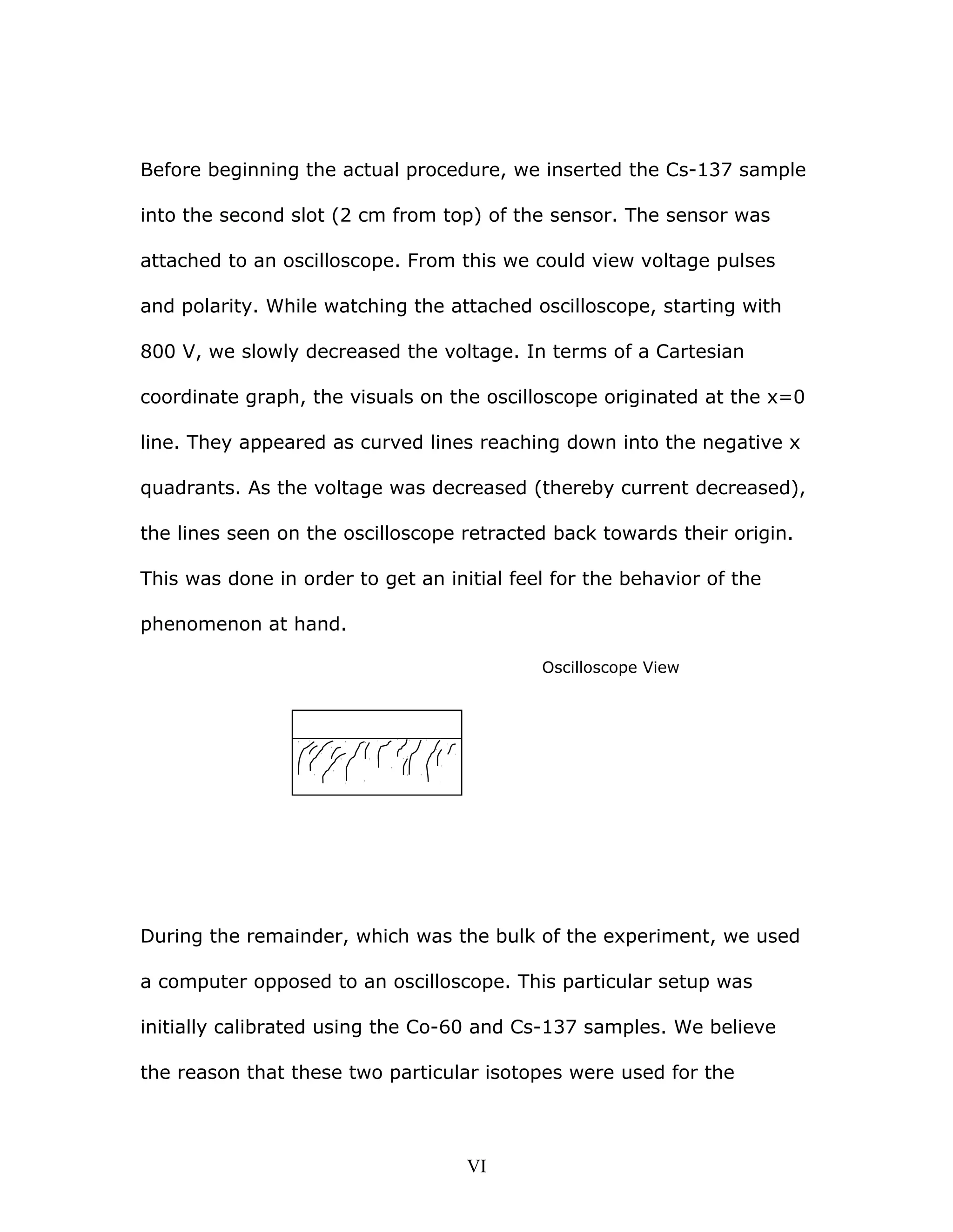
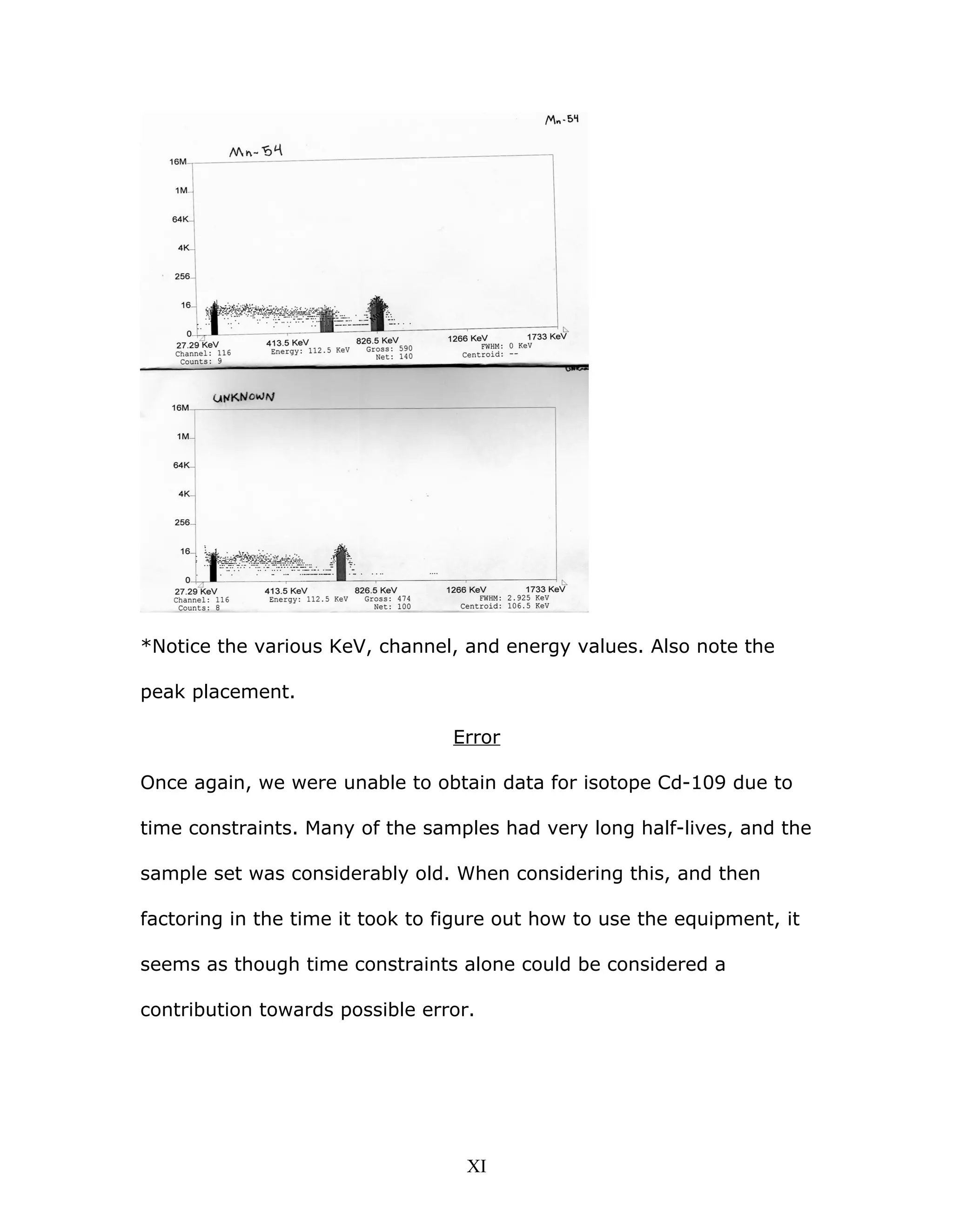
The document summarizes a gamma ray spectroscopy lab experiment. Key findings include:
- Gamma rays from various radioactive samples were measured using a sodium iodide detector and spectrometer interface.
- A linear relationship was found between the gamma ray energies and their channel numbers on the spectrometer.
- All major gamma ray interactions (photoelectric effect, Compton scattering) were observed except pair production which requires higher energies.
- The unknown isotope was identified as Cesium-137 based on its measured energy of 655.5 keV, though the author initially disagreed with this assessment.
X-ray physics is summarized as follows:
(1) X-rays are produced when fast moving electrons are stopped by a target material, with 1% of the electron's kinetic energy converted to X-rays. (2) X-ray generators use a high voltage source to accelerate electrons from a heated cathode filament toward an anode target, producing a bremsstrahlung spectrum of X-rays. (3) Modern X-ray tubes feature a rotating anode to dissipate heat and allow longer exposures without damage to the anode.
This document discusses the attenuation of x-rays as they pass through matter. It defines key terms like quantity, quality, intensity, attenuation, linear attenuation coefficient, half value layer, and mass attenuation coefficient. It describes how the energy of x-rays, the density and atomic number of the absorbing material, and whether radiation is monoenergetic or polyenergetic affect attenuation. Higher energy x-rays or absorbers with lower density/atomic number experience less attenuation. Attenuation follows an exponential curve on semi-log graphs. Photoelectric interactions dominate at low energies while Compton scattering increases at higher energies. These principles underlie the contrast seen in x-ray images.
This document provides an overview of radiation dosimeters and their properties. It discusses the key characteristics of dosimeters including accuracy, precision, linearity, dose rate dependence, energy response, directional dependence, spatial resolution, readout convenience, and ease of use. It also describes common dosimeter systems, focusing on ionization chamber dosimeters which are the recommended standard for beam calibration and reference dosimetry in radiation therapy. Cylindrical and parallel-plate ionization chambers are discussed in detail.
Wilhelm Conrad Roentgen discovered X-rays in 1895 while experimenting with cathode rays. He noted a new type of ray coming from the cathode tube that could pass through materials and photographed his wife's hand. X-rays are produced when high-speed electrons collide with a metal target in a vacuum tube. This produces bremsstrahlung X-rays of varying energies and characteristic X-rays of specific energies related to the target material. Factors like target material, voltage, current, and filtration determine the quantity, quality, and efficiency of the X-ray beam produced.
Analysis of space charge controlled electric field 1Chandan Kumar
The document discusses space charge and its effects on cable insulation failures. Space charge forms due to inhomogeneous resistivity, ionization within dielectrics, charge injection from electrodes, and polarization. Its presence distorts electric fields inside dielectrics, potentially leading to localized breakdown. Simulation results show how voids and trapped charge can enhance electric field stresses. The document also examines space charge limited current in cable insulation and simulates the relationship between current density and voltage for parallel plate electrodes, finding good agreement with analytical solutions. Future work is proposed to further study space charge effects in cables and insulation materials.
X ray machines - conventional and digitalUrfeya Mirza
X-ray machines use high-energy electromagnetic radiation to generate digital or film images of the internal structures of objects. Conventional x-ray machines use film that must be developed, while digital x-ray machines directly convert x-rays to electrical signals and display images digitally. Both use an x-ray tube to generate x-rays, which are controlled via technique factors selected on the machine's control panel. Digital x-ray offers advantages like adjustable images and compatibility with digital record systems.
This document discusses various radiation quantities and units used in medical physics. It defines units like becquerel (Bq), gray (Gy), sievert (Sv), and rem (Roentgen equivalent man) used to measure activity, absorbed dose, equivalent dose, and effective dose. It also discusses concepts like half-life, decay constant, kerma, exposure, and dose conversion factors. International organizations like ICRP and ICRU provide recommendations on radiation quantities, units, and safety limits.
The document discusses various interactions between radiation and matter, including excitation, ionization, and radiative losses. It describes the processes of excitation, ionization, specific ionization, linear energy transfer, scattering, bremsstrahlung, and the four main interactions of x-rays and gamma rays with matter: Rayleigh scattering, Compton scattering, photoelectric absorption, and pair production. The probability and characteristics of each interaction depends on factors like the atomic number of the absorbing material and the energy of the incident radiation. These interactions are important in diagnostic imaging and radiation therapy.
Luminescence is the emission of light from a cool object, in contrast to incandescence which is the emission of light from a hot object. There are three main types of luminescence: phosphorescence involves the absorption and slow re-emission of light, fluorescence involves fast absorption and re-emission of light, and chemiluminescence is the emission of light driven by a chemical reaction. Phosphorescent minerals will glow after exposure to UV light. Fluorescence is seen in scorpion exoskeletons and deep sea organisms. Chemiluminescence, the most common form in living organisms, is used by fireflies, deep sea fish, and microorganisms.
This document discusses various interactions of radiation with matter, including the photoelectric effect, Compton scattering, and pair production. It also covers the linear attenuation coefficient and how it relates to the attenuation of x-rays and gamma rays as they pass through matter. Additionally, it defines terms such as half-value layer, mean free path, fluence, flux, energy fluence, and kerma in the context of radiation physics.
The document discusses the interaction of radiation with matter. It explains that radiation can be electromagnetic or particulate. When electromagnetic radiation like x-rays or gamma rays pass through matter, they can undergo attenuation, absorption, scattering, or transmission. The major interactions that cause attenuation are coherent scattering, the photoelectric effect, the Compton effect, pair production, and photonuclear interactions. It describes each of these interactions in detail and how they transfer energy from the radiation to the absorbing material.
This document discusses the construction and types of x-ray films used in medical imaging. It begins with an overview of the layers that make up an x-ray film, including the adhesive layer, emulsion layer containing silver halide crystals, and protective supercoat layer. The document then discusses the history of film bases and characteristics of modern polyester bases. It describes the functions of duplitized and single emulsion films, advantages and disadvantages of each, and common film types and sizes used for different medical imaging purposes.
This document discusses the dual wave-particle nature of X-rays and their interaction with matter. It notes that X-rays can behave as both waves that propagate through space, as well as particles called photons. The two main interactions discussed are the photoelectric effect and Compton scattering.
The photoelectric effect occurs when a photon ejects an inner shell electron from an atom. This produces characteristic radiation, a photoelectron, and an ionized atom. It is most likely with low-energy photons and high atomic number elements. Compton scattering occurs when a high-energy photon ejects a loosely bound outer shell electron. This produces a recoil electron and scattered photon, with energy distributed between the two. Scattered photons are
This document discusses scintillation detectors and their properties. Scintillation detectors work by emitting light when exposed to radiation, and this light output can be used to measure incident radiation. The key properties of scintillation detectors are that they must have high scintillation efficiency, light yield proportional to deposited energy, short decay time, transparency to emitted wavelengths, and be able to be made in large sizes and desired shapes. Common inorganic scintillators discussed are NaI(Tl), which is widely used due to its availability and high detection efficiency, and BGO, which has high intrinsic efficiency for high gamma energies.
The copper in a stationary anode plays a dual role:
1. It supports the tungsten target.
2. It efficiently removes heat from the tungsten target.
Copper acts as a heat sink, drawing heat away from the tungsten target to prevent it from overheating due to the energy deposited by bombarding electrons.
Filters are used in x-ray imaging to shape the beam and increase the ratio of useful photons for imaging to those that increase patient dose or decrease image contrast. Filters are typically made of metal like aluminum or copper and are placed between the x-ray tube and patient. They absorb the low energy photons that do not penetrate tissue deeply but deposit much radiation in superficial tissues. This provides better tissue penetration by the beam while reducing the skin dose and improving contrast. Different types of filters include inherent, added, compound, and wedge filters which vary in materials and thickness used.
The document discusses beam quality, half value layer (HVL), and filters used in radiation beams. It defines HVL as the thickness of an absorber required to attenuate the beam intensity to half its original value. Beam quality and HVL are influenced by factors like energy, thickness, and density. Different types of filters are used such as inherent, added, combination, and flattening filters to modify the beam spectrum by removing low energy photons through beam hardening. The quality of megavoltage beams is specified by peak energy rather than HVL since they are heavily filtered.
Dr. saleh history of medical physics in saAmmar Felemban
The document discusses the history of medical physics in Saudi Arabia. It notes that the first medical physics departments were established in the late 1970s at King Faisal Specialist Hospital and Riyadh Military Hospital. It then provides details on the pioneers of the field in Saudi Arabia and the growth of medical physics programs and professionals over time. Key events discussed include the establishment of the Saudi Medical Physics Society in 2006 and various conferences held in the country starting in 2002. The document aims to give an overview of the development of medical physics as a specialized field in Saudi Arabia.
This document provides instructions for building and using a Michelson interferometer to precisely measure the wavelength of light. It consists of 3 parts: 1) an overview of how the Michelson interferometer works, 2) technical details of the experimental setup, and 3) a procedure for aligning the mirrors and measuring the wavelength of a He-Ne laser. The goal is to achieve an accuracy of one part in 10,000 by translating one mirror and counting the number of interference fringes that pass a photodiode.
Dosimetry is the process of measuring radiation doses and assigning them to individuals. There are two types of exposure: external, where radiation comes from outside the body, and internal, where radiation is emitted from substances inside the body. Radiation can be measured using personal dosimeters, environmental monitoring, or biological sampling. The main types of radiation are alpha, beta, gamma, and neutrons. Radiation dose is quantified using absorbed dose, equivalent dose, and effective dose. Dosimeters come in passive and active varieties to measure external radiation doses, while internal doses require estimating intake and calculating dose to organs over time. Measurement uncertainty arises from factors like dosimeter calibration and environmental variability.
The document discusses the key components of an X-ray production system, including the cathode, anode, generator, and tube rating charts. The cathode emits electrons via a heated filament towards the anode. X-rays are produced when electrons collide with the anode. Generators supply power to heat the cathode and accelerate electrons. Tube rating charts indicate safe operating limits based on heat loading to prevent damage. Automatic exposure control uses radiation detectors to optimize exposure time based on patient thickness.
The document discusses the concept of relative biological effectiveness (RBE), which is defined as the ratio of absorbed doses of different types of radiation (such as x-rays vs neutrons) required to produce the same biological effect. Higher LET (linear energy transfer) radiation like neutrons and alpha particles have a higher RBE than lower LET radiation like x-rays. The RBE depends on factors like radiation quality, dose, dose rate, and biological system. Radiation with an LET around 100 keV/μm tends to have the highest RBE and lowest oxygen enhancement ratio due to depositing the optimal amount of energy to cause DNA damage. Radiation weighting factors are used to account for different radiation types when assessing health risks.
This document discusses different types of solid state radiation detectors, including scintillation detectors, thermoluminescent dosimeters (TLD), and semiconductor detectors. Scintillation detectors detect radiation via light emission in inorganic crystal materials like NaI or organic crystals like anthracene. TLDs "capture" radiation dose information and release light when heated, allowing dose measurement. Common TLD materials are LiF:Mg,Ti and Li2B4O7:Mn. Semiconductor detectors like silicon and germanium act as solid state ionization chambers and are used for high resolution energy measurement of alpha and beta particles.
Gamma Interactions and Gamma Spectroscopy with Scintillation DetectorsDaniel Maierhafer
The document discusses gamma ray interactions and spectroscopy using scintillation detectors. It describes the three main types of gamma ray interactions: photoelectric absorption, Compton scattering, and pair production. It also discusses energy spectra resulting from these interactions and how detector size affects the response function. The experiment used two sizes of NaI:TL scintillators (2"x2" and 5"x5") along with associated electronics to collect gamma ray spectra from various sources and determine properties like energy resolution and unknown source identification. Procedures and results are presented for analyzing spectra from the sources using a single channel analyzer and multi-channel analyzer.
The document summarizes the design evaluation results for a gamma ray incidence detector housing for a CubeSat mission. Key features of the new design include cork insulation sleeves for thermal insulation and vibration dampening, an internal PCB for signal amplification isolation, and a ceramic interface to electrically isolate the housing from the satellite structure. Testing showed the design met requirements for the satellite's temperature and vibration ranges to ensure proper detector performance during the EXACT mission to study solar flares and measure photon time arrivals.
This document discusses scintillator materials for gamma ray spectroscopy. It describes Lawrence Livermore National Laboratory's efforts to develop new scintillator materials with high energy resolution and stopping power to discriminate gamma ray spectra, while being low cost and having no intrinsic radioactivity. Some promising new materials discussed include single crystal strontium iodide doped with europium, ceramic gadolinium gallium aluminum garnet doped with cerium, and bismuth-loaded polymer plastics. These new materials show energy resolutions that improve on existing sodium iodide and offer potential for lower cost gamma spectroscopy detectors.
Multi-channel Detector Readout Integrated Circuits with ADCs for X-ray and Ga...Gunnar Maehlum
The document describes a family of multi-channel readout integrated circuits (ROICs) developed for X-ray and gamma-ray spectroscopy in space applications. The ROICs called VATAs integrate pre-amplifiers, pulse shaping circuits, discriminators, and 10-bit analog-to-digital converters for each of their 32-64 channels. They deliver digital pulse amplitudes and pixel addresses with low power consumption of 0.2-1.3 mW per channel. The VATAs are being used on several space missions including Astro-H, BepiColombo, and FOXSI to perform radiation detection and spectroscopy of high-energy photons. Test results show the ASICs achieve good energy resolution and low
The Tokai-Mura criticality accident occurred at a nuclear fuel processing plant in Japan in 1999. Workers were preparing a batch of enriched uranium solution when an uncontrolled nuclear chain reaction began, exposing workers and releasing radiation. Two workers died from radiation exposure and over 600 people received doses exceeding an annual public limit. The accident highlighted issues with training and oversight at smaller nuclear facilities outside the mainstream fuel cycle.
This document summarizes an analysis of gamma ray spectroscopy data from experiments on 109Ru and 111Ru nuclei. Key steps included: preparing detectors for experiments at Argonne National Lab; analyzing previous experiment data to find gamma ray energies through correlations; plotting level schematics; and comparing transition intensities. Band crossings near 350-500 keV in 111Ru provided evidence of a possible nuclear structure change from oblate to prolate. The author also traveled to Argonne to perform new experiments using the Gammasphere detector array combined with a "CHICO-like" apparatus.
PEPTIDE LABELLING & GAMMA RAY SPECTROSCOPYupvita pandey
This document summarizes an internship project on peptide labeling and gamma ray spectroscopy. It discusses the discovery of radioactivity, types of radioactive decay, radiolabeling peptides, and their use in diagnostic imaging and cancer therapy. Experiments were described to identify gamma energies from radioactive sources and estimate uranium-238 activity in soil and leaf samples using a gamma detector, with results showing higher activity in soils than leaves.
The characterization of_the_gamma_ray_signal_from_the_central_milk_way_a_comp...Sérgio Sacani
This document analyzes the gamma-ray signal from the central Milky Way that is consistent with emission from annihilating dark matter particles. The authors re-examine Fermi data using cuts on an event parameter to improve gamma-ray maps and more easily separate components. They find the GeV excess is robust and well-fit by a 36-51 GeV dark matter particle annihilating to bottom quarks with a cross section of 1-3×10⁻²⁶ cm3/s. The signal extends over 10 degrees from the Galactic Center and is spherically symmetric, disfavoring explanations from millisecond pulsars or gas interactions.
JEE Physics/ Lakshmikanta Satapathy/ Beta decay of a nucleus and the subsequent emission of a Gamma ray photon with the related concepts of mass defect and excitation energy
Final year project: To Design and Test a low cost Gamma Ray detectorChristopher Mitchell
The document describes designing and testing a low-cost gamma radiation detector using a smartphone camera. The author tests various parameters of the smartphone detector such as calibration with radiation sources, thermal noise, distance effects, shielding effects, timing resolution, and efficiency compared to semiconductor detectors. Although not as efficient as professional detectors, the smartphone detector provides a low-cost option for estimating radiation dose that is more accessible than specialized equipment.
High Sensitivity Gamma Ray Spectroscopy FYPDavid Holland
This document summarizes an investigation into potential impurities in the radiopharmaceutical isotopes samarium-153 and radium-223, which are used to treat bone pain in cancer patients. Samples were analyzed using sodium iodide and germanium detectors. The germanium detector results showed several impurities like actinium-227, thorium-227, protactinium-231 and uranium-235 present in both isotopes. However, further investigation is needed as the detector was not properly calibrated. The presence of impurities could potentially harm patients or cause issues with waste disposal. Samarium-153 and its uses in bone pain treatment are also discussed.
A Low Power & Low Noise Multi-Channel ASIC for X-Ray and Gamma-Ray SpectroscopyGunnar Maehlum
The document describes a family of multi-channel detector readout integrated circuits (ROICs) developed for X-ray and gamma-ray spectroscopy in space applications. The ROICs, called VATAs, integrate analog-to-digital converters for fully digital readout of radiation detectors. VATAs are being used on several space missions including ASTRO-H, BepiColombo, and FOXSI. They have low power dissipation, low noise, and provide digital pulse heights and pixel addresses upon detection of radiation. Test results show the VATAs achieve good energy resolution and meet requirements for noise, power, and temperature range for different space missions.
Gamma rays are a form of electromagnetic radiation emitted from radioactive substances. They have the shortest wavelengths and highest frequencies of any type of electromagnetic wave. Gamma rays are produced during radioactive decay, electron-positron annihilation, and other nuclear processes. Some key applications of gamma rays include use in radiography, cancer treatment, food sterilization, and nuclear weapons.
This is the presentation I gave when defending my Ph.D thesis at SLAC. The title of my defense was "Neutron Star Powered Nebulae: a New View on Pulsar Wind Nebulae with the Fermi Gamma-ray Space Telescope".
Radioactive isotopes emit radiation through radioactive decay as their unstable nuclei break down. There are three main types of radiation emitted: alpha particles, beta particles, and gamma rays. Radioactive isotopes are used in scientific research, analytical applications like radioimmunoassays, and medical diagnostic procedures and therapies. Some key radioactive isotopes used include iodine-131 for thyroid imaging and cancer treatment, technetium-99m for thyroid scans, and strontium-89 or samarium-153 to treat bone metastases.
From my class on nuclear physics for nuclear medicine technologists. This class covers alpha, beta, and gamma decay, plus conversion electrons, Auger electrons, and k-alpha and other X-rays
1) The experiment used gamma-ray spectroscopy to analyze spectra from various radioactive sources. Spectra were recorded at different photomultiplier tube voltages to study resolution and efficiency.
2) Analysis found the number of dynodes in the photomultiplier tube to be 6.5, and resolution R was determined to be inversely proportional to gamma ray energy as expected.
3) Activity of a potassium chloride sample was estimated using detector efficiency calculations, finding 1.7×10^17 40K nuclei, consistent with the expected amount.
The document summarizes an experiment to determine the charge to mass ratio of an electron (e/m) using a Helmholtz coil apparatus. It provides background on the theory and setup of the experiment. Data was collected on the magnetic field and electron beam radius needed to hit targets of different distances. The average calculated e/m ratios were 2.24065 × 10¹¹ c/kg and 2.01474 × 10¹¹ c/kg for magnetic fields lined with and against the Earth's field, respectively. While these differed from the accepted value, the author notes factors like measurement precision that could have contributed to the error.
Ethan conducted a photoelectric effect experiment to calculate Planck's constant. The experiment involved measuring the stopping potential of electrons emitted from a metal surface under monochromatic light of varying wavelengths. Plotting average stopping potential versus the reciprocal of wavelength produced a straight line, from which Planck's constant could be calculated using the slope. Ethan's calculated value of Planck's constant had a 36% error compared to the accepted value, which was within an acceptable range for the experiment.
Detection of gamma radiation is used to study properties of atomic nuclei. Two common detectors are scintillation detectors and semiconductor detectors. Scintillation detectors use scintillation materials that emit visible light when struck by gamma rays, while semiconductor detectors use depletion regions in germanium crystals. Both detectors require amplification and signal processing electronics to analyze the energy deposited by gamma rays. Key measurements involve determining the energy spectrum of gamma ray sources and identifying peaks and edges that reveal information about nuclear properties and interactions.
Experimental Verification of the Kinematic Equations of Special Relativity an...Daniel Bulhosa Solórzano
The document experimentally verifies the kinematic equations of special relativity and determines the mass and charge of the electron. It describes an experiment that measures the momentum and kinetic energy of electrons over a range of speeds. The data is fitted to both the Newtonian and relativistic kinematic models. The relativistic model provides a much better fit and allows determining the electron charge to mass ratio and mass. The values found agree well with accepted values, supporting the validity of special relativity.
Sinusoidal Response of RC & RL CircuitsSachin Mehta
This document describes an experiment on analyzing the sinusoidal responses of RC and RL circuits. RC and RL circuits were constructed using a breadboard, resistors, capacitors, inductors, function generator, oscilloscope and multimeter. Experimental measurements of output voltage, phase shift, and resistor current were taken at various frequencies and compared to theoretical calculations. The results showed close agreement between measured and calculated output voltages, but more discrepancy for RMS voltages, possibly due to experimental or calculation errors.
This document summarizes an experiment that uses optical pumping to measure the nuclear spins and energy levels of rubidium-85 and rubidium-87 isotopes. Helmholtz coils are used to apply a magnetic field to samples, inducing Zeeman splitting of the energy levels. Resonance frequencies are measured and analyzed using the Breit-Rabi equation to determine the nuclear spins match predicted values of 5/2 for Rb-85 and 3/2 for Rb-87. The earth's magnetic field is also measured and found to be consistent with known values. Sources of error are discussed and results show strong agreement with theoretical predictions.
Scattering of Electrons in a Gas (The Franck-Hertz Experiment and the Ionizat...Daniel Bulhosa Solórzano
This document summarizes an experiment to measure the ionization energy of mercury gas and replicate the Franck-Hertz experiment. The experiment used a mercury-filled triode to accelerate electrons into mercury gas. They measured the voltage at which ionization began, finding it to be 9.50 ± 0.85 eV, which agrees with the accepted value of 10.39 eV. They also observed discrete peaks in electron emission from mercury atoms when voltage was varied, supporting Bohr's model that atoms absorb energy in discrete amounts. The average excitation energy was measured to be 4.87 ± 0.36 eV, also consistent with the accepted value of 4.90 eV.
The document summarizes an experiment on analyzing series and parallel RLC circuits. It describes:
1) Calculating the theoretical resonance frequency of a series RLC circuit as 18.8 kHz, but measuring it experimentally as 16.73 kHz, a difference of 11.1%.
2) Plotting the output voltage versus frequency, which reaches a minimum at the theoretical resonance point.
3) Analyzing the phase relationship and impedance characteristics at resonance, finding the voltage and current are in phase.
This document provides a sample question paper for Class XII Physics with instructions and questions. It contains 5 sections (A-E) with a total of 26 multiple choice and numerical questions worth 70 marks. Section A has 5 one-mark questions, Section B has 5 two-mark questions, Section C has 12 three-mark questions, Section D has 1 four-mark question and Section E has 3 five-mark questions. The document also provides important physical constants and formulas required to solve the questions.
This document provides a sample question paper for Class XII Physics with instructions and questions. It contains 5 sections (A-E) with a total of 26 questions of varying marks. Section A contains 5 one-mark questions, Section B contains 5 two-mark questions, Section C contains 12 three-mark questions, Section D contains 1 four-mark question and Section E contains 3 five-mark questions. The document also provides physical constants and formulas that may be required to solve the questions.
Usually, analysis is not considered an easy subject and it can't be understood on its own if you don't have some proper notes and clear concepts so I am here to help you in analysis for clearing few concepts on UV-Visible spectrophotometer, soon will come up with a new set of notes on new topic depending upon the response.
Optical interferometery to detect sound waves as an analogue for gravitationa...Thomas Actn
This document describes an experiment using a Michelson interferometer to detect sound waves as an analogue for gravitational waves. A tuning fork resonating at 440Hz and a piezoelectric crystal were used to generate sound waves near one arm of the interferometer. An Arduino and oscilloscope measured the resulting interference patterns. The Arduino detected the tuning fork frequency accurately but the oscilloscope only detected around half the frequency due to its limited sampling. Multiple constituent frequencies were detected from hand claps, mimicking bursts from supernovae. While the setup could detect these 'fake' gravitational waves, its low sampling rate limited the detectable frequency window.
1) Researchers experimentally generated and detected a radio beam with orbital angular momentum (OAM) and an electromagnetic vortex using a single antenna and spiral reflector.
2) Measurements of beam intensity and interference patterns between two receiving antennas matched numerical simulations and indicated the presence of a vortex and OAM in the radio beam.
3) The results demonstrate for the first time that OAM states can be imparted onto radio beams, opening applications in wireless communications, radar technology, and studies of plasma in astrophysical systems.
This document provides an overview of UV-Visible spectroscopy. It begins with an introduction that describes UV radiation and electronic excitations. It explains how UV spectroscopy works and the types of electronic transitions that are observed. It discusses chromophores, substituent effects, and selection rules. The document then covers using UV spectroscopy for structure determination, including discussing dienes and applying the Woodward-Fieser rules to determine structures of cyclic and acyclic dienes. In the final section, it discusses common errors in applying the Woodward-Fieser rules.
Some possible interpretations from data of the CODALEMA experimentAhmed Ammar Rebai PhD
The purpose of the CODALEMA experiment, installed at the Nan\c{c}ay Radio Observatory (France), is to study the radio-detection of ultra-high energy cosmic rays in the energy range of 10^{16}-10^{18} eV. Distributed over an area of 0.25 km^2, the original device uses in coincidence an array of particle detectors and an array of short antennas, with a centralized acquisition. A new analysis of the observable in energy for radio is presented from this system, taking into account the geomagnetic effect. Since 2011, a new array of radio-detectors, consisting of 60 stand-alone and self-triggered stations, is being deployed over an area of 1.5 km^2 around the initial configuration. This new development leads to specific constraints to be discussed in term of recognition of cosmic rays and in term of analysis of wave-front.
- The document summarizes a student project to design an electronic gauss rifle. It describes the theoretical background of how a gauss rifle works using magnetic fields and inductors to propel a projectile. It also outlines the experimental work done to test different solenoid designs and develop firing circuits. Testing was done to determine optimal design parameters like number of solenoid stacks. The document concludes by providing performance data calculated for a 5-coil gauss rifle prototype.
Laser Pulsing in Linear Compton ScatteringTodd Hodges
This document summarizes a method for calculating the energy spectrum of radiation produced in linear Compton scattering, accounting for the pulsed structure of the incident laser beam. The method involves performing a Lorentz transformation of the Klein-Nishina scattering cross section to calculate the emission from individual electrons in an electron beam, and then summing over all electrons to obtain the total energy spectrum. This approach allows for accurate modeling of effects of electron beam energy spread and emittance. The method is then applied to predict the photon spectrum from a proposed compact inverse Compton scattering x-ray source at Old Dominion University.
The experiment tested two boron trifluoride proportional gas tube neutron detectors and a REM sphere dose meter. The gas tubes detected slow neutrons below 0.5 eV using the 10B(n,α)7Li reaction, while the REM sphere could detect fast neutrons above 0.5 eV after they were slowed. Count rate and pulse height data were taken for the gas tubes at varying voltages to determine optimal operating voltages. Spectral measurements found that gamma rays were effectively blocked. Neutron fluxes were significantly attenuated outside shielding containing boron and cadmium. REM sphere measurements from 1-16 feet from a californium source yielded experimental dose rates from 1.07-77.2 μSv/hr
1. Gamma Ray Spectroscopy Report
Jennifer S. Nalley
Lab Partner: Chris G. Cumby
February 20, 2007
I
2. Abstract
The Gamma Ray Spectroscopy lab allowed us to experimentally verify
the nature, behavior, and patterned phenomenon associated with
gamma ray emission. Using a number of radioactive samples, in
conjunction with a sodium-iodide (NaI) crystal detector and computer
spectrometer interface, we observed and recorded the energies of the
gamma rays emitted by these isotopes. The relationship between
energy and wavelength can be stated as
E = h /λ.
where h is Planks constant. This relationship allows us to state that
electromagnetic radiation with shorter wavelengths, have larger
energies than their shorter wavelength counterparts. This lab deals
with gamma rays, which have very short wavelengths (picometer
scale), which implies a large energy.
Electromagnetic radiation can react with matter in various ways. Best
known of these matter-electromagnetic interactions include the
photoelectric effect, Compton Effect, and pair production. The
photoelectric effect and Compton Scattering differ in that the Compton
Effect deals with a significantly higher energy than that of the
photoelectric effect. In this lab, with the exception of pair production,
each of these interaction results took place. Pair production requires
gamma ray energy equaling two times that of an electron at rest. This
II
3. experiment did not allow energies of this magnitude. Because
Compton Scattering was to be included in this experiment, it is
reasonable that radioactive substances be used, because gamma rays
have extremely high intensity and short wavelength, which suggests
the possibility for a high energy.
During the lab, related phenomenon, showed up in the form(s) of
photo peak energy, Compton edges, and back scattering (Compton
scattering). The initial calibration, which was used as a reference
point, was set using Cs-137 and Co-60. The final data, which was
comprised primarily of each isotopes channel number and energy level
was analyzed and graphed. The linear equation corresponding to the
data points from each isotope was 222.237664.0 += xy . In the
formula, x=channel number, and y= energy in KeV.
Introduction and Theory
The gamma ray spectroscopy lab consisted of an experiment that
allowed us to better understand radioactivity, gamma rays, and how
the energy emitted from gamma rays is distributed. Reading up on
topics, such as Compton Scattering, the Photoelectric Effect, and pair
production, proved to be helpful in the understanding of this lab.
During the lab, we were able to visually observe the energy peaks of
individual isotopes. This was accomplished by the use of purpose
III
4. specific equipment and eight radioactive samples with which we were
provided (one of which was unknown). To briefly state the actual
process within the device, a gamma ray would hit the NaI crystal,
which in turn would eject an electron, which would then return to its
initial state within the crystal, thereby emitting a photon. This happens
because, as something returns to a lower state, it must release energy
to do so. The release of a photon may be considered a disposal of
excess energy. Next, the photon would be caught by the photocathode
of the photomultiplier where once again electrons are ejected, this
time due to the photoelectric effect.
With the samples and equipment, we were able to visually view the
energies of each of the samples respectively, (not including Cd 109,
which we omitted due to lack of time). Generally the energies from the
gamma rays that were emitted from the isotopes showed up on the
screen in such a way that would allow one to determine how, and to
what degree, each of the samples was emitting energy. The plots
shown on the screen were actually the kinetic energies of the
photoelectrons, energies created by the interactions with gamma rays
as described above. In other words, our data was obtained by indirect
means.
As stated above, this experiment, the gamma rays did not have
sufficient energy (1.022 MeV) to give us any readings for pair
IV
5. production. As expected, during Compton Scattering, the electron
absorbed the bulk of the energy, and “scattered” at a 180° angle
(bounced back). Visually, Compton Scattering was recognized by its ill-
defined peak, as it occurred for a wide range of energies. As the name
indicates, it was scattered. The photo peak was a well defined peak
because it was there that the photoelectrons were totally absorbed by
the detector.
After taking into account our calibration, we obtained a channel
number along with a corresponding energy (keV) for each isotope. The
relationship between these two variables served as our data.
Apparatus and Procedure
Equipment:
Included in the main unit:
• NaI –(sodium iodide) crystal detector
• UCS 20 spectrometer interface software
• Photomultiplier
• Radioactive samples (table below)
Isotope Half-life Type
Co-60 5.27 years Gamma
Ba-133 10.5 years Gamma
Co-57 271 days Gamma
Mn-54 313 days Gamma
Cd-109 464 days Gamma
Cs-137 30.2 years Gamma/Beta
Na-22 2.6 years Gamma
*All samples were dated January 2004
*All samples were labels 1.0μ
V
6. Before beginning the actual procedure, we inserted the Cs-137 sample
into the second slot (2 cm from top) of the sensor. The sensor was
attached to an oscilloscope. From this we could view voltage pulses
and polarity. While watching the attached oscilloscope, starting with
800 V, we slowly decreased the voltage. In terms of a Cartesian
coordinate graph, the visuals on the oscilloscope originated at the x=0
line. They appeared as curved lines reaching down into the negative x
quadrants. As the voltage was decreased (thereby current decreased),
the lines seen on the oscilloscope retracted back towards their origin.
This was done in order to get an initial feel for the behavior of the
phenomenon at hand.
Oscilloscope View
During the remainder, which was the bulk of the experiment, we used
a computer opposed to an oscilloscope. This particular setup was
initially calibrated using the Co-60 and Cs-137 samples. We believe
the reason that these two particular isotopes were used for the
VI
7. calibration, relates to the fact that Co-60 and Cc-137 have the shortest
(271 days) and longest (30.2 years) half-lives of the samples
respectively. When the necessary peaks came into view, the
calibration was then set. We followed the recommendation of leaving
the isotope beneath the detector for about 10 minutes for this
calibration. The calibration was set as the table below reads. After
calibration, we kept the voltage constant. Doing otherwise would have
given us skewed data.
Next, individual readings for each isotope were taken. There was a
definite connection between an individual isotopes’ half-life, and the
time it took to get a complete reading for it. Those samples who had
the longer half-lives’, took a significantly longer time to produce an
energy reading. This intuitively makes sense considering the definition
of half-life.
The software used for the main part of the experiment was able to
give numerical values, as well as a visual representation of both the
High Voltage 760
Channels (used to
calibrate)
1611
Energies (used to
calibrate)
1332
VII
8. energy levels and the number of counts per channel for each isotope.
The sodium-iodide detector/ spectrometer to UCS interface gave the
energy counts numerically and visually. A graphical representation for
each isotope’s energy is attached
Data Analysis
After obtaining the data, my instinct was to immediately categorize the
isotopes, hoping to have an easier time in observing any immediate
patterns. This is where it was noticed that the two isotopes used for
the calibration, Cs-137 and Co-60, happened to be the isotopes with
the shortest and longest half-lives respectively. For the duration of the
data analysis, the isotopes were kept in the order of shortest half-life
to longest half-life. The channel number and energy level for each
sample were then entered, and graphed. This is where the most
striking observation was made. For every isotope, there was an
undeniable correlation between any samples channel number and
energy level. When these two variables, (channel number and energy),
were graphed on the same line, this relationship was visually obvious.
When these two variables were then graphed against each other, the
result was what appeared to be a nearly perfect linear fit. The
equation yielded was 222.237664.0 += xy , where y=energy in KeV.
Basically this told us that if a channel number for any particular
VIII
9. sample is noted and plugged into x, the energy can be solved for. The
uncertainty for the channels turned out to be 14.14± , which can also
be considered our propagated error for the energy in KeV, as there is a
linear relationship. Additionally, it is notable that the 2
X appearing
on our graph(s) (actually as 2
R ) is very close to unity. 9999.02
=X .
The unknown isotope was determined to have an energy level of 655.5
KeV. Taking into account the uncertainty mentioned above, we
concluded that our unknown isotope was Cs- 137.
I, for one, was initially confused, and was in disagreement with the
identification of the unknown. While comparing the graph of the
unknown to the others, I noticed that the graph and data for Mn-54
was incredibly similar to that of the unknown, and believed it to be the
IX
11. *Notice the various KeV, channel, and energy values. Also note the
peak placement.
Error
Once again, we were unable to obtain data for isotope Cd-109 due to
time constraints. Many of the samples had very long half-lives, and the
sample set was considerably old. When considering this, and then
factoring in the time it took to figure out how to use the equipment, it
seems as though time constraints alone could be considered a
contribution towards possible error.
XI
12. In addition, there was the risk of over saturating the detector, and
possibility that we did not always allow ample time to allow it to
“clear”.
When we were using the oscilloscope at the beginning of the
experiment, it was noticed that when the box of samples sat as far as
2 feet away from the apparatus, the gamma rays were still being
detected. The great sensitivity of the sensor, (although desirable under
the right conditions), could have contributed to experimental error.
Greater precautions on our part could have been taken.
Conclusion
The quantified energy for gamma rays (and all electromagnetic
radiation) is particularly intriguing when displayed in a visual manner.
In this experiment, there was a definite linear between the channel
number and energy level. Although the procedure for completing this
lab assumed some previous knowledge on the subject, it was
undoubtedly a worthy experiment for exhibiting the nature of gamma
rays and phenomenon related to them. If we were to do the
experiment again in a more meticulous manner, greater accuracy
could have been achieved.
XII
13. References
(1) NUCSAFE Inc- Gamma interactions and id.
http://www.nusafe.com.technology/gamma_interactions_and_spect
roscopy.htm
(2) Nonclassical Physics- Ray Harris pp. 77-93
XIII