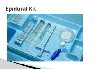
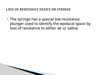
EPIDURAL NEEDLE
 Epidural needles are used to identify and
cannulate the epidural space.
 Two types
1. Tuohy needle
2. Crawford needle
 The Tuohy needle is widely used
Epidural needles
Crawford Needle
1. The needle is 10 cm in length with a shaft of 8 cm
(with 1-cm markings). A 15-cm version exists for
obese patients.
2. The needle wall is thin in order to allow a catheter
to be inserted through it.
3. The needle is provided with a stylet introducer to
prevent occlusion of the lumen by a core of tissue
as the needle is inserted.
4. Some designs allow the wings at the hub to be
added or removed.
5. The commonly used gauges are either 16 G or 18 G.
Components
 The markings on the needle enable the anaesthetist
to determine the distance between the skin and the
epidural space. Hence the length of the catheter left
inside the epidural space can be estimated.
 The shape and design of the bevel enable the
anaesthetist to direct the catheter within the epidural
space.
 A pediatric 19-G, 5-cm long Tuohy needle (with 0.5-cm
markings), allowing the passage of a 21-G nylon
catheter, is available.
 The bluntness of the bevel also minimizes the risk of
accidental dural puncture.
Mechanism of action
 A combined spinal–epidural technique is
possible using a 26-G spinal needle of about 12
cm length with a standard 16-G Tuohy needle.
The Tuohy needle is first positioned in the
epidural space then the spinal needle is
introduced through it into the subarachnoid
space.
 During insertion of the catheter through the
needle, if it is necessary to withdraw the
catheter, the needle must be withdrawn
simultaneously. This is because of the risk of
the catheter being transected by the oblique
bevel.
 In accidental dural puncture, there is a high
incidence of postdural headache due to the
epidural needle’s large bore (e.g. 16 G or 18 G).
 Wrong route errors
Problems in practice and safety
features
Components
1. 90-cm transparent, malleable tube made of nylon. The
16-G version has an external diameter of about 1 mm
and an internal diameter of 0.55 mm.
2. The distal end has two or three side ports with a
closed and rounded tip in order to reduce the risk of
vascular or dural puncture
3. Pediatric designs, 18 G or 19 G, have closer distal side
ports.
4. The distal end of the catheter is marked clearly at 5-cm
intervals, with additional 1-cm markings between 5
and 15 cm
Epidural catheter
5. The proximal end of the catheter is connected
to a Luer lock and a filter. In order to prevent
kinking, some designs incorporate a coil-
reinforced catheter.
6. Some catheters tend to be more rigid than the
normal design. They can be used in patients with
chronic pain to ensure correct placement of the
catheter.
Epidural Kit
 The catheters are designed to pass easily
through their matched gauge epidural needles.
 The markings enable the anaesthetist to place
the desired length of catheter within the
epidural space (usually 3–5 cm).
 An epidural fixing device can be used to
prevent the catheter falling out. The device
clips on the catheter. It has an adhesive flange
that secures it to the skin.
Mechanism of action
 The patency of the catheter should be tested
prior to insertion.
 The catheter can puncture an epidural vessel or
the dura at the time of insertion or even days
later.
 The catheter should not be withdrawn through
the Tuohy needle once it has been threaded
beyond the bevel as that can transect the
catheter. Both needle and catheter should be
removed simultaneously.
Problems in practice and safety
features
 It is almost impossible to predict in which
direction the epidural catheter is heading when
it is advanced.
 Once the catheter has been removed from the
patient, it should be inspected for any signs of
breakage.
 Advancing the catheter too much can cause
knotting
 The hydrophilic filter is a 0.22micron mesh
which acts as a bacterial, viral and foreign body
filter with a priming volume of about 0.7 mL. It
is recommended that the filter should be
changed every 24 h if the catheter is going to
stay in situ for long periods.
THE FILTER
 The syringe has a special low resistance
plunger used to identify the epidural space by
loss of resistance to either air or saline.
LOSS OF RESISTANCE DEVICE OR SYRINGE
Epidural Needle For Anesthesia Technologist

Epidural Needle For Anesthesia Technologist

  • 2.
  • 3.
     Epidural needlesare used to identify and cannulate the epidural space.  Two types 1. Tuohy needle 2. Crawford needle  The Tuohy needle is widely used Epidural needles
  • 5.
  • 6.
    1. The needleis 10 cm in length with a shaft of 8 cm (with 1-cm markings). A 15-cm version exists for obese patients. 2. The needle wall is thin in order to allow a catheter to be inserted through it. 3. The needle is provided with a stylet introducer to prevent occlusion of the lumen by a core of tissue as the needle is inserted. 4. Some designs allow the wings at the hub to be added or removed. 5. The commonly used gauges are either 16 G or 18 G. Components
  • 7.
     The markingson the needle enable the anaesthetist to determine the distance between the skin and the epidural space. Hence the length of the catheter left inside the epidural space can be estimated.  The shape and design of the bevel enable the anaesthetist to direct the catheter within the epidural space.  A pediatric 19-G, 5-cm long Tuohy needle (with 0.5-cm markings), allowing the passage of a 21-G nylon catheter, is available.  The bluntness of the bevel also minimizes the risk of accidental dural puncture. Mechanism of action
  • 8.
     A combinedspinal–epidural technique is possible using a 26-G spinal needle of about 12 cm length with a standard 16-G Tuohy needle. The Tuohy needle is first positioned in the epidural space then the spinal needle is introduced through it into the subarachnoid space.
  • 9.
     During insertionof the catheter through the needle, if it is necessary to withdraw the catheter, the needle must be withdrawn simultaneously. This is because of the risk of the catheter being transected by the oblique bevel.  In accidental dural puncture, there is a high incidence of postdural headache due to the epidural needle’s large bore (e.g. 16 G or 18 G).  Wrong route errors Problems in practice and safety features
  • 10.
    Components 1. 90-cm transparent,malleable tube made of nylon. The 16-G version has an external diameter of about 1 mm and an internal diameter of 0.55 mm. 2. The distal end has two or three side ports with a closed and rounded tip in order to reduce the risk of vascular or dural puncture 3. Pediatric designs, 18 G or 19 G, have closer distal side ports. 4. The distal end of the catheter is marked clearly at 5-cm intervals, with additional 1-cm markings between 5 and 15 cm Epidural catheter
  • 11.
    5. The proximalend of the catheter is connected to a Luer lock and a filter. In order to prevent kinking, some designs incorporate a coil- reinforced catheter. 6. Some catheters tend to be more rigid than the normal design. They can be used in patients with chronic pain to ensure correct placement of the catheter.
  • 12.
  • 13.
     The cathetersare designed to pass easily through their matched gauge epidural needles.  The markings enable the anaesthetist to place the desired length of catheter within the epidural space (usually 3–5 cm).  An epidural fixing device can be used to prevent the catheter falling out. The device clips on the catheter. It has an adhesive flange that secures it to the skin. Mechanism of action
  • 14.
     The patencyof the catheter should be tested prior to insertion.  The catheter can puncture an epidural vessel or the dura at the time of insertion or even days later.  The catheter should not be withdrawn through the Tuohy needle once it has been threaded beyond the bevel as that can transect the catheter. Both needle and catheter should be removed simultaneously. Problems in practice and safety features
  • 15.
     It isalmost impossible to predict in which direction the epidural catheter is heading when it is advanced.  Once the catheter has been removed from the patient, it should be inspected for any signs of breakage.  Advancing the catheter too much can cause knotting
  • 16.
     The hydrophilicfilter is a 0.22micron mesh which acts as a bacterial, viral and foreign body filter with a priming volume of about 0.7 mL. It is recommended that the filter should be changed every 24 h if the catheter is going to stay in situ for long periods. THE FILTER
  • 17.
     The syringehas a special low resistance plunger used to identify the epidural space by loss of resistance to either air or saline. LOSS OF RESISTANCE DEVICE OR SYRINGE