HEMODYNAMIC DISORDERS
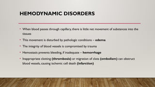
• Whenblood passes through capillary, there is little net movement of substances into the
tissues
• This movement is disturbed by pathologic conditions – edema
• The integrity of blood vessels is compromised by trauma
• Hemostasis prevents bleeding, if inadequate – hemorrhage
• Inappropriate clotting (thrombosis) or migration of clots (embolism) can obstruct
blood vessels, causing ischemic cell death (infarction)
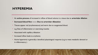
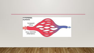
HYPEREMIA
• An activeprocess of increased in inflow of blood volume in a tissue due to arteriolar dilation
• Increased blood flow ------- Due to arteriolar dilatation
• Tissues appear red (erythematous) and warm due to oxygenated blood.
• e.g. Sites of inflammation or exercising muscles
• Associated with capillary dilatation
• Increased inflow leads to erythema
• Active hyperemia is generally a beneficial physiological response (e.g to meet metabolic demand or
in inflammatory )
6.
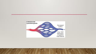
CONGESTION
• It isthe medical conditions that arise due to an abnormal accumulation of fluid, typically
blood or other bodily fluids, in tissues or organs.
• This condition often results from an imbalance in the circulatory system and can lead to
a range of health issues.
8.
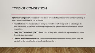
TYPES OF CONGESTION
1)VenousCongestion:This occurs when blood flow out of a particular area is impaired, leading to
an accumulation of blood. It can be due to:
• Heart Failure:The heart's reduced ability to pump blood effectively leads to a backlog in the
veins, particularly in the lungs (pulmonary congestion) or systemic circulation (systemic venous
congestion).
• DeepVeinThrombosis (DVT): Blood clots in deep veins, often in the legs, can obstruct blood
flow and cause congestion.
• ChronicVenous Insufficiency:A condition where veins have trouble sending blood from the
legs back to the heart, leading to swelling and discomfort.
9.
2) Pulmonary Congestion:Specifically related to the lungs, this occurs when fluid
accumulates in the lung tissues and alveoli. Common causes include:
• Left-Sided Heart Failure:When the left side of the heart fails to pump effectively, fluid
backs up into the lungs.
• Pulmonary Edema:A condition where excess fluid collects in the lung tissues, often
due to heart failure or acute respiratory distress.
10.
3) Hepatic Congestion:Thisis when blood backs up in the liver due to:
• Right-Sided Heart Failure: Impaired pumping from the right side of the heart can lead
to fluid buildup in the liver.
• Cirrhosis: Liver scarring can obstruct blood flow through the liver, causing congestion.
11.
4) Portal Hypertension:Increased blood pressure in the portal venous system can lead
to congestion in the spleen and other abdominal organs. It is often associated with liver
disease, especially cirrhosis.
12.
PATHOPHYSIOLOGY:
• Impaired BloodFlow: Congestion often results from problems in blood flow, either
due to obstructive conditions like thrombosis or due to heart failure where the heart's
pumping capacity is diminished.
• Increased Hydrostatic Pressure:When blood flow is obstructed, pressure increases
in the veins, pushing fluid out into surrounding tissues.
• Fluid Accumulation:The fluid that accumulates in tissues can lead to edema, which is
swelling due to excess fluid.
13.
SYMPTOMS:
Symptoms of congestionpathology vary depending on the affected area but can include:
• Swelling: Often in the legs or abdomen.
• Shortness of Breath: Especially if the lungs are affected.
• Pain or Discomfort: In the areas where fluid accumulates.
• Fatigue: Due to reduced efficiency of the heart or other organs.
14.
EDEMA
• Water makes60% of body weight
• 2/3 intracellular
• 1/3 extracellular
• Most in interstitium and very small amount in plasma
• Edema refers to accumulation of extra fluid within tissues interstitium
• Usually in lower limb, but also involve the entire body
• Old age and pregnant women are more likely to get edema
15.
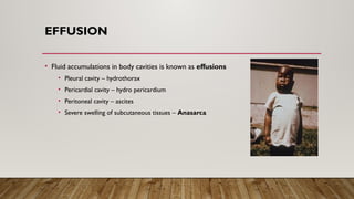
EFFUSION
• Fluid accumulationsin body cavities is known as effusions
• Pleural cavity – hydrothorax
• Pericardial cavity – hydro pericardium
• Peritoneal cavity – ascites
• Severe swelling of subcutaneous tissues – Anasarca
16.
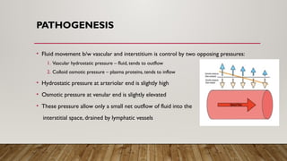
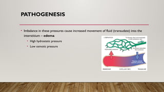
PATHOGENESIS
• Fluid movementb/w vascular and interstitium is control by two opposing pressures:
1. Vascular hydrostatic pressure – fluid, tends to outflow
2. Colloid osmotic pressure – plasma proteins, tends to inflow
• Hydrostatic pressure at arteriolar end is slightly high
• Osmotic pressure at venular end is slightly elevated
• These pressure allow only a small net outflow of fluid into the
interstitial space, drained by lymphatic vessels
17.
PATHOGENESIS
• Imbalance inthese pressures cause increased movement of fluid (transudate) into the
interstitium – edema
• High hydrostatic pressure
• Low osmotic pressure
MECHANISM
Increased Hydrostatic Pressure
•Caused by disorders that impair venous return
• Local increases in IV pressure
• DVT in the lower extremity – local edema distal to affected leg
• CHF – Generalize increases in venous pressure – systemic edema
20.
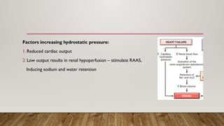
Factors increasing hydrostaticpressure:
1. Reduced cardiac output
2. Low output results in renal hypoperfusion – stimulate RAAS,
Inducing sodium and water retention
21.
MECHANISM
Reduced Plasma OsmoticPressure
• Caused by reduction of plasma albumin concentrations
• Albumin make ½ of total plasma protein
• Albumin lost – Nephrotic syndrome – leak glomerulus, loss of albumin in urine –
generalized edema
• Synthesizes in inadequate amounts – liver cirrhosis and protein malnutrition
22.
MECHANISM
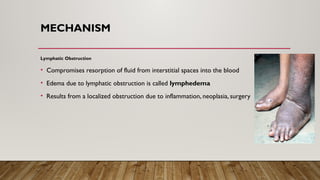
Lymphatic Obstruction
• Compromisesresorption of fluid from interstitial spaces into the blood
• Edema due to lymphatic obstruction is called lymphedema
• Results from a localized obstruction due to inflammation, neoplasia, surgery
23.
MECHANISM
Sodium Retention
• Excessiveretention of salt (with water) increase hydrostatic pressure (expansion of IV
volume) and reducing plasma osmotic pressure
• Diseases that compromise renal function
1. Glomerulonephritis and acute renal failure (low GFR)
2. Hyperaldosteronism – heart failure stimulating RAAS
24.
MECHANISM
Inflammation
• Increased vascularpermeability
• Exudation occur which increased osmotic pressure in interstitium
• Protein in interstitium exert osmotic pressure and attract more and more fluid
25.
SIGNS
• Swelling
• Skinthat hold a dimple or pit after being hold or pressed
• Shiny skin
• Enlarge abdomen
• Sudden weight gain
• Severe leg pain, feel heavy and difficult to walk
• Ulcer may developed on the skin – impair blood flow


![64965 hemodyn[1] edema](https://cdn.slidesharecdn.com/ss_thumbnails/64965hemodyn1-edema-150608143733-lva1-app6892-thumbnail.jpg?width=640&height=640&fit=bounds)


