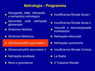
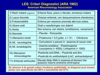
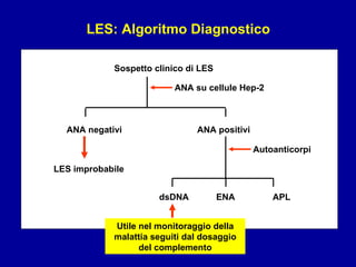
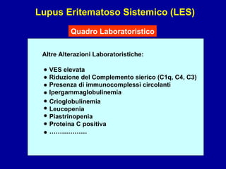
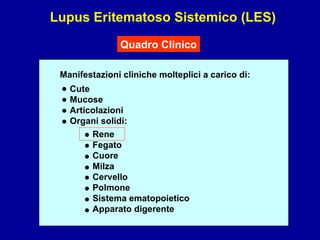
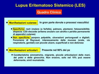
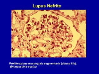
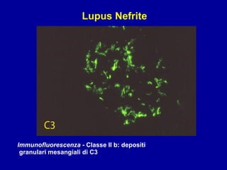
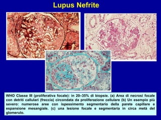
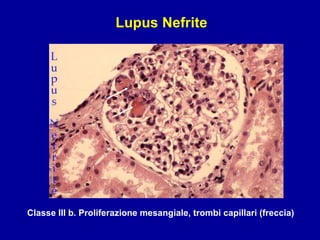
This document discusses renal tubular acidosis (RTA). It begins by explaining the different types of RTA, including proximal (Type 1), distal (Type 2), and combined (Type 3). It then covers the clinical presentation, diagnostic evaluation, and management of RTA. Key points include that children with RTA often present with failure to thrive, polyuria, and polydipsia. Diagnosis involves assessing for a normal anion gap metabolic acidosis along with electrolyte abnormalities. Treatment focuses on bicarbonate replacement and addressing complications like hypercalciuria. With early diagnosis and treatment, most children can see improved growth and development.
Sistemik skleroz (SS), cilt ve iç organların fibrozu ve mikrosirkulasyon bozuklukları ile karakterize, bağ dokusunun sebebi bilinmeyen sistemik bir hastalığıdır.
This document discusses tubulointerstitial diseases, which are disorders involving injury to the kidney tubules and interstitium. Tubulointerstitial diseases include acute tubular necrosis, acute or chronic tubulointerstitial nephritis, and conditions like reflux nephropathy. The document defines various tubulointerstitial diseases like pyelonephritis, which is a kidney infection that may cause symptoms like fever and back pain. Rare diseases like pyeloureteritis cystica are also discussed. The guidelines provide ICD-10 codes for classifying different types of tubulointerstitial and tubular diseases.
1. Acute tubular necrosis is characterized by low epithelial lining, single cell necrosis, and casts in tubule lumens. Regenerative changes include nucleomegaly and mitosis. Ethylene glycol poisoning causes tubule swelling and vacuolization with oxalate crystals.
2. Acute interstitial nephritis shows interstitial inflammation with lymphocytes and macrophages invading tubules. Chronic interstitial nephritis displays tubular atrophy, fibrosis and hyaline casts.
3. Granulomatous interstitial nephritis is defined by granulomas that can be non-necrotizing or necrotizing. Causes include drugs, sarcoidosis
metabolic acidosis develops because of defects in the ability of the renal tubules to perform the normal functions required to maintain acid-base balance.
This 20-year old male presented with a history of delayed growth, puberty, fever, severe joint pain and abdominal pain. Examination found pallor, icterus and tenderness. Labs showed anemia, elevated bilirubin and liver enzymes. Peripheral smear showed normocytic normochromic RBCs with some sickle cells. USG found gallstones and spleen not visualized. Hb electrophoresis showed HbS, consistent with sickle cell anemia.
Tubulointerstitial nephritis involves inflammatory reactions in the renal tubules and interstitium. It can be primary, secondary to other renal diseases, idiopathic, reactive to infections, or infectious itself. Acute tubular necrosis is characterized by tubular epithelial cell death due to ischemia or toxins and can cause acute kidney injury. Chronic tubulointerstitial nephritis develops over months or years and is associated with progressive loss of kidney function. It has many etiologies including drugs, infections, obstructive nephropathy, and autoimmune diseases.
This document provides an overview of renal tubular acidosis (RTA). It discusses the normal renal mechanisms that regulate acid-base balance and the pathophysiology of different types of RTA. The main types of RTA are distal (type 1) RTA, proximal (type 2) RTA, and type 4 RTA related to aldosterone deficiency or resistance. Distal RTA is characterized by impaired acid secretion leading to high urine pH and hypokalemia. Proximal RTA involves bicarbonate wasting and may cause bone disease. Type 4 RTA presents with hyperkalemia due to reduced ammonium excretion. Treatment involves alkali supplementation and potassium management depending on the specific RTA subtype
This document summarizes renal tubular acidosis (RTA), including the different types (proximal, distal, and hyperkalemic), causes, pathogenesis, clinical manifestations, diagnosis, and treatment. There are four main types of RTA - proximal (Type II) involving impaired bicarbonate reabsorption in the proximal tubule, distal (Type I) due to impaired hydrogen ion secretion in the distal tubule, and hyperkalemic (Type IV) resulting from impaired aldosterone production or response leading to impaired potassium and hydrogen ion handling. The document outlines the characteristic features, underlying causes, and approaches to diagnosis and management of metabolic acidosis for each RTA type.
Acute poststreptococcal glomerulonephritisGirish S
Acute poststreptococcal glomerulonephritis is an acute nephritic syndrome that commonly occurs 1-2 weeks following a streptococcal throat or skin infection, where immune complexes deposit in the glomerular basement membrane of the kidneys. It causes gross hematuria, edema, hypertension, and renal insufficiency. On renal biopsy, the glomeruli appear enlarged with diffuse mesangial cell proliferation and polymorphonuclear leukocytes. While most children fully recover within 2 months, severe cases can lead to chronic renal insufficiency or complications from acute renal failure like heart failure, hypertension, or seizures. Treatment focuses on managing acute renal failure and hypertension.
Systemic Lupus Erythematosus (SLE) is a multi-gene autoimmune disease caused by a combination of genetic and environmental factors. It is characterized by abnormal immune responses that result in inflammation and damage to various organs. Diagnosis requires meeting 4 out of 11 classification criteria relating to clinical symptoms and blood markers. Management aims to induce remission of acute flares, maintain improvements to suppress symptoms, and prevent organ damage. Treatment choices depend on the severity and potential reversibility of manifestations. The goal is controlling symptoms without cure since complete sustained remission is rare.
IgA nephropathy is characterized by predominant IgA deposition in the glomerular mesangium. It is the most common cause of glomerulonephritis worldwide. Primary IgA nephropathy occurs most commonly in the second and third decades of life and presents with macroscopic hematuria, asymptomatic hematuria with mild proteinuria, or nephrotic syndrome. Treatment involves controlling blood pressure with ACE inhibitors or ARBs, treating any proteinuria or hematuria, and considering corticosteroids for progressive disease. The pathogenesis involves abnormal IgA immune responses leading to pathogenic IgA deposition in the kidney and subsequent glomerular inflammation and injury.
This document outlines the pathogenesis and management of uremic pruritus. It begins with an outline of the topics to be discussed, including dialysis adequacy and modification, xerosis, microinflammation, nervous conduction, the opioid hypothesis, various drug therapies, physical therapy using UV light, and other treatments. It then discusses various studies that have examined the relationship between increased dialysis adequacy as measured by Kt/V and the severity of pruritus. The document also reviews the role of xerosis, microinflammation mediated by cytokines like TNF-α and IL-1β, the nervous system through substances like substance P, and the opioid hypothesis in the pathogenesis of uremic pruritus.
This document provides an overview of liver pathology, covering the anatomy and histology of the normal liver, as well as pathological conditions including viral hepatitis, cirrhosis, tumors, and other disorders. Key points discussed include the lobular structure of the liver, patterns of inflammation and necrosis seen in viral hepatitis, characteristics of cirrhosis including macronodular and micronodular types, features of hepatocellular carcinoma and cholangiocarcinoma, and abnormalities involving fatty change, vascular disorders, and pigmentation. Liver biopsy is described as the gold standard for diagnosis, with assessment of architecture, inflammation, necrosis, and other features.
Focal segmental glomerulosclerosis (FSGS) is a kidney disease characterized by scarring in the glomeruli. It can be primary/idiopathic or secondary to other causes like viral infections, drugs, ischemia, etc. Patients often present with nephrotic syndrome. Treatment involves controlling blood pressure and proteinuria with ACE inhibitors/ARBs initially. For idiopathic FSGS with nephrotic syndrome, the first line treatment is corticosteroids. Mycophenolate mofetil or calcineurin inhibitors can be used as steroid-sparing alternatives. For steroid-resistant FSGS, calcineurin inhibitors like cyclosporine A are recommended.
Introduzione all'uso di Slideshare. Traduzione di "Slideshare tutorial", presentazione in catalano di Immaculada Vilatersana del Centro di Formazione per Adulti di Matarò, Barcelona.
1. Nefrologia - Programma Nosografia delle nefropatie e semeiotica nefrologica Insufficienza Renale Acuta I • • Generalità sulle nefropatie glomerulari Insufficienza Renale Acuta II • • Vasculiti e microangiopatie trombotiche Sindrome Nefritica • • Sindrome Nefrosica Nefropatie intersiziali • • Glomerulonefriti secondarie I Nefropatie ischemiche • • Insufficienza Renale Cronica Glomerulonefriti secondarie II • • Nefropatie ereditarie La Dialisi • • Rene e gravidanza Il Trapianto Renale • •
2.
3. Lupus Eritematoso Sistemico (LES) Eziologia 1) Non nota 2) Da farmaci Genetici Ambientali - Alto tasso di concordanza tra gemelli monozigoti - 10-15% è presente un familiare con la patologia - Alta frequenza di alcuni genotipi HLA (A1, B8, DR3) - Maggior frequenza in pop. con deficit del complemento Fattori Predisponenti - Sconosciuti - Correlazione con raggi ultravioletti B e UVA - 70% dei pz è fotosensibile Ormonali
4. Lupus Eritematoso Sistemico (LES) Patogenesi Auto-anticorpi pluriclonali diretti contro acidi nucleici e proteine intacellulari (istoni, cromatina, ribonucleoproteine)
5.
6.
7. LES: Ruolo del Laboratorio Anticorpi anti-antigeni nucleari estraibili (anti – ENA) Sono rivolti verso una serie di antigeni proteici estratti dal nucleo • Quelli di maggior rilevanza sono: anti-Sm (Smith antigen); anti-RNP; anti-SSA/Ro; Anti-SSB/La; Anti-Sc170; Anti-Jo1 Anticorpi anti-Sm sono altamente specifici per la diagnosi (criteri ARA) • Anticorpi anti-fosfolipidi (APL) Diretti contro il complesso fosfolipide / beta 2 glicoproteina • Associati al fenomeno LAC • Tecniche ELISA più sensibili ma meno specifiche dell’ immunoblotting • Presenti nel 30-40% dei pz con LES, ma non sono esclusivi di questa patologia, si associano a manifestazioni trombotiche e/o a poliabortività
8. LES: Ruolo del Laboratorio Anticorpi anti-nucleosomi Giocano un ruolo fondamentale nella patogenesi del LES • I nucleosomi sono l’unità fondamentale della cromatina e derivano dall’associazione tra istoni e DNA Sensibilità per la diagnosi di LES superiore a quella degli anticorpi anti-ds-DNA • Anticorpi anti-C1q Diretti contro la regione collagene simile del C1q • Correlati con la presenza di interessamento renale • Prevalenza nel LES: 70% •
9. Sospetto clinico di LES ANA negativi ANA positivi dsDNA ENA APL ANA su cellule Hep-2 LES improbabile Autoanticorpi LES: Algoritmo Diagnostico Utile nel monitoraggio della malattia seguiti dal dosaggio del complemento
10. Lupus Eritematoso Sistemico (LES) Quadro Laboratoristico Altre Alterazioni Laboratoristiche: VES elevata Riduzione del Complemento sierico (C1q, C4, C3) Presenza di immunocomplessi circolanti Ipergammaglobulinemia Crioglobulinemia Leucopenia Piastrinopenia Proteina C positiva ……………… • • • • • • • • •
12. Lupus Eritematoso Sistemico (LES) Quadro Clinico In gran parte dovute a processi vasculitici • • • Manifestazioni cutanee: • Manifestazioni articolari: Presente nel 90% dei pz Caratteristiche simmetriche, migrante, piccole articolazioni delle mani, dei polsi e delle ginocchia. Non erosivo, solo nel 10% può essere deformante, simil reumatoide. • Specifiche: rash (malare (a farfalla), palmare, plantare); fotosensibilità; alopecia; LES discoide (eritema anulare con atrofia e perdita permanente di appendici cutanee) Non specifiche: porpora palpabile, ulcerazioni periungueali e digitali; Fenomeno di Raynaud, interessamento delle mucose (orale, vie respiratorie, genitali) con piccole ulcere, superficiali e non dolorose
13. Lupus Eritematoso Sistemico (LES) Quadro Clinico SNC: cefalea, corea, disordini cognitivi, disturbi edll’umore, depressione, epilessia, eventi cerebrovascolari, fomre demielinizzanti, meningite asettica, psicosi, organic brain syndrome (manifestazione finale) SN periferico: disordini del sistema autonomico, interessamento ei nervi cranci, miastenia gravis, mononeuropatie, polineuropatie. • • • Manifestazioni a carico del sistema nervoso: • Manifestazioni ematologiche: Citopenie (Leucopenia, Piastrinopenia, per lo più secondarie alla comparsa di autoanticorpi), alterazioni dell’omeostasi coagulativa (stati trombofilici acquisiti quali la sindrome da anticrpi antifosfolipidi – APL), linfoadenopatie reattive o neoplastiche • Presente nel 50% dei pz Frequente l’associazione tra la presenza di Anticorpi Antifofolipidi e TIA o Stroke; dibattuta l’associazione tra interesamento del SNC e Autoanticorpi Anti-proteina ribosomiale
14. Lupus Eritematoso Sistemico (LES) Quadro Clinico Pericardite (spesso sintomo d’esordio di malattia); interessamento valvolare con endocardite subacuta non batterica associata a aPL; trombosi arterovenose (presenza di aPL); ipertensione arteriosa (80% dei pz); aterosclerosi accelerata; eventi ischemici arteriosi. • • Manifestazioni cardiovascolari: • Interessamento polmonare: Versamento pleurico (30-60% dei pz); polmonite acuta non batterica; atelectasie; tromboembolie polmonari; emorragie polmonari (vasculite) • • Interessamento muscolo scheletrico: Miosite; miopatia steroidea; necrosi asettica • • Interessamento oculare: Uveite o retinopatia (legate a ipertensione o vasculite) •
16. Lupus Eritematoso Sistemico (LES) Quadro Clinico NEFRITE LUPICA La più frequente Nefropatia Secondaria Una delle manifestazioni più frequenti e più gravi del LES. Si manifesta clinicamente in oltre la metà dei pazienti, può essere la prima manifestazione della malattia, ma può anche comparire tardivamente. Nell’80% dei casi compare entro 3 anni dalla diagnosi. La presentazione clinica è molto variabile in rapporto al tipo e alla estensione delle lesioni renali: da anomalie urinarie isolate (proteinuria, microematuria) fino all’insufficienza renale rapidamente progressiva che porta il pz alla dialisi in poche settimane. Possibili sindromi intermedie come la Sindrome Nefrosica (40 %), la Sindrome Nefritica acuta (30 %). Ipertensione arteriosa frequente nella classi III e IV. • • • • • •
17. Lupus Eritematoso Sistemico (LES) Nefrite Lupica Quadro Morfologico: Considerazioni 1) Tutte le componenti del parenchima renale, glomeruli, interstizio, tubuli, vasi, possono essere colpite anche se il glomerulo è il bersaglio preferenziale degli immunodepositi. 2) Le lesioni istologiche possono essere: a. Attive: potenzialmente reversibili con una terapia adeguata (proliferazione cellulare, necrosi/carioressi, “wire loops”, trombi ialini, “crescents” cellulari, infiltrati infiammatori interstiziali, necrosi tubulare, necrosi fibrinoide e trombosi vascoalre) b. Croniche: non responsive alla terapia, rappresentano l’evoluzione sfavorevole di lesioni attive non trattate o non responsive alla terapia (glomerulosclerosi globale o segmentaria, “crescents” fibrotiche, fibrosi interstiziale, atrofia tubulare, ialinosi arteriolare) 3) L’entità e la qualità del coinvolgimento renale varia da paziente a paziente ed anche nel singolo paziente le lesioni sono estremamente variabili
18. Lupus Eritematoso Sistemico (LES) Classificazione della Nefrite Lupica (WHO revisionata nel 2004) Classe Istologia Clinica Classe I Glomeruli normali al MO con immunodepositi mesangiali all’IF Assente/anomalie urinarie isolate Classe II GN mesangiale Ipercellularità mesangiale con immunodepositi mesangiali (possono esserci pochi e isolati depositi subendoteliali o subepiteliali) Anomalie urinarie isolate, rara sindrome nefrosica o insuff renale Classe III Nefrite lupica focale Tutte le lesioni glomerulari attive o croniche con interessamento di meno del 50% dei glomeruli con distribuzione segmentaria o globale Variabile: anomalie urinarie isolate, sindrome nefrosica o nefritica Classe IV Nefrite lupica diffusa Stesse lesioni della classe III che interessano più del 50% dei glomeruli. IV-S: >50% glom con lesioni segmentarie IV-G: >50% glom con lesioni globali Proteinuria, ematuria. Ipertensione arteriosa e insuff renale Classe V Nefrite lupica membranosa Immunodepositi subepiteliali ed intramembranosi a distribuzione globale o segmentaria con/senza alterazioni mesangiali Proteinuria moderata severa. Rara l’ipertensione arteriosa e l’insuff renale Classe VI GN cronica >90% dei glomeruli con sclerosi globale Insufficienza renale cronica
19. WHO Classe I (lesioni Minime): in <5% di biopsie (in pasienti senza o con lieve nefropatia). Microscopia ottica normale, alla immunoperossidasi estesi depositi di C1c (IgG e C3) lungo le zone mesangiali Lupus Nefrite
20. WHO Classe II (Mesangiale): in 10–25% di biopsie. (a) espansione mesangiale dal centro alla periferia, con scarso aumento della cellularità (b) Estesi depositi mesangiali di IgG (immunoperossidasi); è la più severa alterazione di questa classe. (a) (b Lupus Nefrite
21.
22.
23. WHO Classe III (proliferativa focale): in 20–35% di biopsie. (a) Area di necrosi focale con detriti callulari (freccia) circondata da proliferazione cellulare (b) Un esempio più severo: numerose aree con ispessimento segmentario della parete capillare e espansione mesangiale. (c) una lesione focale e segmentaria in circa metà del glomerulo. Lupus Nefrite
24.
25.
26.
27.
28.
29.
30.
31.
32.
33.
34. WHO Classe V (membranosa): 10-15 % di biopsie. I depositi sono sul versante esterno della parete capillare. Vi sono abbondanti aggregati mesangiali (blu scuro) e lungo la parete dei capillari. Non sono presenti lesioni proliferative. Lupus Nefrite
35. Nefrite Lupica Terapia Specifica Terapia di Supporto Terapia in base al quadro clinico generale 1) Fase d’induzione: steroidi, ciclofosfamide Fase di mantenimento: steroidi, azatioprina 2) Terapia Per le classi IIb-III-IV-V Nuovi Farmaci: Micofenolato Mofetile, Ciclosporina
Editor's Notes
Deriva dal disordine del sistema immunit. Con il concorso di diversi fattori, ambientali, ormonali, raziali e genetici. Alterazioni dell’attività umorale con produzione di autoanticorpi.
Sm = Smith antigen; Lac = lupus anticoagulant Activity (anticoagulante in vitro ma in vivo coagulante, trombosi)