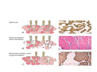
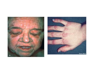
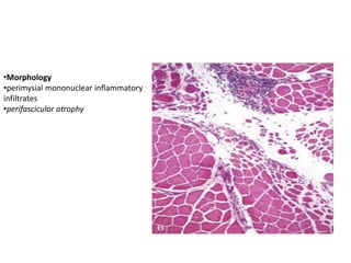
DISEASES OF THE NEUROMUSCULAR
JUNCTION
• Regardless of cause, patients with disorders
that impair the function of neuromuscular
junctions complain of painless weakness and
fatigue
Myasthenia Gravis
• is an autoimmune disease that is usually associated with
autoantibodies directed against Ach receptors
• Pathogenesis
– About 85% of patients have autoantibodies against postsynaptic
ACh receptors
– most of the remaining patients have antibodies against the
sarcolemmal protein musclespecific receptor tyrosine kinase
• There is a strong association between pathogenic anti–ACh
receptor autoantibodies and thymic abnormalities
• Patients with anti–ACh receptor antibodies typically
present with fluctuating weakness that worsens with
exertion and often over the course of the day.
• Diplopia and ptosis due to involvement of
extraocular muscles are common and distinguish
myasthenia gravis from myopathies
• Cases with antibodies against muscle-specific
receptor tyrosine kinase differ from typical cases
by exhibiting more focal muscle involvement
• Diagnosis is based on clinical history, physical
findings, identification of autoantibodies, and
electrophysiologic studies
DISEASES OF SKELETAL MUSCLE
Skeletal Muscle Atrophy
• Causes: Loss of innervation, disuse, cachexia, old
age, and primary myopathies
• Clusters or groups of atrophic fibers -neurogenic
disease.
• Perifascicular atrophy - in dermatomyositis
• Type II fiber atrophy with sparing of type I fibers is
seen with prolonged corticosteroid therapy or
disuse
Inflammatory Myopathies
• Historically, dermatomyositis, polymyositis,
and inclusion body myositis have been
considered the three main primary
inflammatory myopathies
Dermatomyositis
• is a systemic autoimmune disease that typically
presents with proximal muscle weakness and skin
changes.
• Pathogenesis
– is an immunologic disease in which damage to small blood
vessels contributes to muscle injury
– Biopsies of muscle and skin may show deposition of the
complement membrane attack complex (C5b-9) within
capillary beds
• may occur in adults or in children(av=7yr)
• Dermatomyositis is the most common inflammatory
myopathy in children
C/F
• Muscle weakness is slow in onset, symmetric,
and often accompanied by myalgias.
• It typically affects the proximal muscles first
• heliotrope rash associated with periorbital
edema
• a scaling erythematous eruption or dusky red
patches over the knuckles, elbows, and knees
(Gottron papules)
•Morphology
•perimysial mononuclear inflammatory
infiltrates
•perifascicular atrophy
Polymyositis
• is an adult-onset inflammatory myopathy that
shares myalgia and weakness with
dermatomyositis but lacks its distinctive
cutaneous feature
• vascular injury does not have a major role in
the pathogenesis
Inclusion Body Myositis
• is a disease of late adulthood that typically affects
patients older than 50 years
• is the most common inflammatory myopathy in
patients older than age 65 years
• present with slowly progressive muscle weakness
that tends to be most severe in the quadriceps
and the distal upper extremity muscles
• Abnormal cytoplasmic inclusions described as
“rimmed vacuoles”
Toxic Myopathies
• can be caused by prescription or recreational
drugs or by certain hormonal imbalances
• Eg statins, thyroid dysfunction, alcohol etc..
Muscular Dystrophies
• include many inherited disorders of skeletal muscle
that have in common progressive muscle damage that
typically manifests between childhood and adulthood
• with the exception of congenital muscular dystrophies,
these diseases do not present in infancy
• The most common muscular dystrophies are X-linked
and stem from mutations that disrupt the function of a
large structural protein called dystrophin
Duchenne and Becker Muscular
Dystrophy
• The most common early-onset form is referred to
as Duchenne muscular dystrophy
– has a severe progressive phenotype
• Becker muscular dystrophy is a second relatively
common dystrophinopathy that is characterized
by later disease onset and a milder phenotype
• are caused by loss-of-function mutations in the
dystrophin gene on the X chromosome
• Dystrophin is thought to provide mechanical
stability to the myofiber and its cell membrane
during muscle contraction
• C/F of duchenne MD
– Boys with Duchenne muscular dystrophy appear normal at
birth.
– Very early motor milestones are met, but walking is often
delayed
– Despite supportive care, the mean age of death is 25 to 30 years
of age
• Dystrophin is also expressed in the heart and the CNS
• in contrast Becker md , typically presents in later childhood,
adolescence, or adult life; has slower progression; and may
have a near-normal life expectancy
• morphologic changes that muscular dystrophies elicit in skeletal muscle
tissue may differ in severity but do not discriminate between different
forms of dystrophy
END

musculoskeletal.pptx

  • 1.
    DISEASES OF THENEUROMUSCULAR JUNCTION • Regardless of cause, patients with disorders that impair the function of neuromuscular junctions complain of painless weakness and fatigue
  • 2.
    Myasthenia Gravis • isan autoimmune disease that is usually associated with autoantibodies directed against Ach receptors • Pathogenesis – About 85% of patients have autoantibodies against postsynaptic ACh receptors – most of the remaining patients have antibodies against the sarcolemmal protein musclespecific receptor tyrosine kinase • There is a strong association between pathogenic anti–ACh receptor autoantibodies and thymic abnormalities • Patients with anti–ACh receptor antibodies typically present with fluctuating weakness that worsens with exertion and often over the course of the day.
  • 3.
    • Diplopia andptosis due to involvement of extraocular muscles are common and distinguish myasthenia gravis from myopathies • Cases with antibodies against muscle-specific receptor tyrosine kinase differ from typical cases by exhibiting more focal muscle involvement • Diagnosis is based on clinical history, physical findings, identification of autoantibodies, and electrophysiologic studies
  • 4.
  • 5.
    Skeletal Muscle Atrophy •Causes: Loss of innervation, disuse, cachexia, old age, and primary myopathies • Clusters or groups of atrophic fibers -neurogenic disease. • Perifascicular atrophy - in dermatomyositis • Type II fiber atrophy with sparing of type I fibers is seen with prolonged corticosteroid therapy or disuse
  • 7.
    Inflammatory Myopathies • Historically,dermatomyositis, polymyositis, and inclusion body myositis have been considered the three main primary inflammatory myopathies
  • 8.
    Dermatomyositis • is asystemic autoimmune disease that typically presents with proximal muscle weakness and skin changes. • Pathogenesis – is an immunologic disease in which damage to small blood vessels contributes to muscle injury – Biopsies of muscle and skin may show deposition of the complement membrane attack complex (C5b-9) within capillary beds • may occur in adults or in children(av=7yr) • Dermatomyositis is the most common inflammatory myopathy in children
  • 9.
    C/F • Muscle weaknessis slow in onset, symmetric, and often accompanied by myalgias. • It typically affects the proximal muscles first • heliotrope rash associated with periorbital edema • a scaling erythematous eruption or dusky red patches over the knuckles, elbows, and knees (Gottron papules)
  • 11.
  • 12.
    Polymyositis • is anadult-onset inflammatory myopathy that shares myalgia and weakness with dermatomyositis but lacks its distinctive cutaneous feature • vascular injury does not have a major role in the pathogenesis
  • 13.
    Inclusion Body Myositis •is a disease of late adulthood that typically affects patients older than 50 years • is the most common inflammatory myopathy in patients older than age 65 years • present with slowly progressive muscle weakness that tends to be most severe in the quadriceps and the distal upper extremity muscles • Abnormal cytoplasmic inclusions described as “rimmed vacuoles”
  • 14.
    Toxic Myopathies • canbe caused by prescription or recreational drugs or by certain hormonal imbalances • Eg statins, thyroid dysfunction, alcohol etc..
  • 15.
    Muscular Dystrophies • includemany inherited disorders of skeletal muscle that have in common progressive muscle damage that typically manifests between childhood and adulthood • with the exception of congenital muscular dystrophies, these diseases do not present in infancy • The most common muscular dystrophies are X-linked and stem from mutations that disrupt the function of a large structural protein called dystrophin
  • 16.
    Duchenne and BeckerMuscular Dystrophy • The most common early-onset form is referred to as Duchenne muscular dystrophy – has a severe progressive phenotype • Becker muscular dystrophy is a second relatively common dystrophinopathy that is characterized by later disease onset and a milder phenotype • are caused by loss-of-function mutations in the dystrophin gene on the X chromosome • Dystrophin is thought to provide mechanical stability to the myofiber and its cell membrane during muscle contraction
  • 17.
    • C/F ofduchenne MD – Boys with Duchenne muscular dystrophy appear normal at birth. – Very early motor milestones are met, but walking is often delayed – Despite supportive care, the mean age of death is 25 to 30 years of age • Dystrophin is also expressed in the heart and the CNS • in contrast Becker md , typically presents in later childhood, adolescence, or adult life; has slower progression; and may have a near-normal life expectancy
  • 18.
    • morphologic changesthat muscular dystrophies elicit in skeletal muscle tissue may differ in severity but do not discriminate between different forms of dystrophy
  • 19.