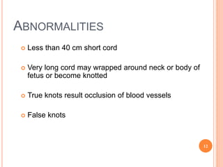
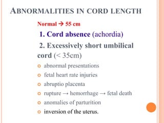
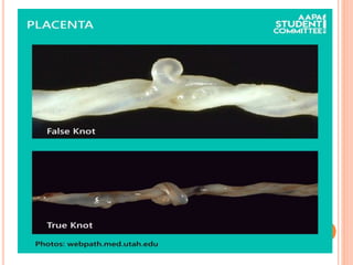
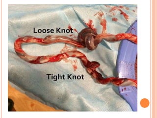
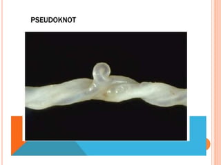
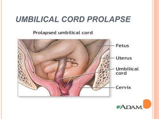
UMBILICAL CORD
 Presented by:
Mrs. L. Kamala Devi
Principal
HAMM Nursing School
Hojai, Assam
DEVELOPMENT OF THE UMBILICAL CORD
 The umbilical cord is formed when the body stalk and
the ductus omphalo-entericus (vitelline duct, a long
narrow tube that joins the yolk sac to the midgut lumen
of the developing fetus) as well as the umbilical coelom (
part of mesoderm)are enveloped by the spreading
amnion between the 4th and 8th week. Finally, when the
membranes of the amniotic cavity come into contact
with those of the chorionic cavity and the two extra-
embryonic mesoderm layers that cover both
membranes, fuse. With the flexing movements of the
embryo, the amnion encircles the body stalk, the ductus
omphalo-entericus and the umbilical vessels, thus
circumscribing the elements of the umbilical cord.
A. body stalk
B. stem of umbilical vesicles
1.Amniotic cavity
2.Umbilical vesicle
3.Chorionic cavity
4.Villous chorion
5.Allantois
Body stalk at around the
3rd week
Formation of the umbilical cord at
around the 3.5th week
A. Body stalk
B. Stem of umbilical vesicle
C. Umbilical cord
 1.Amniotic cavity
2.Umbilical vesicle
3.Chorionic cavity
4. Chorion leave
Umbilical cord :The body stalk and
the yolk stalk are now united and
form the umbilical cord. Through
increasing secretion of amniotic fluid
the chorionic cavity becomes
obliterated. Here at around the 4-5th
week: The chorionic cavity is reduced
in size
Umbilical vesicle in the chorionic cavity: Flexing of
the embryo at around the 8th week with expansion
of the amnion that encircles the body stalk and the
ductus omphalo-entericus, the umbilical coelom
and the umbilical vessels
The developing fetus during 10th week
of intrauterine life.
 In the early stage (at around the 8th week) the umbilical
cord is in the form of a very thick and short section with
the following structures:
 The ductus omphalo-entericus which connects the
primitive intestines with the umbilical vesicle and two
vitelline vessels (vasa omphalomesenterica, 2 arteries and
2 veins). The umbilical vesicle is located in the chorionic
cavity (exocoelom = extra-embryonic coelom).
 The body stalk with the allantois, the umbilical vessels (2
arteries and 1 vein). During the development it gets shifted
ventrally in order to finally fuse with the stem of the
umbilical vesicle.
 The umbilical coelom that connects the extra-embryonic
coelom with the intra-embryonic coelom
1. Ductus omphalo-entericus
2.Extra-embryonic coelom
(umbilcal coelom)
3. Allantois
4. Umbilical vein
5. Umbilical arteries
6. Amnion
7.Intestinal tube(physiologic umbilical hernia)
8.Vasa omphalomesenterica
Transversal section of the primitive
umbilical cord after approximately 8
weeks
Transversal section of the primitive
umbilical cord with physiologic
umbilical hernia at around the 3rd
month
 Further development promotes both lengthening as well as
the reduction of some structures.
 Lengthening:
The amniotic cavity forms a covering around the ductus
omphalo-entericus and the body stalk, which elongates. The
newly formed umbilical cord continues to lengthen to allow
for fetal movements and coils in the amniotic cavity.
 Reduction:
Numerous elements degenerate in the 3rd month.
 the omphalo-enteric duct (it can remain in the form of a
Meckel's diverticulum)
 the umbilical vesicle of the allantois (it is obliterated in
order to form the umbilical ligament, lying medially in
adults)
 the vitelline circulation system in the extra-embryonic
region.
 the umbilical coelom, which clumps and disappears.
 Finally, only the body stalk remains with its umbilical
vessels (2 arteries, 1 vein), which are surrounded by an
amniotic epithelial layer. The connective tissue of the
body stalk and the amnion (that stems from the extra-
embryonic mesoblast) go over into a common umbilical
cord connective tissue, the so-called "Wharton jelly", an
elastic and resistant tissue that protects the umbilical
vessels from possible mechanical pressure and creasing.
UMBILICAL CORD
 Length of cord varies from no cord (achordia) to 300 cm
with up to 3 cm diameters(average 55cm) sufficient to
allow delivery of baby without traction to placenta.
 -Transmits umbilical blood vessels
 -Two arteries from internal iliac artery, unoxygenated
blood and one vein from Ductus venosus having
oxygenated blood.
 the extracellular matrix → Wharton jelly.
11
ABNORMALITIES
 Less than 40 cm short cord
 Very long cord may wrapped around neck or body of
fetus or become knotted
 True knots result occlusion of blood vessels
 False knots
12
THE UMBILICAL CORD PATHOLOGY
A. Abnormalities of development
B. Accidental pathology
A. ABNORMALITIES OF DEVELOPMENT
 abnormalities of cord insertion
-marginal insertion
-velamentous insertion
 abnormalities in cord length
 tumors of umbilical cord
 vascular anomalies (single
umbilical artery)
.
Marginal
insertion
2% - 15% .
 associated with preterm
labour (?).
 US.
Battledore
placenta
ABNORMALITIES OF CORD INSERTION
VELAMENTOUS INSERTION
ABNORMALITIES OF CORD INSERTION
Velamentous insertion –
vasa praevia
Fetal vessels run in the membranes below the presenting
fetal part.
Spontaneous / artificial rupture of membranes - rupture
the vessels - fetal exsanguination – Benkiser
syndrome.
 Hypoxia if the vessels are compressed between baby
and birth canal.
Fetal mortality - 33-100%, if not dg. prenatally.
ABNORMALITIES IN CORD LENGTH
Normal  55 cm
1. Cord absence (achordia)
2. Excessively short umbilical
cord (< 35cm)
 abnormal presentations
 fetal heart rate injuries
 abruptio placenta
 rupture → hemorrhage → fetal death
 anomalies of parturition
 inversion of the uterus.
ABNORMALITIES IN CORD
LENGTH
3. Excessive length (cord length >
70cm)
 vascular occlusion (thrombi)
 true knots
 cord prolapse
 loops of the cord.
B. ACCIDENTAL PATHOLOGY
 loops
 knots
 prolapse
 thrombosis
 ruptures
 eventualities which lead to umbilical
vessels compression and fetal distress.
Umbilical cord knots
 True knots - distinguished from false
knots (varicosities or accumulations of
Wharton's jelly) ► no clinical
significance
 True knots result from active fetal
movements (1.1 % of births).
UMBILICAL CORD PROLAPSE
Types of umbilical cord prolapse
1. occult cord prolapse
2. overt cord prolapse
3. funic presentation = cord
presentation = procubitus.
Overt cord prolapse is always associated with rupture
of the membranes and displacement of the cord into the
vagina, often throughout the introitus.
UMBILICAL CORD PROLAPSE
Ruptured membranes
 occult cord prolapse (descent of the
umbilical cord alongside)
 overt cord prolapse (umbilical cord
past the presenting part).
UMBILICAL CORD PROLAPSE
NO ruptured membranes
Funic presentation = cord
presentation = procubitus →
one or more loops of umbilical
cord between the fetal presenting
part and the cervix,.
If the cervix is opened the cord can be
easily palpated through the
membranes.
Umbilical cord

Umbilical cord

  • 1.
    UMBILICAL CORD  Presentedby: Mrs. L. Kamala Devi Principal HAMM Nursing School Hojai, Assam
  • 2.
    DEVELOPMENT OF THEUMBILICAL CORD  The umbilical cord is formed when the body stalk and the ductus omphalo-entericus (vitelline duct, a long narrow tube that joins the yolk sac to the midgut lumen of the developing fetus) as well as the umbilical coelom ( part of mesoderm)are enveloped by the spreading amnion between the 4th and 8th week. Finally, when the membranes of the amniotic cavity come into contact with those of the chorionic cavity and the two extra- embryonic mesoderm layers that cover both membranes, fuse. With the flexing movements of the embryo, the amnion encircles the body stalk, the ductus omphalo-entericus and the umbilical vessels, thus circumscribing the elements of the umbilical cord.
  • 4.
    A. body stalk B.stem of umbilical vesicles 1.Amniotic cavity 2.Umbilical vesicle 3.Chorionic cavity 4.Villous chorion 5.Allantois Body stalk at around the 3rd week Formation of the umbilical cord at around the 3.5th week
  • 5.
    A. Body stalk B.Stem of umbilical vesicle C. Umbilical cord  1.Amniotic cavity 2.Umbilical vesicle 3.Chorionic cavity 4. Chorion leave Umbilical cord :The body stalk and the yolk stalk are now united and form the umbilical cord. Through increasing secretion of amniotic fluid the chorionic cavity becomes obliterated. Here at around the 4-5th week: The chorionic cavity is reduced in size Umbilical vesicle in the chorionic cavity: Flexing of the embryo at around the 8th week with expansion of the amnion that encircles the body stalk and the ductus omphalo-entericus, the umbilical coelom and the umbilical vessels
  • 6.
    The developing fetusduring 10th week of intrauterine life.
  • 7.
     In theearly stage (at around the 8th week) the umbilical cord is in the form of a very thick and short section with the following structures:  The ductus omphalo-entericus which connects the primitive intestines with the umbilical vesicle and two vitelline vessels (vasa omphalomesenterica, 2 arteries and 2 veins). The umbilical vesicle is located in the chorionic cavity (exocoelom = extra-embryonic coelom).  The body stalk with the allantois, the umbilical vessels (2 arteries and 1 vein). During the development it gets shifted ventrally in order to finally fuse with the stem of the umbilical vesicle.  The umbilical coelom that connects the extra-embryonic coelom with the intra-embryonic coelom
  • 8.
    1. Ductus omphalo-entericus 2.Extra-embryoniccoelom (umbilcal coelom) 3. Allantois 4. Umbilical vein 5. Umbilical arteries 6. Amnion 7.Intestinal tube(physiologic umbilical hernia) 8.Vasa omphalomesenterica Transversal section of the primitive umbilical cord after approximately 8 weeks Transversal section of the primitive umbilical cord with physiologic umbilical hernia at around the 3rd month
  • 9.
     Further developmentpromotes both lengthening as well as the reduction of some structures.  Lengthening: The amniotic cavity forms a covering around the ductus omphalo-entericus and the body stalk, which elongates. The newly formed umbilical cord continues to lengthen to allow for fetal movements and coils in the amniotic cavity.  Reduction: Numerous elements degenerate in the 3rd month.  the omphalo-enteric duct (it can remain in the form of a Meckel's diverticulum)
  • 10.
     the umbilicalvesicle of the allantois (it is obliterated in order to form the umbilical ligament, lying medially in adults)  the vitelline circulation system in the extra-embryonic region.  the umbilical coelom, which clumps and disappears.  Finally, only the body stalk remains with its umbilical vessels (2 arteries, 1 vein), which are surrounded by an amniotic epithelial layer. The connective tissue of the body stalk and the amnion (that stems from the extra- embryonic mesoblast) go over into a common umbilical cord connective tissue, the so-called "Wharton jelly", an elastic and resistant tissue that protects the umbilical vessels from possible mechanical pressure and creasing.
  • 11.
    UMBILICAL CORD  Lengthof cord varies from no cord (achordia) to 300 cm with up to 3 cm diameters(average 55cm) sufficient to allow delivery of baby without traction to placenta.  -Transmits umbilical blood vessels  -Two arteries from internal iliac artery, unoxygenated blood and one vein from Ductus venosus having oxygenated blood.  the extracellular matrix → Wharton jelly. 11
  • 12.
    ABNORMALITIES  Less than40 cm short cord  Very long cord may wrapped around neck or body of fetus or become knotted  True knots result occlusion of blood vessels  False knots 12
  • 13.
    THE UMBILICAL CORDPATHOLOGY A. Abnormalities of development B. Accidental pathology
  • 14.
    A. ABNORMALITIES OFDEVELOPMENT  abnormalities of cord insertion -marginal insertion -velamentous insertion  abnormalities in cord length  tumors of umbilical cord  vascular anomalies (single umbilical artery)
  • 15.
    . Marginal insertion 2% - 15%.  associated with preterm labour (?).  US. Battledore placenta
  • 16.
    ABNORMALITIES OF CORDINSERTION VELAMENTOUS INSERTION
  • 17.
    ABNORMALITIES OF CORDINSERTION Velamentous insertion – vasa praevia Fetal vessels run in the membranes below the presenting fetal part. Spontaneous / artificial rupture of membranes - rupture the vessels - fetal exsanguination – Benkiser syndrome.  Hypoxia if the vessels are compressed between baby and birth canal. Fetal mortality - 33-100%, if not dg. prenatally.
  • 18.
    ABNORMALITIES IN CORDLENGTH Normal  55 cm 1. Cord absence (achordia) 2. Excessively short umbilical cord (< 35cm)  abnormal presentations  fetal heart rate injuries  abruptio placenta  rupture → hemorrhage → fetal death  anomalies of parturition  inversion of the uterus.
  • 19.
    ABNORMALITIES IN CORD LENGTH 3.Excessive length (cord length > 70cm)  vascular occlusion (thrombi)  true knots  cord prolapse  loops of the cord.
  • 20.
    B. ACCIDENTAL PATHOLOGY loops  knots  prolapse  thrombosis  ruptures  eventualities which lead to umbilical vessels compression and fetal distress.
  • 21.
    Umbilical cord knots True knots - distinguished from false knots (varicosities or accumulations of Wharton's jelly) ► no clinical significance  True knots result from active fetal movements (1.1 % of births).
  • 25.
    UMBILICAL CORD PROLAPSE Typesof umbilical cord prolapse 1. occult cord prolapse 2. overt cord prolapse 3. funic presentation = cord presentation = procubitus. Overt cord prolapse is always associated with rupture of the membranes and displacement of the cord into the vagina, often throughout the introitus.
  • 26.
  • 27.
    UMBILICAL CORD PROLAPSE Rupturedmembranes  occult cord prolapse (descent of the umbilical cord alongside)  overt cord prolapse (umbilical cord past the presenting part).
  • 28.
    UMBILICAL CORD PROLAPSE NOruptured membranes Funic presentation = cord presentation = procubitus → one or more loops of umbilical cord between the fetal presenting part and the cervix,. If the cervix is opened the cord can be easily palpated through the membranes.