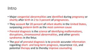
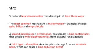
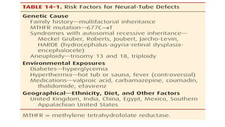
Prenatal Diagnosis
Themba Hospital FCOG(SA) Part 1 Tutorials
By Dr N.E Manana
Intro
• Major congenital abnormalities are identified during pregnancy or
shortly after birth in 2 to 3 percent of pregnancies.
• They account for 20 percent of infant deaths in the United States,
surpassing preterm birth as the most common cause
• Prenatal diagnosis is the science of identifying malformations,
disruptions, chromosomal abnormalities, and other genetic
syndromes in the fetus
• The goal of prenatal diagnosis is to provide accurate information
regarding short- and long-term prognosis, recurrence risk, and
potential therapy and to thereby improve counseling
Intro
• Structural fetal abnormalities may develop in at least three ways.
• The most common mechanism is malformation—Examples include
spina bifida and omphalocele
• A second mechanism is deformation, an example is limb contractures
that develop with oligohydramnios from bilateral renal agenesis
• A third type is disruption, An example is damage from an amnionic
band, which can cause a limb-reduction defect
NEURAL-TUBE DEFECTS
• These defects include anencephaly, spina bifida, cephalocele, and
other rare spinal fusion (schisis) abnormalities
• Neural-tube defects (NTDs) are the second most common class of
birth defect after cardiac anomalies, and their reported frequency is
approximately 0.9 per 1000 births
• More than 40 years ago, Brock and associates (1972, 1973) observed
that pregnancies complicated by NTDs had higher levels of alpha-
fetoprotein (AFP) in both maternal serum and amnionic fluid.
• This formed the basis for the first maternal serum screening test for a
fetal defect
• Table 14.1
Maternal Serum Alpha-Fetoprotein Screening
(MSAFP)
• Figure 14.1
Maternal Serum Alpha-Fetoprotein Screening
• Several factors influence maternal serum AFP :
1. Maternal weight—The AFP concentration is adjusted for the
maternal volume of distribution.
2. Gestational age— In general, the MoM should be recalculated if the
biparietal diameter differs from the stated gestational age by more
than 1week.
3. Race/ethnicity—African American women have at least 10-percent
higher serum AFP concentrations but are at lower risk for fetal NTDs.
4. Diabetes—Serum levels may be 10 to 20 percent lower in women
with insulin-treated diabetes, despite a three- to fourfold increased risk
for NTDs
5. Multifetal gestation—Higher screening threshold values are used
• Figure 14.3
• Figure 14.4
Management of the Fetus with Spina Bifida
• The optimal mode of delivery for a fetus with open spina bifida remains
controversial.
• Some have recommended cesarean delivery before the onset of labor,
positing that it may reduce the risk of mechanical trauma and spinal
infection
• Open fetal surgery to repair NTDs has been the subject of several clinical
studies
• Fetal surgery resulted in improved motor outcomes and reduced need for
ventriculoperitoneal shunt placement at age 2 to 3 years.
• However, surgery itself was associated with significant maternal and fetal
risks.
DOWN SYNDROME AND OTHER
ANEUPLOIDIES
• The risk of fetal trisomy increases with maternal age
• Until the mid-1980s, prenatal diagnostic testing for fetal aneuploidy was
offered for “advanced maternal age.”
• However, age alone is a poor screening test, because approximately 70
percent of Down syndrome pregnancies are in women younger than 35
years.
• Nearly 30 years ago, Merkatz and associates (1984) observed that
pregnancies with Down syndrome were characterized by lower maternal
serum AFP levels at 15 to 20 week
• During the past two decades, there have been major advances in the area
of aneuploidy screening
• Table 14.5
First-Trimester Screening
• The most commonly used protocol involves measurement of
sonographic nuchal translucency and two maternal serum analytes.
• This is performed between 11 and 14 weeks’ gestation.
• This is the maximum thickness of the subcutaneous translucent area
between the skin and soft tissue overlying the fetal spine at the back
of the neck
• An increased NT thickness itself is not a fetal abnormality, but rather
is a marker that confers increased risk
• Approximately one third of fetuses with increased nuchal
translucency thickness will have a chromosome abnormality, nearly
half of which are Down syndrome
• Figure 14.5
Serum Analytes
• Two analytes used for first-trimester aneuploidy screening are human
chorionic gonadotropin—either intact or free β-hCG— and pregnancy-
associated plasma protein A (PAPP-A)
• In cases of fetal Down syndrome, the first-trimester serum free β-hCG level
is higher, approximately 2.0 MoM, and the PAPP-A level is lower,
approximately 0.5 MoM
• If gestational age is correct, the use of these serum markers—without NT
measurement—results in detection rates for fetal Down syndrome up to 67
percent at a false-positive rate of 5 percent
• Aneuploidy detection is significantly greater if these first-trimester analytes
are either:
(1) combined with the sonographic NT measurement or
(2) combined with second-trimester analytes, which is termed serum
integrated screening
Second-Trimester Screening
• Pregnancies with fetal Down syndrome are characterized by lower
maternal serum AFP levels—approximately 0.7 MoM, higher hCG
levels—approximately 2.0 MoM, and lower unconjugated estriol
levels—approximately 0.8 MoM
• This triple test can detect 61 to 70 percent of Down syndrome
• Levels of all three markers are decreased in the setting of trisomy 18,
with a detection rate similar to that for Down syndrome at a false-
positive rate of only 0.5 percent
• Levels of a fourth marker—dimeric inhibin alpha—are elevated in
Down syndrome, with an average value of 1.8 MoM
• The quad test is the most commonly used second-trimester serum
screening test for aneuploidy
Combined First- and Second-Trimester
Screening
• Combined screening strategies enhance aneuploidy detection
• Three types of screening strategies are available:
1. Integrated screening combines results of first- and second trimester
tests
2. Sequential screening discloses the results of first-trimester screening
to women at highest risk, who are then offered invasive testing
Cell-Free Fetal DNA Screening
• Using massively parallel sequencing or chromosome selective
sequencing to isolate cell-free fetal DNA from maternal plasma, fetal
Down syndrome and other autosomal trisomies may be detected as
early as 10 weeks’ gestation
• Recent trials of these techniques in high-risk pregnancies have yielded
detection rates for trisomies 21, 18, and 13 of approximately 98
percent at a false-positive rate of 0.5 percent or less
• If an abnormal result is identified, genetic counseling should be
performed, and invasive prenatal diagnostic testing should be offered
to confirm the results.
Sonographic Screening
• Table 14.6
• Table 14.7
• Figure 14.6
First-Trimester Sonographic Findings
• Unlike second-trimester soft signs, which may be readily visible
during a standard sonogram, first-trimester findings associated with
aneuploidy require specialized training.
• The fetal NT is unique in that it has become a component of
aneuploidy screening offered to all women
• Other first-trimester findings associated with an increased risk for
fetal Down syndrome include an absent fetal nasal bone, wider
fronto-maxillary facial angle— indicating a flat facial profile, tricuspid
regurgitation, and abnormal ductus venosus flow
PREGNANCIES AT INCREASED RISK FOR
GENETIC DISORDERS
• Table 14.9
PRENATAL AND PREIMPLANTATION
DIAGNOSTIC TESTING
• Invasive procedures used in prenatal diagnosis—amniocentesis,
chorionic villus sampling, and fetal blood sampling— enable a vast
array of sophisticated genetic diagnoses to be made before birth
• Preimplantation genetic diagnosis permits similar diagnoses to be
made in oocytes or embryos before implantation
• Improvements in aneuploidy screening tests during the past decade
as described in the preceding section have resulted in a significant
decrease in the number of prenatal diagnostic procedures
Thank you

4. prenatal dx.pptx

  • 1.
    Prenatal Diagnosis Themba HospitalFCOG(SA) Part 1 Tutorials By Dr N.E Manana
  • 2.
    Intro • Major congenitalabnormalities are identified during pregnancy or shortly after birth in 2 to 3 percent of pregnancies. • They account for 20 percent of infant deaths in the United States, surpassing preterm birth as the most common cause • Prenatal diagnosis is the science of identifying malformations, disruptions, chromosomal abnormalities, and other genetic syndromes in the fetus • The goal of prenatal diagnosis is to provide accurate information regarding short- and long-term prognosis, recurrence risk, and potential therapy and to thereby improve counseling
  • 3.
    Intro • Structural fetalabnormalities may develop in at least three ways. • The most common mechanism is malformation—Examples include spina bifida and omphalocele • A second mechanism is deformation, an example is limb contractures that develop with oligohydramnios from bilateral renal agenesis • A third type is disruption, An example is damage from an amnionic band, which can cause a limb-reduction defect
  • 4.
    NEURAL-TUBE DEFECTS • Thesedefects include anencephaly, spina bifida, cephalocele, and other rare spinal fusion (schisis) abnormalities • Neural-tube defects (NTDs) are the second most common class of birth defect after cardiac anomalies, and their reported frequency is approximately 0.9 per 1000 births • More than 40 years ago, Brock and associates (1972, 1973) observed that pregnancies complicated by NTDs had higher levels of alpha- fetoprotein (AFP) in both maternal serum and amnionic fluid. • This formed the basis for the first maternal serum screening test for a fetal defect
  • 5.
  • 6.
    Maternal Serum Alpha-FetoproteinScreening (MSAFP) • Figure 14.1
  • 7.
    Maternal Serum Alpha-FetoproteinScreening • Several factors influence maternal serum AFP : 1. Maternal weight—The AFP concentration is adjusted for the maternal volume of distribution. 2. Gestational age— In general, the MoM should be recalculated if the biparietal diameter differs from the stated gestational age by more than 1week. 3. Race/ethnicity—African American women have at least 10-percent higher serum AFP concentrations but are at lower risk for fetal NTDs. 4. Diabetes—Serum levels may be 10 to 20 percent lower in women with insulin-treated diabetes, despite a three- to fourfold increased risk for NTDs 5. Multifetal gestation—Higher screening threshold values are used
  • 8.
  • 9.
  • 10.
    Management of theFetus with Spina Bifida • The optimal mode of delivery for a fetus with open spina bifida remains controversial. • Some have recommended cesarean delivery before the onset of labor, positing that it may reduce the risk of mechanical trauma and spinal infection • Open fetal surgery to repair NTDs has been the subject of several clinical studies • Fetal surgery resulted in improved motor outcomes and reduced need for ventriculoperitoneal shunt placement at age 2 to 3 years. • However, surgery itself was associated with significant maternal and fetal risks.
  • 11.
    DOWN SYNDROME ANDOTHER ANEUPLOIDIES • The risk of fetal trisomy increases with maternal age • Until the mid-1980s, prenatal diagnostic testing for fetal aneuploidy was offered for “advanced maternal age.” • However, age alone is a poor screening test, because approximately 70 percent of Down syndrome pregnancies are in women younger than 35 years. • Nearly 30 years ago, Merkatz and associates (1984) observed that pregnancies with Down syndrome were characterized by lower maternal serum AFP levels at 15 to 20 week • During the past two decades, there have been major advances in the area of aneuploidy screening
  • 12.
  • 13.
    First-Trimester Screening • Themost commonly used protocol involves measurement of sonographic nuchal translucency and two maternal serum analytes. • This is performed between 11 and 14 weeks’ gestation. • This is the maximum thickness of the subcutaneous translucent area between the skin and soft tissue overlying the fetal spine at the back of the neck • An increased NT thickness itself is not a fetal abnormality, but rather is a marker that confers increased risk • Approximately one third of fetuses with increased nuchal translucency thickness will have a chromosome abnormality, nearly half of which are Down syndrome
  • 14.
  • 15.
    Serum Analytes • Twoanalytes used for first-trimester aneuploidy screening are human chorionic gonadotropin—either intact or free β-hCG— and pregnancy- associated plasma protein A (PAPP-A) • In cases of fetal Down syndrome, the first-trimester serum free β-hCG level is higher, approximately 2.0 MoM, and the PAPP-A level is lower, approximately 0.5 MoM • If gestational age is correct, the use of these serum markers—without NT measurement—results in detection rates for fetal Down syndrome up to 67 percent at a false-positive rate of 5 percent • Aneuploidy detection is significantly greater if these first-trimester analytes are either: (1) combined with the sonographic NT measurement or (2) combined with second-trimester analytes, which is termed serum integrated screening
  • 16.
    Second-Trimester Screening • Pregnancieswith fetal Down syndrome are characterized by lower maternal serum AFP levels—approximately 0.7 MoM, higher hCG levels—approximately 2.0 MoM, and lower unconjugated estriol levels—approximately 0.8 MoM • This triple test can detect 61 to 70 percent of Down syndrome • Levels of all three markers are decreased in the setting of trisomy 18, with a detection rate similar to that for Down syndrome at a false- positive rate of only 0.5 percent • Levels of a fourth marker—dimeric inhibin alpha—are elevated in Down syndrome, with an average value of 1.8 MoM • The quad test is the most commonly used second-trimester serum screening test for aneuploidy
  • 17.
    Combined First- andSecond-Trimester Screening • Combined screening strategies enhance aneuploidy detection • Three types of screening strategies are available: 1. Integrated screening combines results of first- and second trimester tests 2. Sequential screening discloses the results of first-trimester screening to women at highest risk, who are then offered invasive testing
  • 18.
    Cell-Free Fetal DNAScreening • Using massively parallel sequencing or chromosome selective sequencing to isolate cell-free fetal DNA from maternal plasma, fetal Down syndrome and other autosomal trisomies may be detected as early as 10 weeks’ gestation • Recent trials of these techniques in high-risk pregnancies have yielded detection rates for trisomies 21, 18, and 13 of approximately 98 percent at a false-positive rate of 0.5 percent or less • If an abnormal result is identified, genetic counseling should be performed, and invasive prenatal diagnostic testing should be offered to confirm the results.
  • 19.
  • 20.
  • 21.
  • 22.
    First-Trimester Sonographic Findings •Unlike second-trimester soft signs, which may be readily visible during a standard sonogram, first-trimester findings associated with aneuploidy require specialized training. • The fetal NT is unique in that it has become a component of aneuploidy screening offered to all women • Other first-trimester findings associated with an increased risk for fetal Down syndrome include an absent fetal nasal bone, wider fronto-maxillary facial angle— indicating a flat facial profile, tricuspid regurgitation, and abnormal ductus venosus flow
  • 23.
    PREGNANCIES AT INCREASEDRISK FOR GENETIC DISORDERS • Table 14.9
  • 24.
    PRENATAL AND PREIMPLANTATION DIAGNOSTICTESTING • Invasive procedures used in prenatal diagnosis—amniocentesis, chorionic villus sampling, and fetal blood sampling— enable a vast array of sophisticated genetic diagnoses to be made before birth • Preimplantation genetic diagnosis permits similar diagnoses to be made in oocytes or embryos before implantation • Improvements in aneuploidy screening tests during the past decade as described in the preceding section have resulted in a significant decrease in the number of prenatal diagnostic procedures
  • 25.

Editor's Notes

  • #3 It encompasses routine screening tests for aneuploidy and neural-tube defects, invasive diagnostic tests such as chorionic villus sampling and amniocentesis, additional screening and diagnostic tests offered to those at risk for specific genetic disorders, and the diagnosis of structural malformations with specialized sonography and other fetal imaging techniques
  • #4 malformation—an intrinsic abnormality “programmed” in development, regardless of whether a precise genetic etiology is known. Examples include spina bifida and omphalocele deformation, by which a fetus develops abnormally because of extrinsic mechanical forces imposed by the uterine environment. An example is limb contractures that develop with oligohydramnios from bilateral renal agenesis disruption, which is a more severe change in form or function that occurs when genetically normal tissue is modified as the result of a specific insult. An example is damage from an amnionic band, which can cause a limb-reduction defect Multiple structural or developmental abnormalities may also present together as a syndrome, sequence, or association. A syndrome is a cluster of several anomalies or defects that have the same cause—for example, trisomy 18. A sequence describes anomalies that all developed sequentially from one initial insult. An example is the Pierre-Robin sequence in which micrognathia causes posterior displacement of the tongue—glossoptosis— which leads to a posterior rounded cleft in the palate. An association is a group of specific abnormalities that occur together frequently but do not seem to be linked etiologically. Diagnosis of the VACTERL association, for example, includes three or more of the following: vertebral defects, anal atresia, cardiac defects, tracheoesophageal fistula, renal anomalies, and limb abnormalities. Because of overlap of anomaly patterns, it is readily apparent that classification of fetal malformations is challenging, and reclassification is required periodically
  • #6 Prevention Most women at increased risk for NTDs benefit from 4 mg folic acid taken daily before conception and through the first trimester. This is particularly important if a woman has one or more prior affected children or if either the pregnant woman or her partner has such a defect. Folic acid supplementation may not decrease the risk for NTDs in those with valproic acid exposure, pregestational diabetes, first-trimester fever or hot tub exposure, or defects associated with a genetic syndrome (American College of Obstetricians and Gynecologists, 2013b). The policy of routine fortification of cereal grains with folic acid, which has been in place in the United States since 1998, provides approximately 200 additional micrograms of folic acid daily and may reduce the first occurrence of NTDs in low-risk women by approximately 20 percent (Honein, 2001). It is recommended that all women at low risk take 400 μg of folic acid orally every day before conception and through the first trimester, to reduce the NTD risk by as much as 80 percent
  • #7 Alpha-fetoprotein (AFP) is a glycoprotein synthesized by the fetal yolk sac and later by the fetal gastrointestinal tract and liver. It is the major serum protein in the embryo and fetus and is thus analogous to albumin As shown in Figure 14-1, its concentration increases steadily in both fetal serum and amnionic fluid until 13 weeks’ gestation, after which, levels rapidly decline. Conversely, AFP is found in steadily increasing quantities in maternal serum after 12 weeks The normal concentration gradient between fetal plasma and maternal serum ison the order of 50,000:1. Defects in fetal integument, such as neural-tube and ventral wall defects, permit AFP to leak into the amnionic fluid, resulting in dramatically increased maternal serum AFP levels. It was shown more than 30 years ago that maternal serum AFP concentrations at 16 to 18 weeks exceeded 2.5 multiples of the median (MoM) in a large proportion of women carrying fetuses with either anencephaly or spina bifida Since the mid-1980s, MSAFP concentration has been routinely measured as a screening test for NTDs Maternal serum AFP screening is generally performed from 15 through 20 weeks, within a protocol that includes quality control, counseling, and follow-up. AFP is measured in nanograms per milliliter and reported as multiples of the median (MoM) of the unaffected population. Using MoM normalizes the distribution of AFP levels and permits comparison of results from different laboratories and populations. Using a maternal serum AFP level of 2.0 or 2.5 MoM as the upper limit of normal, most laboratories report a detection rate—test sensitivity— of at least 90 percent for anencephaly and 80 percent for spina bifida at a screen-positive rate of 3 to 5 percent The positive predictive value—the proportion with AFP elevation that have an affected fetus—is only 2 to 6 percent. This is explained by the overlap in AFP distributions in affected and unaffected pregnancies
  • #8 At Parkland Hospital, an AFP level is considered elevated in a twin pregnancy if greater than 3.5 MoM, but other laboratories use 4.0 or even 5.0 MoM. According to the American College of Obstetricians and Gynecologists (2013b), all pregnant women should be offered screening for NTDs. Women who present for care early in pregnancy often have the option of several different screening tests for aneuploidy, as discussed subsequently. Those who elect second-trimester multiple marker serum screening will have a maternal serum AFP level measured as a component. Those who elect first-trimester screening or chorionic villus sampling may receive neural-tube defect screening either with serum AFP at 15 to 20 weeks or with sonography
  • #9 Targeted Sonography More than 25 years ago, Nicolaides and colleagues (1986) described frontal bone scalloping—the lemon sign, and anterior curvature of the cerebellum with effacement of the cisterna magna—the banana sign—in second-trimester fetuses with open spina bifida These investigators also frequently noted a small biparietal diameter and ventriculomegaly in such casesthe size and location of spinal defects (Chap. 10, p. 202). Using these findings, experienced investigators have described nearly 100-percent detection of open NTDs (Norem, 2005; Sepulveda, 1995). Overall NTD risk may be reduced by at least 95 percent when no spine or cranial abnormality is observed Most centers use targeted sonography as the primary method of evaluating maternal serum AFP level elevation. The American College of Obstetricians and Gynecologists (2013b) recommends that women be counseled regarding the risks and benefits of targeted sonography and amniocentesis, the risk associated with the degree of AFP level elevation or other risk factors, and the quality and findings of the sonographic examination before making a decision. Watson and coworkers (1991) reported that 99 percent of fetuses with open spina bifida had one or more of these findings. In addition to these cranial findings, transverse and sagittal images of the spine are increasingly used to characterize
  • #10 Amniocentesis Although amniocentesis for amnionic fluid AFP measurement was once considered the standard for open NTD diagnosis, it has been replaced in most centers by targeted sonography If the amnionic fluid AFP level was elevated, an assay for acetylcholinesterase was performed, and if positive, was considered diagnostic of an NTD Acetylcholinesterase leaks directly from exposed neural tissue into the amnionic fluid. The overall sensitivity of amniocentesis is approximately 98 percent for open NTDs, with a false-positive rate of 0.4 percentOther fetal abnormalities associated with elevated amnionic fluid AFP levels and positive assay for acetylcholinesterase include ventral wall defects, esophageal atresia, fetal teratoma, cloacal exstrophy, and skin abnormalities such as epidermolysis bullosa.
  • #11 Although some have found improved motor function in children delivered operatively, others have not identified benefit in short- or longterm outcomes (Lewis, 2004; Luthy, 1991; Merrill, 1998). The American College of Obstetricians and Gynecologists (2013b) recommends that the route of delivery for the fetus with spina bifida should be individualized
  • #12 Other significant risk factors include a prior pregnancy with autosomal trisomy or triploidy or a woman or her partner with a numerical chromosomal abnormality or structural chromosomal rearrangement, such as a balanced translocation aneuploidy screening: The addition of other serum analytes to second-trimester screening has improved Down syndrome detection rates to approximately 80 percent for the quadruple marker test (Table 14-5). 2. First-trimester screening at 11 to 14 weeks’ gestation, using the fetal nuchal translucency measurement together with serum analytes, has achieved Down syndrome detection rates comparable to those for second-trimester screening in women younger than 35 years (American College of Obstetricians and Gynecologists, 2013c). 3. Combinations of first- and second-trimester screening yield Down syndrome detection rates as high as 90 to 95 percent (Malone, 2005b). 4. Maternal serum cell-free fetal DNA testing for trisomy 21, 18, and 13 has become available as a screening test for high-risk pregnancies, with a 98-percent detection rate and a false-positive rate of 0.5 percent (American College of Obstetricians and Gynecologists, 2012b; Bianchi, 2012; Palomaki, 2011, 2012). With the exception of cell-free fetal DNA testing, each firstand/or second-trimester aneuploidy screening test is based on a composite likelihood ratio, and the maternal age-related risk is multiplied by this ratio. This principle also applies to modification of the Down syndrome risk by selected sonographic findings
  • #13 Counseling Because technology advances have resulted in improved aneuploidy detection with available screening tests, the American College of Obstetricians and Gynecologists (2013c) recommends that all women who present for prenatal care before 20 weeks be offered screening. Available screening paradigms are shown in Table 14-5. A positive screening test result indicates increased risk, but it is not diagnostic of aneuploidy. Conversely, a negative screening test indicates that the risk is not increased, but it does not guarantee a normal fetus. Although Down syndrome is the focus of most aneuploidy screening protocols, it accounts for only half of all fetal chromosomal abnormality cases. Invasive diagnostic tests such as chorionic villus sampling and amniocentesis are safe and effective Regardless of age, all women are counseled regarding the differences between screening and diagnostic tests, and they are given the option of invasive diagnostic testing
  • #14 The NT measurement is expressed as a multiple of the gestational age-specific median, similar to serum markers used for aneuploidy screening Increased NT thickness is also associated with other aneuploidies, genetic syndromes, and various birth defects, especially fetal cardiac anomalies (Atzei, 2005; Simpson, 2007). Because of this, if the NT measurement is 3.5 mm or greater, the patient should be offered targeted sonography, with or without fetal echocardiography, in addition to fetal karyotyping The NT must be imaged and measured with a high degree of precision for aneuploidy detection to be accurate. This has led to standardized training, certification, and ongoing quality review programs. In the United States, training, credentialing, and monitoring are available through the Nuchal Translucency Quality Review (NTQR) program (www.ntqr.org)
  • #15 It is measured in the sagittal plane, when the crown-rump length measures between 38 and 84 mm. Specific criteria for NT measurement are listed in Table 10-3 (p. 196). As shown in Table 14-5, as an isolated marker, NT detects 64 to 70 percent of fetuses with Down syndrome at a falsepositive rate of 5 percent, and it has maximal sensitivity at 11 weeks (Malone, 2005b). The risk conferred by an increased NT thickness is independent of that of serum analytes, and combining NT with serum analyte values results in greatly improved aneuploidy detection Thus, NT is generally used as an isolated marker only in screening for multifetal gestations, in which serum screening is not as accurate or may not be available (American College of Obstetricians and Gynecologists, 2013c). An exception is that if the NT measurement is increased to 3 to 4 mm, then the aneuploidy risk is unlikely to be normalized using serum analyte assessment, and invasive testing should be offered
  • #16 With trisomy 18 and trisomy 13, levels of both analytes are lower In twin pregnancies, serum free β-hCG and PAPP-A levels are approximately doubled compared with singleton values (Vink, 2012). Even with specific curves, a normal dichorionic cotwin will tend to normalize screening results, and thus, the aneuploidy detection rate is at least 15-percent lower Combined First-Trimester Screening The most commonly used screening protocol combines the NT measurement with serum hCG and PAPP-A. Using this protocol, Down syndrome detection rates in large prospective trials range from 79 to 87 percent, at a false-positive rate of 5 percent The detection rate is approximately 5-percent higher if performed at 11 compared with 13 weeks The detection rate for trisomies 18 and 13 is approximately 90 percent, at a 2-percent false-positive rate Maternal age does affect the performance of first-trimester aneuploidy screening tests. In prospective trials, combined first-trimester screening resulted in Down syndrome detection rates of 67 to 75 percent in women younger than 35 years at delivery, which are 10-percent lower than the overall detection rates in these studies (Malone, 2005b; Wapner, 2003). Among women older than 35 at delivery, however, Down syndrome detection rates were 90 to 95 percent, albeit at a higher falsepositive rate of 15 to 22 percent
  • #17 The addition of dimeric inhibin to the other three markers is the quadruple or quad test, which has a trisomy 21 detection rate of approximately 80 percent at a false-positive rate of 5 percen As with first-trimester screening, aneuploidy detection rates will be slightly lower in younger women and higher in women older than 35 years at delivery. If second-trimester serum screening is used in twin pregnancies, aneuploidy detection rates are significantly lower As a stand-alone test, it is generally used if women do not begin care until the secondtrimester or if first-trimester screening is not available. As subsequently discussed, combining the quad test with first-trimester screening yields even greater aneuploidy detection rates
  • #18 Integrated screening combines results of first- and secondtrimester tests. This includes a combined measurement of fetal NT and serum analyte levels at 11 to 14 weeks’ gestation plus quadruple markers at 15 to 20 weeks. An aneuploidy risk is then calculated from these seven parameters. As expected, integrated screening has the highest Down syndrome detection rate—94 to 96 percent at a false-positive rate of 5 percent Sequential screening There are two testing strategies in this category: • With stepwise sequential screening, women with firsttrimester screen results that confer risk for Down syndrome above a particular threshold are offered invasive testing, and the remaining women receive second-trimester screening. The threshold is set at approximately 1 percent, because in a screened population, the 1 percent at highest risk includes approximately 70 percent of Down syndrome pregnancies (Cuckle, 2005). This method of screening may achieve up to a 95-percent detection rate With contingent sequential screening, women are divided into high-, moderate-, and low-risk groups. Those at highest risk, for example, the top 1 percent, are offered invasive testing. Women at moderate risk, who comprise 15 to 20 percent of the population, undergo second-trimester screening. The remaining 80 to 85 percent, who are at or below a 1:1000 risk, receive negative screening test results and have no further testing (Cuckle, 2005). Thus, most of those screened are provided with results almost immediately while still maintaining a high detection rate. This rate ranges from 88 to 94 percent Integrated and sequential screening strategies require coordination between the provider and laboratory to ensure that the second sample is obtained during the appropriate gestational age window, sent to the same laboratory, and linked to the first-trimester results
  • #19 This novel technology has recently become clinically available as a screening test, but it is not considered a replacement diagnostic test. Pretest counseling is recommended The American College of Obstetricians and Gynecologists (2012b) currently recommends that the test may be offered to the following groups: • Women 35 years or older at delivery • Those with sonographic findings indicating increased risk for fetal aneuploidy • Those with a prior pregnancy complicated by trisomy 21, 18, or 13 • Patient or partner carries a balanced robertsonian translocation indicating increased risk for fetal trisomy 21 or 13 • Those with an abnormal first-, second-, or combined firstand second-trimester screening test result for aneuploidy
  • #20 Major abnormalities and minor sonographic markers contribute significantly to aneuploidy detection. As shown in Table 14-6, with few exceptions, the aneuploidy risk associated with any major abnormality is high enough to warrant offering an invasive test for fetal karyotype and/or chromosomal microarray analysis Importantly, a fetus with one abnormality may have others that are less likely to be detected sonographically or even undetectable sonographically but that greatly affect the prognosis nonetheless Most fetuses with aneuploidy that is likely to be lethal in utero—such as trisomy 18 and 13 and triploidy—usually have sonographic abnormalities that can be seen by the second trimester. However, only 25 to 30 percent of second-trimester fetuses with Down syndrome will have a major malformation that can be identified sonographically
  • #21 For more than two decades, investigators have recognized that the sonographic detection of aneuploidy, particularly Down syndrome, may be improved by minor markers that are collectively referred to as “soft signs.” Minor markers are normal variants rather than fetal abnormalities, and in the absence of aneuploidy or an associated abnormality, they do not significantly affect prognosis Unfortunately, at least 10 percent of unaffected pregnancies will have one of these soft signs, significantly limiting their utility for general population screening The incorporation of minor markers into second-trimester screening protocols has been studied primarily in high-risk populations. In this setting, detection rates of 50 to 75 percent for Down syndrome have been reported (American College of Obstetricians and Gynecologists, 2013c). With the exception of increased nuchal skinfold thickness, the identification of an isolated second-trimester marker in an otherwise low-risk pregnancy is not generally considered sufficient to warrant “highrisk” status
  • #23 Each has also been associated with an increased risk for trisomies 18 and 13 and other aneuploidies. However, these signs have not become widely adopted for routine use in the United States. Fetal Nasal Bone. In approximately two thirds of fetuses with Down syndrome, the nasal bone is not visible at the 11- to 14-week examination (Cicero, 2004; Rosen, 2007; Sonek, 2006). Currently, this is the only first-trimester marker, other than NT, for which the Nuchal Translucency Quality Review Program has established a training program. Criteria for adequate assessment include that the fetus occupies most of the image; that there be a 45-degree angle of insonation with the fetal profile; that the profile be well defined in the midsagittal plane, with the tip of the nose and the third and fourth ventricles visible; and that the nasal bone brightness be greater than or equal to that of the overlying skin
  • #24 Couples with a personal or family history of a heritable genetic disorder should be offered genetic counseling. They should be given an estimated risk of having an affected infant and provided information concerning benefits and limitations of available prenatal testing options
  • #25 In a study of more than 160,000 pregnant women 35 years and older, patient acceptance of amniocentesis procedures decreased from 56 to 36 percent between 2001 and 2008, while that of chorionic villus sampling decreased from 36 to 24 percent (Nakata, 2010). Fetal blood sampling procedures have also decreased, but for different reasons. Namely, amniocentesis with fluorescence in situ hybridization (FISH) has decreased the need for rapid karyotyping from fetal blood (Chap. 13, p. 276); the number of DNA-based tests performed on amnionic fluid has greatly expanded; and fetal middle cerebral artery Doppler studies have improved the accuracy of fetal anemia detection