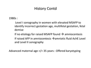
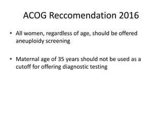
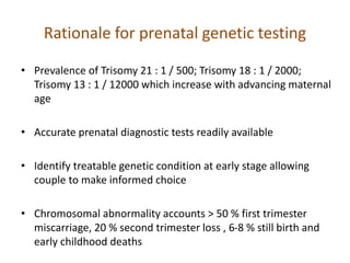
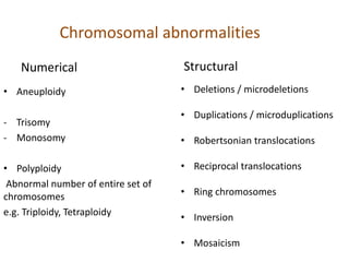
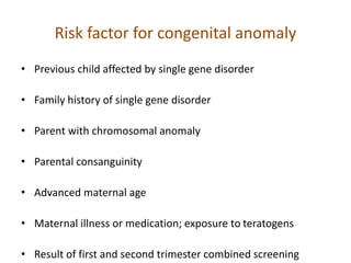
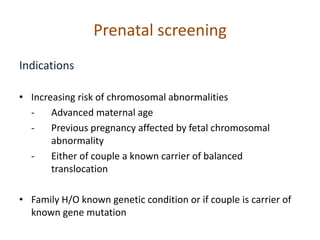
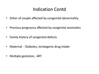
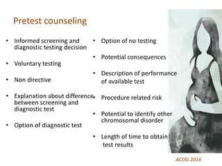
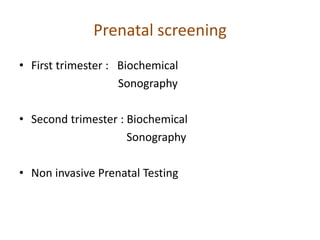
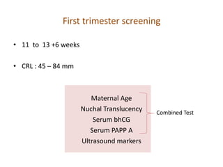
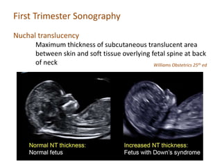
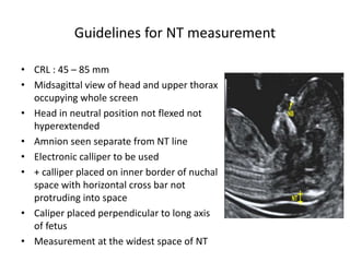
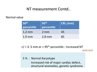
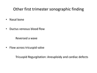
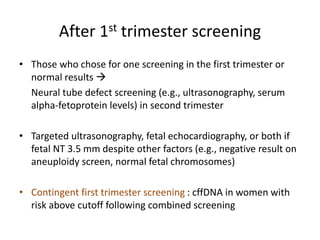
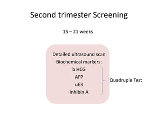
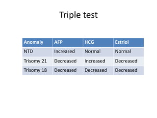
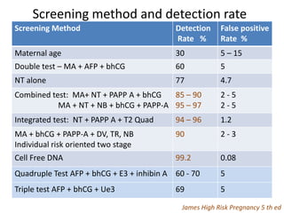
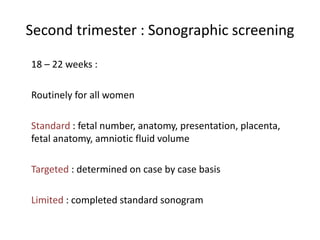
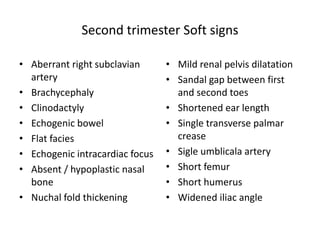
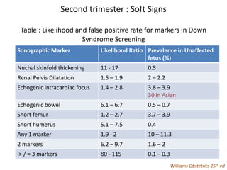
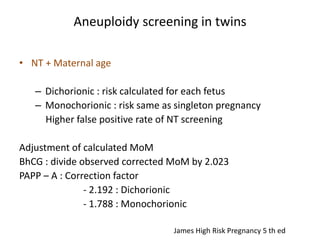
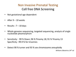
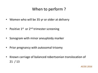
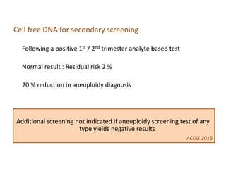
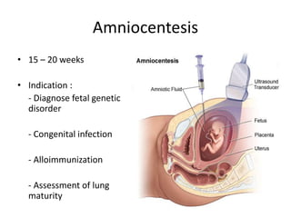
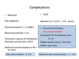
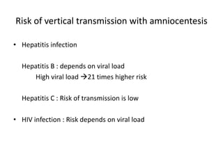
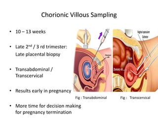
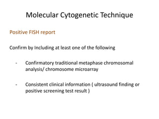
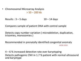
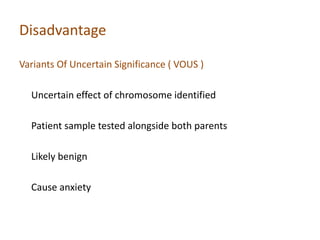
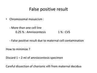
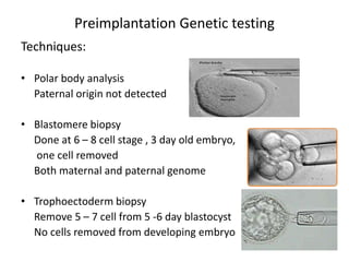
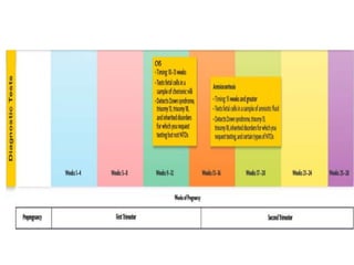
Prenatal Genetic Diagnosis
Kritika Jha
MD Resident
2079 / 11 / 10
1 – 2 pm
Contents
• Introduction
• Screening : First trimester
Second Trimester
• Diagnostic test: Invasive
Non Invasive
Preimplantation Genetic testing
Newer diagnostic tests
Introduction
Genetic testing is offered for a condition or carrier status
when there is no prior evidence of its presence in an
individual
James High Risk Pregnancy 5th ed
Intended to determine, with as much certainty as possible
whether a specific genetic disorder or condition is present
ACOG Practice Bulletin : Prenatal Diagnosis of genetic disorder
2016
Introduction
Historical perspective
1972, 1973
Higher level of Alpha Fetoprotein in maternal serum and
amniotic fluid found in pregnancy with neural tube defect
1977
Widespread serum screening of MSAFP
History Contd
1980s :
Level I sonography in women with elevated MSAFP to
identify incorrect gestation age, multifetal gestation, fetal
demise
If no etiology for raised MSAFP found  amniocentesis
If raised AFP in amniocentesis amniotic fluid AchE Level
and Level II sonography
Advanced maternal age >/= 35 years : Offered karyotyping
ACOG Reccomendation 2016
• All women, regardless of age, should be offered
aneuploidy screening
• Maternal age of 35 years should not be used as a
cutoff for offering diagnostic testing
Rationale for prenatal genetic testing
• Prevalence of Trisomy 21 : 1 / 500; Trisomy 18 : 1 / 2000;
Trisomy 13 : 1 / 12000 which increase with advancing maternal
age
• Accurate prenatal diagnostic tests readily available
• Identify treatable genetic condition at early stage allowing
couple to make informed choice
• Chromosomal abnormality accounts > 50 % first trimester
miscarriage, 20 % second trimester loss , 6-8 % still birth and
early childhood deaths
Chromosomal abnormalities
Numerical
• Aneuploidy
- Trisomy
- Monosomy
• Polyploidy
Abnormal number of entire set of
chromosomes
e.g. Triploidy, Tetraploidy
Structural
• Deletions / microdeletions
• Duplications / microduplications
• Robertsonian translocations
• Reciprocal translocations
• Ring chromosomes
• Inversion
• Mosaicism
Risk factor for congenital anomaly
• Previous child affected by single gene disorder
• Family history of single gene disorder
• Parent with chromosomal anomaly
• Parental consanguinity
• Advanced maternal age
• Maternal illness or medication; exposure to teratogens
• Result of first and second trimester combined screening
Prenatal screening
Indications
• Increasing risk of chromosomal abnormalities
- Advanced maternal age
- Previous pregnancy affected by fetal chromosomal
abnormality
- Either of couple a known carrier of balanced
translocation
• Family H/O known genetic condition or if couple is carrier of
known gene mutation
Indication Contd
• Either of couple affected by congenital abnormality
• Previous pregnancy affected by congenital anomalies
• Family history of congenital defects
• Maternal - Diabetes, teratogenic drug intake
• Multiple gestation, ART
Pretest counseling
• Informed screening and
diagnostic testing decision
• Voluntary testing
• Non directive
• Explanation about difference
between screening and
diagnostic test
• Option of diagnostic test
• Option of no testing
• Potential consequences
• Description of performance
of available test
• Procedure related risk
• Potential to identify other
chromosomal disorder
• Length of time to obtain
test results
ACOG 2016
Prenatal screening
• First trimester : Biochemical
Sonography
• Second trimester : Biochemical
Sonography
• Non invasive Prenatal Testing
First trimester screening
• 11 to 13 +6 weeks
• CRL : 45 – 84 mm
Maternal Age
Nuchal Translucency
Serum bhCG
Serum PAPP A
Ultrasound markers
Combined Test
Maternal age and risk of trisomy in fetus
Maternal Age
years
Gestation
weeks
Risk of Trisomy
21
Risk of Trisomy
18
Risk of Trisomy
13
20 12 1 / 1000 1 / 2500 1 / 8000
20 39 / term 1/ 1500 1 / 18000 1 / 42000
35 12 1 / 250 1 / 600 1 / 1800
35 39 / term 1 / 350 1 / 4000 1 / 10000
Serum Biochemical marker
Beta hCG Median MoM PAPP-A Median MoM
Normal 1 Normal 1
Trisomy 21 2 Trisomy 21 0.5
Trisomy 18 0.2 Trisomy 18 0.2
Trisomy 13 0.5 Trisomy 13 0.3
Arias High Risk Pregnancy
First trimester Sonography
11 – 14 weeks
Assessment in at risk pregnancy
40 % detection rate for structural abnormalities
First Trimester Sonography
Nuchal translucency
Maximum thickness of subcutaneous translucent area
between skin and soft tissue overlying fetal spine at back
of neck Williams Obstetrics 25th ed
Guidelines for NT measurement
• CRL : 45 – 85 mm
• Midsagittal view of head and upper thorax
occupying whole screen
• Head in neutral position not flexed not
hyperextended
• Amnion seen separate from NT line
• Electronic calliper to be used
• + calliper placed on inner border of nuchal
space with horizontal cross bar not
protruding into space
• Caliper placed perpendicular to long axis
of fetus
• Measurement at the widest space of NT
NT measurement Contd..
Normal value
>/ = 3. 5 mm or > 95th percentile : Increased NT
ACOG 2016
5 % : Normal Karyotype
Increased risk of major cardiac defect,
structural anomalies, genetic syndrome
50th
percentile
95th
percentile
CRL (mm)
1.2 mm 2 mm 45
1.9 mm 2.8 mm 85
Follow up of increased NT
Increased NT > 95 th centile
Karyotype / CVS
Abnormal Normal
US at 16 weeks
Nuchal edema > 5 mm Normal
20 week scan
Echocardiography Abnormal Normal
James High Risk Pregnancy 5th ed
Sonographic screening
Table : Aneuploidy Risk with major fetal anomalies
Abnormality Birth Prevalence Aneuploidy
Risk (%)
Common aneuploidy
Cystic hygroma 1/5000 50 - 70 45 X; 21; 18; 13; triploidy
Nonimmune
Hydrops
1/1500 - 4000 10 - 20 21, 18, 13, 45X, triploidy
Venticulomegaly 1/1000 - 2000 5 – 25 13, 18, 21, triploidy
Cleft lip / palate 1/ 1000 5 - 15 18, 13
Cardiac defect 5 – 8 / 1000 10 - 30 21; 18; 13; 45X; 22q 11.2
microdeletion
Oesophageal atresia 1/ 4000 10 18, 21
Duodenal atresia 1/ 10000 31 21
Holoprosencephaly
Dandy walker
syndrome
1/ 10000 - 15000 30 - 40 13, 18, 21, 22, triploidy
Fig : A – Holoprosencephaly; B – Encephalocele; C- Acrania; D – Single Atrium;
E –Endocardial Cushion defect; F – Polycystic Kidney; G – Giant Bladder; H – Cystic Hygroma
I – Hydrops Fetalis
Sonography screening Cont
Screening for major fetal anomalies
• Aneuploidy screening not recommended for major
abnormality
 Fetal risk not normalized with normal screening result
 False negative screening result
 Risk of genetic syndrome not identified through screening
test
• Targeted sonography indicated
ACOG 2016
Other first trimester sonographic finding
• Nasal bone
• Ductus venosus blood flow
Reversed a wave
• Flow across tricuspid valve
Tricuspid Regurgitation: Aneuploidy and cardiac defects
After 1st trimester screening
• Those who chose for one screening in the first trimester or
normal results 
Neural tube defect screening (e.g., ultrasonography, serum
alpha-fetoprotein levels) in second trimester
• Targeted ultrasonography, fetal echocardiography, or both if
fetal NT 3.5 mm despite other factors (e.g., negative result on
aneuploidy screen, normal fetal chromosomes)
• Contingent first trimester screening : cffDNA in women with
risk above cutoff following combined screening
Second trimester Screening
15 – 21 weeks
Detailed ultrasound scan
Biochemical markers:
b HCG
AFP
uE3
Inhibin A
Quadruple Test
Triple test
Anomaly AFP HCG Estriol
NTD Increased Normal Normal
Trisomy 21 Decreased Increased Decreased
Trisomy 18 Decreased Decreased Decreased
Quadruple Test
Types of Screening
• Integrated screening
NT and PAPP A at 11- 14 weeks + Quadruple test at 15 – 21
weeks
• Sequential screening
Sequential screening
Stepwise screening:
First trimester screen results
High risk:
Genetic counseling and invasive testing
Low risk :
Second trimester screening
Sequential screening
• Contingent screening
Low, intermediate, or high risk based on first-trimester screening
results.
High risk (1:30): Invasive testing
Intermediate risk (1:30 – 1: 1500) : Second trimester
screening
Low risk ( < 1: 1500) : No further testing
Higher detection rate (91 %) , 5 % false positive rate
Cost effective, reduces number of second-trimester screening tests
being performed
Screening method and detection rate
Screening Method Detection
Rate %
False positive
Rate %
Maternal age 30 5 – 15
Double test – MA + AFP + bhCG 60 5
NT alone 77 4.7
Combined test: MA+ NT + PAPP A + bhCG
MA + NT + NB + bhCG + PAPP-A
85 – 90
95 – 97
2 - 5
2 - 5
Integrated test: NT + PAPP A + T2 Quad 94 – 96 1.2
MA + bhCG + PAPP-A + DV, TR, NB
Individual risk oriented two stage
90 2 - 3
Cell Free DNA 99.2 0.08
Quadruple Test AFP + bhCG + E3 + inhibin A 60 - 70 5
Triple test AFP + bhCG + Ue3 69 5
James High Risk Pregnancy 5 th ed
Second trimester : Sonographic screening
18 – 22 weeks :
Routinely for all women
Standard : fetal number, anatomy, presentation, placenta,
fetal anatomy, amniotic fluid volume
Targeted : determined on case by case basis
Limited : completed standard sonogram
Second trimester Soft signs
• Aberrant right subclavian
artery
• Brachycephaly
• Clinodactyly
• Echogenic bowel
• Flat facies
• Echogenic intracardiac focus
• Absent / hypoplastic nasal
bone
• Nuchal fold thickening
• Mild renal pelvis dilatation
• Sandal gap between first
and second toes
• Shortened ear length
• Single transverse palmar
crease
• Sigle umblicala artery
• Short femur
• Short humerus
• Widened iliac angle
Second trimester : Soft Signs
Table : Likelihood and false positive rate for markers in Down
Syndrome Screening
Sonographic Marker Likelihood Ratio Prevalence in Unaffected
fetus (%)
Nuchal skinfold thickening 11 - 17 0.5
Renal Pelvis Dilatation 1.5 – 1.9 2 – 2.2
Echogenic intracardiac focus 1.4 – 2.8 3.8 – 3.9
30 in Asian
Echogenic bowel 6.1 – 6.7 0.5 – 0.7
Short femur 1.2 – 2.7 3.7 – 3.9
Short humerus 5.1 – 7.5 0.4
Any 1 marker 1.9 - 2 10 – 11.3
2 markers 6.2 – 9.7 1.6 – 2
> / = 3 markers 80 - 115 0.1 – 0.3
Williams Obstetrics 25th ed
Specialised examination :
Fetal Echocardiography
Indication
• Suspected fetal cardiac
anomaly
• Chromosomal abnormality
• Fetal arrythmia
• Thick nuchal translucency
• Monochorionic twin
• First degree relative to the
fetus with congenital
cardiac defect
• In vitro fertilisation
• Maternal anti Ro , anti La
antibodies
• Maternal metabolic disease
- pregestational diabetes ,
Phenylketonuria
Williams Obstetrics 25th ed
Aneuploidy screening in twins
• NT + Maternal age
– Dichorionic : risk calculated for each fetus
– Monochorionic : risk same as singleton pregnancy
Higher false positive rate of NT screening
Adjustment of calculated MoM
BhCG : divide observed corrected MoM by 2.023
PAPP – A : Correction factor
- 2.192 : Dichorionic
- 1.788 : Monochorionic
James High Risk Pregnancy 5 th ed
Non Invasive Prenatal Testing
Cell free DNA Screening
• Not gestational age dependent
• After 9 – 10 weeks
• Results : 7 – 10 days
• Whole genome sequencing, targeted sequencing, analysis of single
nucleotide pleomorphism
• Sensitivity : 99 % Down; 96 % Trisomy 18; 91 % Trisomy 13
Specificity : 99.9 % for trisomies
• Detect 90 % turner and 93 % sex chromosome aneuploidy
Williams Obstetrics 24th ed
When to perform ?
• Women who will be 35 yr or older at delivery
• Positive 1st or 2nd trimester screening
• Sonogram with minor aneuploidy marker
• Prior pregnancy with autosomal trisomy
• Known carriage of balanced robertsonian translocation of
21 / 13
ACOG 2016
Cell free DNA for secondary screening
Following a positive 1st / 2nd trimester analyte based test
Normal result : Residual risk 2 %
20 % reduction in aneuploidy diagnosis
Additional screening not indicated if aneuploidy screening test of any
type yields negative results
ACOG 2016
Limitation
• Normal result : 2 % residual risk of chromosomal abnormality
• False positive results in vanishing twin
• Possibility of detection of maternal genetic aberration and
malignancy
• Results may not reflect fetal DNA complement, may indicate
placental mosaicism, maternal mosaicism
• Specific for individual aneuploidy
Limitation Contd..
• Limited efficacy in twin pregnancy
• Assay failure, high variance
• Increased cost
• Low fetal fraction ( no-call result )
- Genetic counselling, offer amniocentesis, targeted
sonography
- Repeat screening : Screen failure > 40 %
Diagnostic test
• Amniocentesis
• Chorionic villous sampling
• Cell free fetal DNA ( NIPD )
• Pre-implantation Genetic Diagnosis
• Newer diagnostic test
Indications for Diagnostic Procedure
• High risk aneuploidy screening result
• Structural anomalies or markers of fetal aneuploidy identified
at dating scan or fetal anomaly screening
• History of pregnancy affected by aneuploidy or genetic
disease
• Family history of genetic disease with high risk of recurrence
in fetus
Amniocentesis
• 15 – 20 weeks
• Indication :
- Diagnose fetal genetic
disorder
- Congenital infection
- Alloimmunization
- Assessment of lung
maturity
Fig : Steps of Amniocentesis
Complications
• Maternal
Pain abdomen
Chorioamnionitis ( < 1/ 1000 )
Blood stained fluid < 1 %
Premature rupture of membrane:
Perinatal survival rate > 90 %
Maternal isommunization in Rh –
ve cases
• Fetal
Abortion 0.1– 0.3 % ( 1 % - twins)
• Early Amniocentesis:
Not recommended
Increased risk of pregnancy loss
2.5 %
Talipes equino varus / club foot
Failure rate more
Cell culture failure : 0. 5 % Maternal cell contamination : 1 – 2 %
Risk of vertical transmission with amniocentesis
• Hepatitis infection
Hepatitis B : depends on viral load
High viral load 21 times higher risk
Hepatitis C : Risk of transmission is low
• HIV infection : Risk depends on viral load
Chorionic Villous Sampling
• 10 – 13 weeks
• Late 2nd / 3 rd trimester:
Late placental biopsy
• Transabdominal /
Transcervical
• Results early in pregnancy
• More time for decision making
for pregnancy termination
Fig : Tranabdominal Fig : Transcervical
COMPLICATIONS
• Vaginal spotting / bleeding : 32 %
• Procedure related loss : 0.2 - 2 %
Transcervical > transabdominal
• Amniotic fluid leakage/ infection < 0.5 %
• Limb reduction defect and oromandibular limb hypogenesis :
6/ 10000
Risk increases if performed < 10 weeks
Cell culture failure : 1 %
Other invasive procedures
• Fetal Blood sampling
Diagnostic and therapeutic
• Fetal Tissue biopsy
Rare metabolic diseases and muscular dystrophies
Molecular Cytogenetic Technique
• Conventional Karyotyping : 7- 10 days ( 3.5 – 10 Mb)
Diagnostic accuracy : 99 % for aneuploidy
Detect Balanced chromosomal alteration
• FISH : 1- 2 Mb
Direct (Interphase)
Targeted diagnosis : 24 – 48 hours
Cultured cell (Metaphase) : 7 -14 days
Detect microdeletion / duplication
Positive FISH report
Confirm by Including at least one of the following
- Confirmatory traditional metaphase chromosomal
analysis/ chromosome microarray
- Consistent clinical information ( ultrasound finding or
positive screening test result )
Molecular Cytogenetic Technique
• Chromosomal Microarray Analysis
> 50 – 200 kb
Results : 3 – 5 days 10 – 14 days
Compares sample of patient DNA with control sample
Detects copy number variation ( microdeletion, duplication,
trisomies, monosomies )
Recommended in prenatally identified congenital anomaly
ACOG 2016
4 – 6 % increased detection rate over karyotyping
Detects pathogenic CNV in 1.7 % patient with normal ultrasound
and karyotype
Disadvantages of CMA
12 % normal copy number variation seen
Detect chromosome alteration whose association with
disease risk is unknown (VOUS )
Incidental finding eg; microdeletion for Duchenne muscular
dystrophy
Variants Of Uncertain Significance ( VOUS )
Uncertain effect of chromosome identified
Patient sample tested alongside both parents
Likely benign
Cause anxiety
Disadvantage
False positive result
• Chromosomal mosaicism :
- More than one cell line
0.25 % : Amniocentesis 1 % : CVS
- False positive result due to maternal cell contamination
How to minimize ?
Discard 1 – 2 ml of amniocentesis specimen
Careful dissection of chorionic villi from maternal decidua
• Molecular DNA Testing
Specific mutation based on ultrasound finding / family
history
Results : 3 – 14 days
• Quantitative Fluorescence – Polymerase Chain Reaction
48 – 56 hours
Molecular Cytogenetic Technique
Carrier screening for genetic disorder
• Genetic counselling to couples with personal or family history
of heritable genetic disorder
• Types :
Ethnicity based
Panethnic
Expanded carrier Screening
• South east Asia : Thalassemia α and β type
Thalassemia
• Alpha Thalassemia : routine carrier screening not offered
Hb electrophoresis : normal
Microcytic anemia in absence of iron deficiency
• Beta Thassemia
Hb electrophoresis : Hb A2, HbF, HbA level
Fetal Diagnosis : Invasive ( CVS/ Amniocentesis )
NIPT (Cell Free DNA )
Preimplantation Genetic testing
Includes both screening and diagnosis ( PGS and PGD)
• Preimplantation Genetic testing Monogenic (single gene
disorder) : PGT – M
• Preimplantation Genetic Testing for Structural rearrangement
: PGT – SR
• Preimplantation Genetic Testing for Aneuploidy :
PGT – A
Confirmation by CVS / amniocentesis is recommended
Techniques:
• Polar body analysis
Paternal origin not detected
• Blastomere biopsy
Done at 6 – 8 cell stage , 3 day old embryo,
one cell removed
Both maternal and paternal genome
• Trophoectoderm biopsy
Remove 5 – 7 cell from 5 -6 day blastocyst
No cells removed from developing embryo
Preimplantation Genetic testing
Disadvantages
• Errors :
Allele fail to amplify / Partial amplification of allele
• Lower number of available embryos for transfer
• Rates of miscarriage not reduced
Advantage
Decreased rate of multiple gestation
Preimplantation Genetic testing
New development in genetic testing
• Targeted genetic testing : Next generation Sequencing
Many genes sequenced together in a single test
Highly skilled analysis and time consuming ( 8 weeks )
• Whole Exome Sequencing and Whole Genome Sequencing
85 % disease causing variants occur in exomes
Identify exact gene alteration
Considered for evaluation of fetus with a likely genetic disorder
in which CMA failed to detect
Clinical utility for prenatal cases limited
• Non Invasive Prenatal Diagnosis (NIPD )
Indication
- Determine sex of baby when mother is carrier for
serious X linked disorder
- Aid in management of pregnancies at risk for
autosomal recessive condition.
Dexamethasone : in CAH to prevent virilization of
affected females
- Asessment of Rh D genotype
Take Home Message
• All women, regardless of age, should be offered genetic
screening
• Decision to offer screening or invasive testing should not be
based on age alone but should take into account patient
preferences
• Counselling regarding genetic testing, its implication in present
and future pregnancy
• First trimester combined screening is most sensitive
Take Home Message
• Prenatal genetic diagnosis is not available for all genetic
disorder
• Screening for multiple pregnancy by age and nuchal
translucency
• Test can not always predict clinical outcome
• Not all abnormality detected are pathogenic, findings of
undetermined significance can be bothersome
References
• James High Risk Pregnancy 5 th ed
• Williams Obstetrics 25 th ed
• Arias Practical Guide to High Risk Pregnancy and Delivery
5th ed
• ACOG Practice bulletin on Prenatal Genetic Testing 2016
THANK YOU

prenatal genet.pptx

  • 1.
    Prenatal Genetic Diagnosis KritikaJha MD Resident 2079 / 11 / 10 1 – 2 pm
  • 2.
    Contents • Introduction • Screening: First trimester Second Trimester • Diagnostic test: Invasive Non Invasive Preimplantation Genetic testing Newer diagnostic tests
  • 3.
    Introduction Genetic testing isoffered for a condition or carrier status when there is no prior evidence of its presence in an individual James High Risk Pregnancy 5th ed Intended to determine, with as much certainty as possible whether a specific genetic disorder or condition is present ACOG Practice Bulletin : Prenatal Diagnosis of genetic disorder 2016
  • 4.
    Introduction Historical perspective 1972, 1973 Higherlevel of Alpha Fetoprotein in maternal serum and amniotic fluid found in pregnancy with neural tube defect 1977 Widespread serum screening of MSAFP
  • 5.
    History Contd 1980s : LevelI sonography in women with elevated MSAFP to identify incorrect gestation age, multifetal gestation, fetal demise If no etiology for raised MSAFP found  amniocentesis If raised AFP in amniocentesis amniotic fluid AchE Level and Level II sonography Advanced maternal age >/= 35 years : Offered karyotyping
  • 6.
    ACOG Reccomendation 2016 •All women, regardless of age, should be offered aneuploidy screening • Maternal age of 35 years should not be used as a cutoff for offering diagnostic testing
  • 7.
    Rationale for prenatalgenetic testing • Prevalence of Trisomy 21 : 1 / 500; Trisomy 18 : 1 / 2000; Trisomy 13 : 1 / 12000 which increase with advancing maternal age • Accurate prenatal diagnostic tests readily available • Identify treatable genetic condition at early stage allowing couple to make informed choice • Chromosomal abnormality accounts > 50 % first trimester miscarriage, 20 % second trimester loss , 6-8 % still birth and early childhood deaths
  • 8.
    Chromosomal abnormalities Numerical • Aneuploidy -Trisomy - Monosomy • Polyploidy Abnormal number of entire set of chromosomes e.g. Triploidy, Tetraploidy Structural • Deletions / microdeletions • Duplications / microduplications • Robertsonian translocations • Reciprocal translocations • Ring chromosomes • Inversion • Mosaicism
  • 9.
    Risk factor forcongenital anomaly • Previous child affected by single gene disorder • Family history of single gene disorder • Parent with chromosomal anomaly • Parental consanguinity • Advanced maternal age • Maternal illness or medication; exposure to teratogens • Result of first and second trimester combined screening
  • 10.
    Prenatal screening Indications • Increasingrisk of chromosomal abnormalities - Advanced maternal age - Previous pregnancy affected by fetal chromosomal abnormality - Either of couple a known carrier of balanced translocation • Family H/O known genetic condition or if couple is carrier of known gene mutation
  • 11.
    Indication Contd • Eitherof couple affected by congenital abnormality • Previous pregnancy affected by congenital anomalies • Family history of congenital defects • Maternal - Diabetes, teratogenic drug intake • Multiple gestation, ART
  • 12.
    Pretest counseling • Informedscreening and diagnostic testing decision • Voluntary testing • Non directive • Explanation about difference between screening and diagnostic test • Option of diagnostic test • Option of no testing • Potential consequences • Description of performance of available test • Procedure related risk • Potential to identify other chromosomal disorder • Length of time to obtain test results ACOG 2016
  • 13.
    Prenatal screening • Firsttrimester : Biochemical Sonography • Second trimester : Biochemical Sonography • Non invasive Prenatal Testing
  • 14.
    First trimester screening •11 to 13 +6 weeks • CRL : 45 – 84 mm Maternal Age Nuchal Translucency Serum bhCG Serum PAPP A Ultrasound markers Combined Test
  • 15.
    Maternal age andrisk of trisomy in fetus Maternal Age years Gestation weeks Risk of Trisomy 21 Risk of Trisomy 18 Risk of Trisomy 13 20 12 1 / 1000 1 / 2500 1 / 8000 20 39 / term 1/ 1500 1 / 18000 1 / 42000 35 12 1 / 250 1 / 600 1 / 1800 35 39 / term 1 / 350 1 / 4000 1 / 10000
  • 16.
    Serum Biochemical marker BetahCG Median MoM PAPP-A Median MoM Normal 1 Normal 1 Trisomy 21 2 Trisomy 21 0.5 Trisomy 18 0.2 Trisomy 18 0.2 Trisomy 13 0.5 Trisomy 13 0.3 Arias High Risk Pregnancy
  • 17.
    First trimester Sonography 11– 14 weeks Assessment in at risk pregnancy 40 % detection rate for structural abnormalities
  • 18.
    First Trimester Sonography Nuchaltranslucency Maximum thickness of subcutaneous translucent area between skin and soft tissue overlying fetal spine at back of neck Williams Obstetrics 25th ed
  • 19.
    Guidelines for NTmeasurement • CRL : 45 – 85 mm • Midsagittal view of head and upper thorax occupying whole screen • Head in neutral position not flexed not hyperextended • Amnion seen separate from NT line • Electronic calliper to be used • + calliper placed on inner border of nuchal space with horizontal cross bar not protruding into space • Caliper placed perpendicular to long axis of fetus • Measurement at the widest space of NT
  • 20.
    NT measurement Contd.. Normalvalue >/ = 3. 5 mm or > 95th percentile : Increased NT ACOG 2016 5 % : Normal Karyotype Increased risk of major cardiac defect, structural anomalies, genetic syndrome 50th percentile 95th percentile CRL (mm) 1.2 mm 2 mm 45 1.9 mm 2.8 mm 85
  • 21.
    Follow up ofincreased NT Increased NT > 95 th centile Karyotype / CVS Abnormal Normal US at 16 weeks Nuchal edema > 5 mm Normal 20 week scan Echocardiography Abnormal Normal James High Risk Pregnancy 5th ed
  • 22.
    Sonographic screening Table :Aneuploidy Risk with major fetal anomalies Abnormality Birth Prevalence Aneuploidy Risk (%) Common aneuploidy Cystic hygroma 1/5000 50 - 70 45 X; 21; 18; 13; triploidy Nonimmune Hydrops 1/1500 - 4000 10 - 20 21, 18, 13, 45X, triploidy Venticulomegaly 1/1000 - 2000 5 – 25 13, 18, 21, triploidy Cleft lip / palate 1/ 1000 5 - 15 18, 13 Cardiac defect 5 – 8 / 1000 10 - 30 21; 18; 13; 45X; 22q 11.2 microdeletion Oesophageal atresia 1/ 4000 10 18, 21 Duodenal atresia 1/ 10000 31 21 Holoprosencephaly Dandy walker syndrome 1/ 10000 - 15000 30 - 40 13, 18, 21, 22, triploidy
  • 23.
    Fig : A– Holoprosencephaly; B – Encephalocele; C- Acrania; D – Single Atrium; E –Endocardial Cushion defect; F – Polycystic Kidney; G – Giant Bladder; H – Cystic Hygroma I – Hydrops Fetalis
  • 24.
    Sonography screening Cont Screeningfor major fetal anomalies • Aneuploidy screening not recommended for major abnormality  Fetal risk not normalized with normal screening result  False negative screening result  Risk of genetic syndrome not identified through screening test • Targeted sonography indicated ACOG 2016
  • 25.
    Other first trimestersonographic finding • Nasal bone • Ductus venosus blood flow Reversed a wave • Flow across tricuspid valve Tricuspid Regurgitation: Aneuploidy and cardiac defects
  • 26.
    After 1st trimesterscreening • Those who chose for one screening in the first trimester or normal results  Neural tube defect screening (e.g., ultrasonography, serum alpha-fetoprotein levels) in second trimester • Targeted ultrasonography, fetal echocardiography, or both if fetal NT 3.5 mm despite other factors (e.g., negative result on aneuploidy screen, normal fetal chromosomes) • Contingent first trimester screening : cffDNA in women with risk above cutoff following combined screening
  • 27.
    Second trimester Screening 15– 21 weeks Detailed ultrasound scan Biochemical markers: b HCG AFP uE3 Inhibin A Quadruple Test
  • 28.
    Triple test Anomaly AFPHCG Estriol NTD Increased Normal Normal Trisomy 21 Decreased Increased Decreased Trisomy 18 Decreased Decreased Decreased
  • 29.
  • 30.
    Types of Screening •Integrated screening NT and PAPP A at 11- 14 weeks + Quadruple test at 15 – 21 weeks • Sequential screening
  • 31.
    Sequential screening Stepwise screening: Firsttrimester screen results High risk: Genetic counseling and invasive testing Low risk : Second trimester screening
  • 32.
    Sequential screening • Contingentscreening Low, intermediate, or high risk based on first-trimester screening results. High risk (1:30): Invasive testing Intermediate risk (1:30 – 1: 1500) : Second trimester screening Low risk ( < 1: 1500) : No further testing Higher detection rate (91 %) , 5 % false positive rate Cost effective, reduces number of second-trimester screening tests being performed
  • 33.
    Screening method anddetection rate Screening Method Detection Rate % False positive Rate % Maternal age 30 5 – 15 Double test – MA + AFP + bhCG 60 5 NT alone 77 4.7 Combined test: MA+ NT + PAPP A + bhCG MA + NT + NB + bhCG + PAPP-A 85 – 90 95 – 97 2 - 5 2 - 5 Integrated test: NT + PAPP A + T2 Quad 94 – 96 1.2 MA + bhCG + PAPP-A + DV, TR, NB Individual risk oriented two stage 90 2 - 3 Cell Free DNA 99.2 0.08 Quadruple Test AFP + bhCG + E3 + inhibin A 60 - 70 5 Triple test AFP + bhCG + Ue3 69 5 James High Risk Pregnancy 5 th ed
  • 34.
    Second trimester :Sonographic screening 18 – 22 weeks : Routinely for all women Standard : fetal number, anatomy, presentation, placenta, fetal anatomy, amniotic fluid volume Targeted : determined on case by case basis Limited : completed standard sonogram
  • 35.
    Second trimester Softsigns • Aberrant right subclavian artery • Brachycephaly • Clinodactyly • Echogenic bowel • Flat facies • Echogenic intracardiac focus • Absent / hypoplastic nasal bone • Nuchal fold thickening • Mild renal pelvis dilatation • Sandal gap between first and second toes • Shortened ear length • Single transverse palmar crease • Sigle umblicala artery • Short femur • Short humerus • Widened iliac angle
  • 36.
    Second trimester :Soft Signs Table : Likelihood and false positive rate for markers in Down Syndrome Screening Sonographic Marker Likelihood Ratio Prevalence in Unaffected fetus (%) Nuchal skinfold thickening 11 - 17 0.5 Renal Pelvis Dilatation 1.5 – 1.9 2 – 2.2 Echogenic intracardiac focus 1.4 – 2.8 3.8 – 3.9 30 in Asian Echogenic bowel 6.1 – 6.7 0.5 – 0.7 Short femur 1.2 – 2.7 3.7 – 3.9 Short humerus 5.1 – 7.5 0.4 Any 1 marker 1.9 - 2 10 – 11.3 2 markers 6.2 – 9.7 1.6 – 2 > / = 3 markers 80 - 115 0.1 – 0.3 Williams Obstetrics 25th ed
  • 37.
    Specialised examination : FetalEchocardiography Indication • Suspected fetal cardiac anomaly • Chromosomal abnormality • Fetal arrythmia • Thick nuchal translucency • Monochorionic twin • First degree relative to the fetus with congenital cardiac defect • In vitro fertilisation • Maternal anti Ro , anti La antibodies • Maternal metabolic disease - pregestational diabetes , Phenylketonuria Williams Obstetrics 25th ed
  • 38.
    Aneuploidy screening intwins • NT + Maternal age – Dichorionic : risk calculated for each fetus – Monochorionic : risk same as singleton pregnancy Higher false positive rate of NT screening Adjustment of calculated MoM BhCG : divide observed corrected MoM by 2.023 PAPP – A : Correction factor - 2.192 : Dichorionic - 1.788 : Monochorionic James High Risk Pregnancy 5 th ed
  • 39.
    Non Invasive PrenatalTesting Cell free DNA Screening • Not gestational age dependent • After 9 – 10 weeks • Results : 7 – 10 days • Whole genome sequencing, targeted sequencing, analysis of single nucleotide pleomorphism • Sensitivity : 99 % Down; 96 % Trisomy 18; 91 % Trisomy 13 Specificity : 99.9 % for trisomies • Detect 90 % turner and 93 % sex chromosome aneuploidy Williams Obstetrics 24th ed
  • 40.
    When to perform? • Women who will be 35 yr or older at delivery • Positive 1st or 2nd trimester screening • Sonogram with minor aneuploidy marker • Prior pregnancy with autosomal trisomy • Known carriage of balanced robertsonian translocation of 21 / 13 ACOG 2016
  • 41.
    Cell free DNAfor secondary screening Following a positive 1st / 2nd trimester analyte based test Normal result : Residual risk 2 % 20 % reduction in aneuploidy diagnosis Additional screening not indicated if aneuploidy screening test of any type yields negative results ACOG 2016
  • 42.
    Limitation • Normal result: 2 % residual risk of chromosomal abnormality • False positive results in vanishing twin • Possibility of detection of maternal genetic aberration and malignancy • Results may not reflect fetal DNA complement, may indicate placental mosaicism, maternal mosaicism • Specific for individual aneuploidy
  • 43.
    Limitation Contd.. • Limitedefficacy in twin pregnancy • Assay failure, high variance • Increased cost • Low fetal fraction ( no-call result ) - Genetic counselling, offer amniocentesis, targeted sonography - Repeat screening : Screen failure > 40 %
  • 45.
    Diagnostic test • Amniocentesis •Chorionic villous sampling • Cell free fetal DNA ( NIPD ) • Pre-implantation Genetic Diagnosis • Newer diagnostic test
  • 46.
    Indications for DiagnosticProcedure • High risk aneuploidy screening result • Structural anomalies or markers of fetal aneuploidy identified at dating scan or fetal anomaly screening • History of pregnancy affected by aneuploidy or genetic disease • Family history of genetic disease with high risk of recurrence in fetus
  • 47.
    Amniocentesis • 15 –20 weeks • Indication : - Diagnose fetal genetic disorder - Congenital infection - Alloimmunization - Assessment of lung maturity
  • 48.
    Fig : Stepsof Amniocentesis
  • 49.
    Complications • Maternal Pain abdomen Chorioamnionitis( < 1/ 1000 ) Blood stained fluid < 1 % Premature rupture of membrane: Perinatal survival rate > 90 % Maternal isommunization in Rh – ve cases • Fetal Abortion 0.1– 0.3 % ( 1 % - twins) • Early Amniocentesis: Not recommended Increased risk of pregnancy loss 2.5 % Talipes equino varus / club foot Failure rate more Cell culture failure : 0. 5 % Maternal cell contamination : 1 – 2 %
  • 50.
    Risk of verticaltransmission with amniocentesis • Hepatitis infection Hepatitis B : depends on viral load High viral load 21 times higher risk Hepatitis C : Risk of transmission is low • HIV infection : Risk depends on viral load
  • 51.
    Chorionic Villous Sampling •10 – 13 weeks • Late 2nd / 3 rd trimester: Late placental biopsy • Transabdominal / Transcervical • Results early in pregnancy • More time for decision making for pregnancy termination Fig : Tranabdominal Fig : Transcervical
  • 52.
    COMPLICATIONS • Vaginal spotting/ bleeding : 32 % • Procedure related loss : 0.2 - 2 % Transcervical > transabdominal • Amniotic fluid leakage/ infection < 0.5 % • Limb reduction defect and oromandibular limb hypogenesis : 6/ 10000 Risk increases if performed < 10 weeks Cell culture failure : 1 %
  • 53.
    Other invasive procedures •Fetal Blood sampling Diagnostic and therapeutic • Fetal Tissue biopsy Rare metabolic diseases and muscular dystrophies
  • 54.
    Molecular Cytogenetic Technique •Conventional Karyotyping : 7- 10 days ( 3.5 – 10 Mb) Diagnostic accuracy : 99 % for aneuploidy Detect Balanced chromosomal alteration • FISH : 1- 2 Mb Direct (Interphase) Targeted diagnosis : 24 – 48 hours Cultured cell (Metaphase) : 7 -14 days Detect microdeletion / duplication
  • 56.
    Positive FISH report Confirmby Including at least one of the following - Confirmatory traditional metaphase chromosomal analysis/ chromosome microarray - Consistent clinical information ( ultrasound finding or positive screening test result ) Molecular Cytogenetic Technique
  • 57.
    • Chromosomal MicroarrayAnalysis > 50 – 200 kb Results : 3 – 5 days 10 – 14 days Compares sample of patient DNA with control sample Detects copy number variation ( microdeletion, duplication, trisomies, monosomies ) Recommended in prenatally identified congenital anomaly ACOG 2016 4 – 6 % increased detection rate over karyotyping Detects pathogenic CNV in 1.7 % patient with normal ultrasound and karyotype
  • 58.
    Disadvantages of CMA 12% normal copy number variation seen Detect chromosome alteration whose association with disease risk is unknown (VOUS ) Incidental finding eg; microdeletion for Duchenne muscular dystrophy
  • 59.
    Variants Of UncertainSignificance ( VOUS ) Uncertain effect of chromosome identified Patient sample tested alongside both parents Likely benign Cause anxiety Disadvantage
  • 60.
    False positive result •Chromosomal mosaicism : - More than one cell line 0.25 % : Amniocentesis 1 % : CVS - False positive result due to maternal cell contamination How to minimize ? Discard 1 – 2 ml of amniocentesis specimen Careful dissection of chorionic villi from maternal decidua
  • 61.
    • Molecular DNATesting Specific mutation based on ultrasound finding / family history Results : 3 – 14 days • Quantitative Fluorescence – Polymerase Chain Reaction 48 – 56 hours Molecular Cytogenetic Technique
  • 62.
    Carrier screening forgenetic disorder • Genetic counselling to couples with personal or family history of heritable genetic disorder • Types : Ethnicity based Panethnic Expanded carrier Screening • South east Asia : Thalassemia α and β type
  • 63.
    Thalassemia • Alpha Thalassemia: routine carrier screening not offered Hb electrophoresis : normal Microcytic anemia in absence of iron deficiency • Beta Thassemia Hb electrophoresis : Hb A2, HbF, HbA level Fetal Diagnosis : Invasive ( CVS/ Amniocentesis ) NIPT (Cell Free DNA )
  • 64.
    Preimplantation Genetic testing Includesboth screening and diagnosis ( PGS and PGD) • Preimplantation Genetic testing Monogenic (single gene disorder) : PGT – M • Preimplantation Genetic Testing for Structural rearrangement : PGT – SR • Preimplantation Genetic Testing for Aneuploidy : PGT – A Confirmation by CVS / amniocentesis is recommended
  • 65.
    Techniques: • Polar bodyanalysis Paternal origin not detected • Blastomere biopsy Done at 6 – 8 cell stage , 3 day old embryo, one cell removed Both maternal and paternal genome • Trophoectoderm biopsy Remove 5 – 7 cell from 5 -6 day blastocyst No cells removed from developing embryo Preimplantation Genetic testing
  • 66.
    Disadvantages • Errors : Allelefail to amplify / Partial amplification of allele • Lower number of available embryos for transfer • Rates of miscarriage not reduced Advantage Decreased rate of multiple gestation Preimplantation Genetic testing
  • 67.
    New development ingenetic testing • Targeted genetic testing : Next generation Sequencing Many genes sequenced together in a single test Highly skilled analysis and time consuming ( 8 weeks ) • Whole Exome Sequencing and Whole Genome Sequencing 85 % disease causing variants occur in exomes Identify exact gene alteration Considered for evaluation of fetus with a likely genetic disorder in which CMA failed to detect Clinical utility for prenatal cases limited
  • 68.
    • Non InvasivePrenatal Diagnosis (NIPD ) Indication - Determine sex of baby when mother is carrier for serious X linked disorder - Aid in management of pregnancies at risk for autosomal recessive condition. Dexamethasone : in CAH to prevent virilization of affected females - Asessment of Rh D genotype
  • 70.
    Take Home Message •All women, regardless of age, should be offered genetic screening • Decision to offer screening or invasive testing should not be based on age alone but should take into account patient preferences • Counselling regarding genetic testing, its implication in present and future pregnancy • First trimester combined screening is most sensitive
  • 71.
    Take Home Message •Prenatal genetic diagnosis is not available for all genetic disorder • Screening for multiple pregnancy by age and nuchal translucency • Test can not always predict clinical outcome • Not all abnormality detected are pathogenic, findings of undetermined significance can be bothersome
  • 72.
    References • James HighRisk Pregnancy 5 th ed • Williams Obstetrics 25 th ed • Arias Practical Guide to High Risk Pregnancy and Delivery 5th ed • ACOG Practice bulletin on Prenatal Genetic Testing 2016
  • 73.

Editor's Notes

  • #20 Mesenchymal edema, distended jugular lymphatics. Downs : collagen content of dermis abnormal, hydrophilic property- accumulation of fluid.gene encoding endothelium inlvolved in both cardiac and lymphatic development
  • #21 Hyperextension : increases and flexion decreases. Biggest of 3 – 5 measurement taken. Asked to cough or tap in abdomen: to move from amnion
  • #22 Twin : > 20 % discordant  TTTS . > 3 mm , > 99th
  • #23 Increased NT : cardiac abnormality : 5 – 10 %. Early echo : 13 – 14 wk for at risk pregnancy, sn – 50 %, has to evaluate at 18 – 22 weeks Sensitivity to detect cardiac defect : 15 – 56 %
  • #24 Choroid plexus cyst : no associated with fetal abnormality
  • #41 Short femur : < 2.5 th percentile or < 90 % shorter based on BPD. Echogenic bowel : 0.5 % -- swallowed blood, cmv infection, cystic fibrosis. Echogenic cardiac focus : 30 % asian. NT > 6 mm * 10 risk.
  • #43 MC - TTS  increased NT. NT taken
  • #47 Fetal fraction : 10 % of total. Low < 4 % -- lower in early pregnancy and maternal increased wt
  • #53 3 cm lateral to probe needle is inserted at angl 45 in same plane. Freehanded or needle guided. Probe parrale to needle.
  • #54 Doubled in twin and obese. Twin : multineedle or single needle . Use indigo carmine, indigocyanine or fluorescein dye.
  • #56 Also single gene disorder if positive family hx 5 – 10 mg : conventional, 15 mg : molecular genetics. Double needle : 19 – 20 g inside 17 – 18 g : second entry avoided if inadequate sample. Tvs in retroverted uterus, post placenta. Flexible polyethylene catheter caseds in metallic obturator. Aspirate using partially filled saline syringe or biopsy forceps
  • #57 Inadequate sample : 94 % adequate. Placental mosaicism : 1 %
  • #65 Amniocenetesis : 90 % normal in cvs positive : genetic counselling; placental mosaicism, risk of chromosomal / structural abnormality; trisomy rescue; uniparental disomy Mosaicism : 1 – cytotrophoblast, 2 : mesodermal villous stroma; 3 – both. 2 ,3 – fetal abnormality