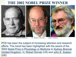
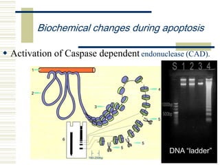
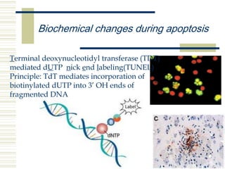
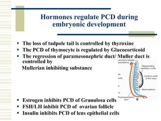
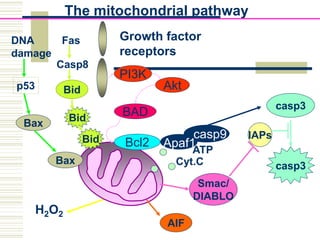
Programmed cell death and
embryonic development
Dept. of Histology & Embryology
Xiangya School of Medicine, CSU
Definition of Programmed Cell Death
 programmed cell death -llec fo epyt a
dezilaiceps sesu llec eht hcihw ni htaed
edicius llec a ;flesti llik ot yrenihcam ralullec
lortnoc ot snaozatem selbane taht msinahcem
netaerht taht sllec etanimile dna rebmun llec
lavivrus s'lamina eht
1885 Flemming first mentioned cell death in rabbit ovarian
follicle (chromatolysis) is physiological, and described
chromatin was condensed into half-moon shape (crescent);
1914 Gräper: Chromatolysis of all cells eliminated from
organs; phagocytosis of neighboring cells; exploring the
phenomenon the organ construction;
1951 Glucksmann reviewed cell death in ontogenesis; a
normal part of animal development, and detailed the
process of chromatolysis;
1964 Lockshin & Williams proposed the concept of
"programmed cell-death" in their research thesis related
to insect (silkworm moth) metamorphosis
Important events in the history
of cell death
1966 Saunders: “The death clock is ticking;” cell death is a
suicide
1972 Kerr, Wyllie, Currie: Apoptosis is general in morphology
and among animals
Important events in the history
of cell death
Sydney Brenner of the Salk Institute used the nematode Caenorhabditis elegans,
which became a multicellular model experimental system, to follow cell division and
differentiation from the fertilized egg to the adult via microscopic observation. He
demonstrated that a specific gene mutation, induced by ethyl methane sulfonate,
could be linked to a specific effect in nematode organ development. His work on
nematodes created an experimental system that laid the foundation for
the study of apoptosis.
John Sulston of the Wellcome Trust Institute in England mapped cell lineages,
where every cell division and differentiation could be followed in the development of
C. elegans. There are only 959 cells in an adult nematode (1090 to 959 cells, 131
cell lost). He showed that specific cells lineages (nerves) undergo programmed cell
death, as an integral part of the normal differentiation process.
Robert Horvitz of MIT discovered and characterized key genes controlling cell
death in C. elegans. He identified the first two bona fide "death genes", ced-3 and
ced-4. Functional ced-3 & ced-4 genes are a prerequisite for cell death to be
executed. Another gene, ced-9, protects against cell death by interacting with ced-3
and ced-4. He has shown how these genes interact with each other in the
cell death process and that corresponding genes (a ced-3-like gene) exists in
humans.
The Nobel Prize in Physiology or Medicine 2002
Sydney Brenner H. Robert Horvitz John Sulston
THE 2002 NOBEL PRIZE WINNER
PCD has been the subject of increasing attention and research
efforts. This trend has been highlighted with the award of the
2002 Nobel Prize in Physiology or Medicine to Sydney Brenner
(United Kingdom), H. Robert Horvitz (US) and John E. Sulston
(UK)
The creation of the conception of
PCD
 Carroll Williams was always known for his colorful
phraseology, and as graduate students we always
tried to emulate him. Because computers were just
beginning to be talked about at the time,
programmed cell death seemed to be a particularly
modern and colorful way of describing what we
saw. It was a metaphor stating what I thought was
pretty obvious — if a biological process occurs at a
defined location and time, then it must in some
fashion be programmed or written into the genetics
of the organism-----from Williams’ student .
PCD and APOPTOSIS
 Apoptosis is used as a synonym for PCD
 Apo: apart
 Ptosis: fallen
 Shedding of leaves from trees
 During embriogenesis ------ occurs as PCD
 Post-embrional life------- as apoptosis
PCD and its significance during
embryonic development
 Optimization of system matching
To regulate the cell number by eliminating wasted, useless,
unwanted, or crippled cells!
For example, 80% nerve cells, 70-95% oocyte died in this way.
 Sculpting of tissues
Such as the formation of the fingers and toes of
the fetus in chicken, mouse or human.
Arrowheads indicate PCD cells. PCD in duck or tortoise
Indicated by arrow
PCD and its significance during
embryonic development
Shortening and rotation
of the myocardial portion
of the OFT(outflow tract
(OFT)) coincident with
cardiomyocyte apoptosis
 Sculpting of tissues
Extensive postnatal apoptosis
occurs in
vessels that regress after birth,
ie, ductus arteriosus and the
umbilical arteries
 Removal of transitional or
useless structures
 Deletion of
transitional
structures
Evolution of aortic arches
 Deletion of transitional structures
Pronephros-
pronephric tubule and
duct
Mesonephros-
mesonephric tubule
and duct
Tadpole tail removed by apoptosis during development
 Deletion of transitional structures
•Autoreactive T cells with the
potential to attack "self" are
removed by apoptosis
Self antigen
recognizing cell
 Essential for the removal of
cells that threaten homeostasis
Morphological changes during apoptosis
Cell shrinkage
Apoptotic B Cells
Hoechst stainingHE staining
Morphological features ofMorphological features of
apoptosisapoptosis
Membrane blebbing
macrophage
Apoptoti
c bodies
Biochemical changes during apoptosis
 Phosphatidylserine inside out
annexin V, a Ca+ dependent PS binding protein
Annexin V staining
flow cytometry
 Activation of Caspase dependent endonuclease (CAD).
Biochemical changes during apoptosis
180-200bp
DNA “ladder”
Biochemical changes during apoptosis
Terminal deoxynucleotidyl transferase (TDT)
mediated dUTP nick end labeling(TUNEL)
Principle: TdT mediates incorporation of
biotinylated dUTP into 3’ OH ends of
fragmented DNA
Biochemical changes during apoptosis
Proteins in favor for apoptosis ↑
 Death receptor superfamily, such as TNFR1
, Fas/APO-1, DR,3,4 and 5, and so on
 Caspase family (cysteine-containing aspartate-specific
protease): Caspase 1-10,13. 11 and 12 species specific
 Apaf-1 (apoptotic protease activating factor-1)
 Mitochondrial membrane releasing factors: Smac/
DIABLO
Proteins against apoptosis ↑
Bcl-2 family (B cell lymphoma/Leukemia-2, Bcl-2), but BAX
subfamily in favor of apoptosis
Cell Death Pathways
Death
Execution
IAPs
Smac/
DIABLO
AIF
Endonuclease
activation
DNA broken
Death
 Harmful factor→ changes of ER Ca2+ stable status
and accumulation of incorrect folding proteins→pro-
caspase-12→ caspase-12→BAX
activated→mitochondria →Cyt C and Apaf1
release→ caspase9 activated→caspase-3
activated→apoptosis
Endoplasmic reticulum (ER) pathway
Hormones regulate PCD during
embryonic development
 The loss of tadpole tail is controlled by thyroxine
 The PCD of thymocyte is regulated by Glucocorticoid
 The regression of paramesonephric duct/ Muller duct is
controlled by
Mullerian inhibiting substance
 Estrogen inhibits PCD of Granulosa cells
 FSH/LH inhibit PCD of ovarian follicle
 Insulin inhibits PCD of lens epithelial cells
Growth factors/cytokins regulate
PCD during embryonic
development
Lack of NGF,FGF ,TGF,G-CSF leads to
PCD
Necrosis
Necrosis vs. Apoptosis
 Cellular condensation
 Membranes remain intact
 Requires ATP
 Cell is phagocytosed, no
tissue reaction
 Ladder-like DNA
fragmentation
 In vivo, individual cells
appear affected
 Active death
• Cellular swelling
• Membranes are broken
• ATP is depleted
• Cell lyses, eliciting an
inflammatory reaction
• DNA fragmentation is
random
• In vivo, whole areas of
the tissue are affected
• Passive death
Necrosis Apoptosis
Questions
 Can you imagine how it likes about DNA gel
electrophoresis from extracts of necrotic tissue?
TUNEL also labels necrotic cells?
 Autophagy, 3rd cell death
31
Receptor pathway (physiological):
Death receptors:
(FAS, TNF-R, etc)
FAS ligand TNF
Death
domains
Adaptor proteins
Pro-caspase 8 (inactive) Caspase 8 (active)
Pro-execution caspase (inactive)
Execution caspase (active)
DeathMITOCHONDRIA
zymogen enzyme
H2O2
Growth factor
receptors
casp9
Bcl2
PI3K
Akt
BAD
Apaf1
Cyt.C
ATP
The mitochondrial pathway
casp3
casp3
IAPs
Smac/
DIABLO
AIF
Bax
Bax
p53
Fas
Casp8
Bid
Bid
Bid
DNA
damage

2013programmed cell dealth r

  • 1.
    Programmed cell deathand embryonic development Dept. of Histology & Embryology Xiangya School of Medicine, CSU
  • 2.
    Definition of ProgrammedCell Death  programmed cell death -llec fo epyt a dezilaiceps sesu llec eht hcihw ni htaed edicius llec a ;flesti llik ot yrenihcam ralullec lortnoc ot snaozatem selbane taht msinahcem netaerht taht sllec etanimile dna rebmun llec lavivrus s'lamina eht
  • 3.
    1885 Flemming firstmentioned cell death in rabbit ovarian follicle (chromatolysis) is physiological, and described chromatin was condensed into half-moon shape (crescent); 1914 Gräper: Chromatolysis of all cells eliminated from organs; phagocytosis of neighboring cells; exploring the phenomenon the organ construction; 1951 Glucksmann reviewed cell death in ontogenesis; a normal part of animal development, and detailed the process of chromatolysis; 1964 Lockshin & Williams proposed the concept of "programmed cell-death" in their research thesis related to insect (silkworm moth) metamorphosis Important events in the history of cell death
  • 4.
    1966 Saunders: “Thedeath clock is ticking;” cell death is a suicide 1972 Kerr, Wyllie, Currie: Apoptosis is general in morphology and among animals Important events in the history of cell death
  • 5.
    Sydney Brenner ofthe Salk Institute used the nematode Caenorhabditis elegans, which became a multicellular model experimental system, to follow cell division and differentiation from the fertilized egg to the adult via microscopic observation. He demonstrated that a specific gene mutation, induced by ethyl methane sulfonate, could be linked to a specific effect in nematode organ development. His work on nematodes created an experimental system that laid the foundation for the study of apoptosis. John Sulston of the Wellcome Trust Institute in England mapped cell lineages, where every cell division and differentiation could be followed in the development of C. elegans. There are only 959 cells in an adult nematode (1090 to 959 cells, 131 cell lost). He showed that specific cells lineages (nerves) undergo programmed cell death, as an integral part of the normal differentiation process. Robert Horvitz of MIT discovered and characterized key genes controlling cell death in C. elegans. He identified the first two bona fide "death genes", ced-3 and ced-4. Functional ced-3 & ced-4 genes are a prerequisite for cell death to be executed. Another gene, ced-9, protects against cell death by interacting with ced-3 and ced-4. He has shown how these genes interact with each other in the cell death process and that corresponding genes (a ced-3-like gene) exists in humans. The Nobel Prize in Physiology or Medicine 2002
  • 6.
    Sydney Brenner H.Robert Horvitz John Sulston THE 2002 NOBEL PRIZE WINNER PCD has been the subject of increasing attention and research efforts. This trend has been highlighted with the award of the 2002 Nobel Prize in Physiology or Medicine to Sydney Brenner (United Kingdom), H. Robert Horvitz (US) and John E. Sulston (UK)
  • 7.
    The creation ofthe conception of PCD  Carroll Williams was always known for his colorful phraseology, and as graduate students we always tried to emulate him. Because computers were just beginning to be talked about at the time, programmed cell death seemed to be a particularly modern and colorful way of describing what we saw. It was a metaphor stating what I thought was pretty obvious — if a biological process occurs at a defined location and time, then it must in some fashion be programmed or written into the genetics of the organism-----from Williams’ student .
  • 8.
    PCD and APOPTOSIS Apoptosis is used as a synonym for PCD  Apo: apart  Ptosis: fallen  Shedding of leaves from trees  During embriogenesis ------ occurs as PCD  Post-embrional life------- as apoptosis
  • 9.
    PCD and itssignificance during embryonic development  Optimization of system matching To regulate the cell number by eliminating wasted, useless, unwanted, or crippled cells! For example, 80% nerve cells, 70-95% oocyte died in this way.
  • 10.
     Sculpting oftissues Such as the formation of the fingers and toes of the fetus in chicken, mouse or human. Arrowheads indicate PCD cells. PCD in duck or tortoise Indicated by arrow PCD and its significance during embryonic development
  • 11.
    Shortening and rotation ofthe myocardial portion of the OFT(outflow tract (OFT)) coincident with cardiomyocyte apoptosis  Sculpting of tissues
  • 12.
    Extensive postnatal apoptosis occursin vessels that regress after birth, ie, ductus arteriosus and the umbilical arteries  Removal of transitional or useless structures
  • 13.
  • 14.
     Deletion oftransitional structures Pronephros- pronephric tubule and duct Mesonephros- mesonephric tubule and duct
  • 15.
    Tadpole tail removedby apoptosis during development  Deletion of transitional structures
  • 16.
    •Autoreactive T cellswith the potential to attack "self" are removed by apoptosis Self antigen recognizing cell  Essential for the removal of cells that threaten homeostasis
  • 17.
    Morphological changes duringapoptosis Cell shrinkage Apoptotic B Cells Hoechst stainingHE staining
  • 18.
    Morphological features ofMorphologicalfeatures of apoptosisapoptosis Membrane blebbing
  • 19.
  • 20.
    Biochemical changes duringapoptosis  Phosphatidylserine inside out annexin V, a Ca+ dependent PS binding protein Annexin V staining flow cytometry
  • 21.
     Activation ofCaspase dependent endonuclease (CAD). Biochemical changes during apoptosis 180-200bp DNA “ladder”
  • 22.
    Biochemical changes duringapoptosis Terminal deoxynucleotidyl transferase (TDT) mediated dUTP nick end labeling(TUNEL) Principle: TdT mediates incorporation of biotinylated dUTP into 3’ OH ends of fragmented DNA
  • 23.
    Biochemical changes duringapoptosis Proteins in favor for apoptosis ↑  Death receptor superfamily, such as TNFR1 , Fas/APO-1, DR,3,4 and 5, and so on  Caspase family (cysteine-containing aspartate-specific protease): Caspase 1-10,13. 11 and 12 species specific  Apaf-1 (apoptotic protease activating factor-1)  Mitochondrial membrane releasing factors: Smac/ DIABLO Proteins against apoptosis ↑ Bcl-2 family (B cell lymphoma/Leukemia-2, Bcl-2), but BAX subfamily in favor of apoptosis
  • 24.
  • 25.
     Harmful factor→changes of ER Ca2+ stable status and accumulation of incorrect folding proteins→pro- caspase-12→ caspase-12→BAX activated→mitochondria →Cyt C and Apaf1 release→ caspase9 activated→caspase-3 activated→apoptosis Endoplasmic reticulum (ER) pathway
  • 26.
    Hormones regulate PCDduring embryonic development  The loss of tadpole tail is controlled by thyroxine  The PCD of thymocyte is regulated by Glucocorticoid  The regression of paramesonephric duct/ Muller duct is controlled by Mullerian inhibiting substance  Estrogen inhibits PCD of Granulosa cells  FSH/LH inhibit PCD of ovarian follicle  Insulin inhibits PCD of lens epithelial cells
  • 27.
    Growth factors/cytokins regulate PCDduring embryonic development Lack of NGF,FGF ,TGF,G-CSF leads to PCD
  • 28.
  • 29.
    Necrosis vs. Apoptosis Cellular condensation  Membranes remain intact  Requires ATP  Cell is phagocytosed, no tissue reaction  Ladder-like DNA fragmentation  In vivo, individual cells appear affected  Active death • Cellular swelling • Membranes are broken • ATP is depleted • Cell lyses, eliciting an inflammatory reaction • DNA fragmentation is random • In vivo, whole areas of the tissue are affected • Passive death Necrosis Apoptosis
  • 30.
    Questions  Can youimagine how it likes about DNA gel electrophoresis from extracts of necrotic tissue? TUNEL also labels necrotic cells?  Autophagy, 3rd cell death
  • 31.
  • 32.
    Receptor pathway (physiological): Deathreceptors: (FAS, TNF-R, etc) FAS ligand TNF Death domains Adaptor proteins Pro-caspase 8 (inactive) Caspase 8 (active) Pro-execution caspase (inactive) Execution caspase (active) DeathMITOCHONDRIA zymogen enzyme
  • 33.
    H2O2 Growth factor receptors casp9 Bcl2 PI3K Akt BAD Apaf1 Cyt.C ATP The mitochondrialpathway casp3 casp3 IAPs Smac/ DIABLO AIF Bax Bax p53 Fas Casp8 Bid Bid Bid DNA damage