- A 53-year-old man presented with 4.5 hours of chest pain radiating to both arms. His EKG showed downsloping ST depression in leads V1-V4 and tall R waves in V3. Posterior leads V7-V9 demonstrated ST elevation.
- He underwent immediate cardiac catheterization, which revealed a total occlusion of the LCA. He received stenting of the LCA.
A 65-year-old man presented to the emergency department with chest pain radiating to his left arm and jaw for 2.5 hours. His EKG showed tall R waves in leads V1-V3 and downsloping ST depression in V1-V4, suggestive of a posterior wall myocardial infarction (MI). Echo revealed akinesia of the posterior left ventricular wall. He received fibrinolytic therapy, which resolved his symptoms. However, he later developed cerebral hemorrhage. The EKG findings, in combination with his risk factors and history, suggested a posterior MI despite the unconventional presentation on EKG.
- A 72-year-old woman presented with chest pain and was found to have cardiovascular risk factors including diabetes, hypertension, and dyslipidemia.
- Her EKG showed normal sinus rhythm, tall R waves in leads V1-V3, and ST segment depression in leads V1-V4. Coronary angiography revealed an occlusion of the proximal left circumflex artery and other lesions.
- Angioplasty and stenting were performed to reopen the occluded vessel and resolve the chest pain. The EKG and angiogram findings were consistent with an inferior-posterior myocardial infarction.
A 52-year-old male presented with chest pain. His ECG showed evolving inferior wall myocardial infarction. ST depression is more frequently seen in lead aVL than other leads for inferior MI. A 51-year-old female presented with prior chest pain and is now pain-free. Her ECG shows Wellens' syndrome type I pattern and she should be monitored closely in the ICCU. Fragmented QRS complexes can indicate ischemia or scar tissue and are associated with worse cardiac outcomes.
This document provides an overview of the various causes of ST elevation on electrocardiograms (ECGs). It discusses electrolyte abnormalities, left bundle branch block, early repolarization, ventricular hypertrophy, aneurysms, infections or injuries, Osborne waves, non-occlusive vasospasm, and Brugada syndrome. Specific ECG patterns are presented for each cause. Key factors to evaluate for each condition include the shape, amplitude, and distribution of ST segments and T waves. Scoring systems like Sgarbossa criteria are also described which can help determine if ST elevation in the setting of left bundle branch block is likely due to acute myocardial infarction.
ST segment elevations can be seen in acute myocardial infarction (AMI) but also have other causes. Non-AMI causes of ST elevation include left bundle branch block, left ventricular hypertrophy, pericarditis, Brugada syndrome, and early repolarization. The morphology, distribution, and magnitude of ST elevations, as well as other ECG features, can help differentiate AMI from other causes of ST elevation. It can be challenging to diagnose AMI using ECG criteria alone, as around half of AMI cases present without typical ST elevation patterns.
Managing Ventricular Arrhythmia In First In Man Studies A Nadaadelnada
The document discusses the increasing need to manage ventricular arrhythmia encountered during early clinical drug development as riskier compounds are advanced. It outlines factors contributing to this issue and strategies for intensive cardiac monitoring in first-in-man studies to ensure safety while accurately assessing potential drug-related arrhythmia events given baseline rates in healthy volunteers. Future directions are highlighted, including expert guidance documents and initiatives to better understand cardiac safety in early development.
Connected aircraft squadron electrocardiographic sign is a new strong index for monitoring and follows up the tachypneic patients with specific T- waves changes in special leads in several cardiorespiratory patients.
This document contains 14 cases with ECG findings and questions. It discusses various cardiac conditions that can present with abnormal ECG patterns, including STEMI, arrhythmias, congenital heart defects, electrolyte imbalances, and more. The key takeaways are: lead reversals can change the appearance of STEMI, sinus bradycardia U waves require specific criteria to diagnose hypokalemia, treating hyperkalemia requires membrane stabilizers followed by agents causing potassium influx, tricuspid atresia is the most common cyanotic congenital heart defect, lead issues can cause pacing problems in STEMI patients, inferior MI with RBBB could indicate distant LAD ischemia, mirror imaging limb leads results in a normal E
Hypocalcemia induced camel-hump t-wave, tee-pee sign, and bradycardia in a ca...YasserMohammedHassan1
Rationale: Electrocardiographic is a fundamental tool for a cardiologist, critical care physician, and emergency medicine specialist. The electrolyte imbalance is a very important entity in clinical medicine management. Camel-hump T-wave and the Tee-Pee sign, recently; Wavy triple and Wavy double signs of hypocalcemia (Yasser’s sign) are electrocardiographic findings linked to electrolyte deficiencies. Patient concerns: A middle-aged male car-painter patient presented to the emergency department with atypical severe twisting chest pain, hypocalcemia, hypokalemia, and hypernatremia.
Diagnosis: Hypocalcemia-induced Camel-hump T-wave, Tee Pee sign, Wavy double sign of hypocalcemia (Yasser’s sign), and bradycardia in a car- painter. Interventions: Electrocardiography, arterial blood gases, oxygenation, and echocardiography. Lessons: The dramatic reversal of Camel-hump T-Wave, Tee-Pee sign, Wavy double sign of hypocalcemia (Yasser’s sign) after calcium gluconate injection interpret that these signs were due to hypocalcemia. The twisting chest pain and its limited disappearance immediately after calcium gluconate injection indicate the pain can be named as “chest tetany”. Non-atropine bradycardia response is evidence that the management of the cause of bradycardia sometimes is essential e.g. hypocalcemia in the current case. Outcomes: There was a dramatic response of both clinical and electrocardiography including Camel-hump T-wave, Tee Pee sign, the wavy double sign of hypocalcemia, and bradycardia.
Electrocardiographic passing phenomenon (flying phenomenon or yasser’s phenom...YasserMohammedHassan1
The document summarizes a presentation on electrocardiographic passing phenomena given at a cardiology conference. It includes 10 case studies demonstrating different types of passing phenomena, such as accelerated junctional rhythm, bigeminy, and sinus arrhythmia. It discusses the historical discovery of these phenomena, how they are analyzed and classified, common target diseases they may indicate but not necessarily be related to, and how reassuring patients can be an effective therapy. Statistics on the study subjects are provided, such as average age and percentages of occupations and main complaints. Serial electrocardiograms are presented for each case study and management primarily involved reassurance without medical intervention.
- Early repolarization is seen in 2-5% of the population and is characterized by J-point elevation and ST-segment slurring or notching, typically in the anterior chest leads.
- While traditionally considered benign, studies have linked early repolarization patterns, especially in the inferior leads, to fatal arrhythmias and sudden cardiac death.
- The prognostic significance may depend on the location and amplitude of the J-point elevation, with inferior locations and elevations over 0.2mV carrying higher risk.
- Early repolarization should not be diagnosed without clinical context and can be difficult to distinguish from conditions like pericarditis based on ECG patterns alone.
Charge syndrome hallmarked with wpws and pda; 20 years post repairing-yasser ...YasserMohammedHassan1
CHARGE syndrome or Hall-Hittner syndrome is a pleiotropic disorder, in which the name is derived from the abbreviation epitomizing its six clinical criteria: ocular coloboma, cardiac defects, choanal atresia, growth or developmental retardation, genital hypoplasia, and ear anomalies or deafness. Wolff-Parkinson-White syndrome is the most frequent pattern of ventricular pre-excitation. Patent ductus arteriosus is one of the most frequent congenital heart diseases due to failure of closure of the ductus arteriosus within 72 hours of birth. CHARGE syndrome, Wolff-Parkinson-White syndrome, and patent ductus arteriosus are so difficult to be present in a single entity.
This document discusses heart failure with preserved systolic function, also known as heart failure with normal ejection fraction. Some key points:
- This condition represents 20-50% of all heart failure cases. However, relatively little is known about its morbidity and mortality.
- Median prevalence in studies was 36%, with a range from 13-74%. Rates of readmission were similar to heart failure with low ejection fraction but mortality was lower at 9% versus 18%.
- Causes include inaccurate diagnosis, episodic systolic dysfunction, diastolic dysfunction from various conditions like hypertension, cardiomyopathy, and aging.
- Diastolic dysfunction refers to abnormally elevated ventricular filling pressures despite normal or
The document discusses differentiating ST elevation myocardial infarction (STEMI) from other causes of ST elevation on an electrocardiogram (ECG). It provides examples of three sample ECGs, describing ECG 1 as showing typical inferior STEMI patterns, ECG 2 as most consistent with pericarditis given its global and concave ST elevation, and ECG 3 showing minimal changes consistent with benign early repolarization. Key factors for differentiation include the magnitude, morphology, distribution of ST elevation, and comparison to previous ECGs. The document emphasizes analyzing ST elevation in the full clinical context and pursuing safe care when in doubt.
1. Ventricular tachycardia (VT) is an important cause of sudden cardiac arrest and can manifest as premature ventricular contractions (PVCs) or sustained VT.
2. VT can be idiopathic, focal, or associated with structural heart disease such as ischemic or non-ischemic cardiomyopathy.
3. Treatment depends on the type and cause of VT but may include medications, implantable cardioverter defibrillators (ICDs), and catheter ablation which can be curative for some idiopathic VT cases.
Sonja discuss the problems with our current paradigm for diagnosing occlusive myocardial infarction by relying predominantly upon ST segment elevation. Watch Sonja present this information at: https://youtu.be/-AkP3I93e8Y
Brugada Syndrome is a genetic cardiac condition characterized by abnormal ECG patterns and risk of sudden cardiac death. It is caused by mutations in the SCN5A gene which encodes cardiac sodium channels. Patients typically present with syncope or sudden death and have a distinctive pattern on ECG of ST segment elevation in leads V1-V3. Risk stratification focuses on history of symptoms like syncope as inducible arrhythmias on electrophysiological study have poor predictive value. Treatment involves medical management with quinidine or device therapy with an ICD for high-risk patients. Lifestyle modifications like avoiding fever or medications that affect sodium channels are also recommended.
Benign early repolarization is seen in 2-5% of the population, usually young physically fit individuals, and is characterized by J-point notching and concave-up ST elevation less than 3mm that decreases with increased heart rate. However, some recent studies have questioned whether it is truly benign, finding an association with increased risk of cardiac events. The diagnosis requires clinical context and exclusion of other possibilities, as features alone do not confirm benign early repolarization.
Movable-weaning off an electrocardiographic phenomenon in hypocalcemia (chang...YasserMohammedHassan1
Movable-weaning off an electrocardiographic phenomenon in hypocalcemia (changeable phenomenon or Yasser’s phenomenon of hypocalcemia) is defined according to the author's opinion in the study as a novel electrocardiographic phenomenon characterized by serial dynamic changes in present in all cases of either Wavy triple or double electrocardiographic signs (Yasser signs) of hypocalcemia. Movable-weaning off an electrocardiographic phenomenon is a guide for both Wavy triple or double electrocardiographic signs (Yasser signs) of hypocalcemia. Don’t be angry if the staring electrocardiography or the last one was normal.
This document discusses STEMI (ST-elevation myocardial infarction). It defines STEMI as irreversible necrosis of heart muscle due to prolonged ischemia. The pathophysiology section explains how ischemia develops and the factors that determine infarct size. Clinical presentation includes symptoms like chest pain and potential physical exam findings. The workup involves an ECG, cardiac biomarkers, and potentially cardiac imaging. Treatment involves aspirin, nitrates, beta blockers, and anticoagulants with the goals of relieving symptoms and initiating reperfusion therapy.
This document provides an overview of 12 lead EKG interpretation and STEMI management. It introduces the presenters, Michael Grzyb and Jeffrey Rupple, and their EMS experience. It reviews benefits of prehospital 12-lead EKG, including recognizing AMI, identifying reperfusion candidates, and reducing time to treatment. Key components of EKG acquisition and interpretation are summarized, including lead placement, indicative leads, and identifying ST elevation indicative of AMI. Coronary artery anatomy and evolution of STEMI on EKG are also reviewed.
The document discusses electrocardiograms (ECGs) in the context of acute coronary syndrome. It begins by describing the normal conduction system and the 12 standard ECG leads. It then explains how ECGs are recorded and the positioning of limb and precordial leads. The document discusses ST segments, T waves, and how to evaluate for ST elevations. It defines acute coronary syndrome and describes the classifications of ST-elevation MI, non-ST-elevation MI, and unstable angina based on ECG and cardiac enzyme findings. Specific ECG patterns for lateral, inferior, septal, and posterior wall MIs are also shown.
ST segment elevations can be seen in acute myocardial infarction (AMI) but also have other causes. Non-AMI causes are more common and include left bundle branch block, left ventricular hypertrophy, pericarditis, early repolarization, and Brugada syndrome. The morphology, distribution, and other ECG features can help differentiate AMI from other causes of ST elevation. It is important to consider other diagnoses fully before attributing ST elevation to AMI.
1) The study aimed to identify the sensitivity and specificity of J-waves and ST-segment changes on ECGs in diagnosing penetrating cardiac injuries in stable trauma patients.
2) They found that J-waves had a sensitivity of 44% and specificity of 85% in detecting cardiac injuries, while ST-segment changes were less specific with a sensitivity of 67% and specificity of 54%.
3) The presence of a J-wave on ECG was a significant indicator of a hemopericardium and suggested further investigation and management at a trauma center may be needed.
1) This document presents a case study of a 61-year-old female patient with shortness of breath, lung cancer with brain metastasis, and bilateral lower limb swelling. Examinations found irregular heart rhythm and crackles in her lungs.
2) Imaging tests including CXR, ventilation scan, perfusion scan, CT scan showed pulmonary thromboembolism with bilateral deep vein thrombosis and SVC thrombosis.
3) The document then discusses using data from the PIOPED II study to determine the sensitivity and specificity of V/Q scintigraphic tests for diagnosing pulmonary embolism. It finds that for acute PE, V/Q scans have a sensitivity of 77.4% and specificity of 97
1. The document discusses the nursing process and its key steps: assessment, nursing diagnosis, planning, implementation, and evaluation. It emphasizes that critical thinking is essential for each step.
2. Key aspects of critical thinking discussed include reflection, intuition, problem-solving, decision-making, and developing critical thinking skills and attitudes.
3. The nursing process provides a systematic framework for nurses to gather data, analyze it, identify issues, design goals and interventions, take action, and evaluate outcomes. It requires ongoing assessment, modification of the care plan as needed, and reevaluation until goals are met.
This document discusses patients' rights in intensive care units. It begins with an introduction to patients' rights as basic rules of conduct between patients and medical caregivers. The talk will discuss some of the ethical considerations around patients' rights while in intensive care, including issues around death, consent, age of consent, research, costs and access. It notes frameworks for ethical principles and discusses New Zealand's National Health and Disability Commissioner, which outlines 10 rights for all people interacting with health services. The document discusses how these rights apply in intensive care settings in New Zealand, as well as responsibilities, particular problems like consent and organ donation, international variations, and how to address ethical dilemmas.
Campbell University is proposing to build a new 70,000 square foot Nursing and Health Science Facility to address North Carolina's growing healthcare needs. The facility will house new Nursing, Physical Therapy, and Medical Research programs. North Carolina faces shortages in nurses and physical therapists, and its aging population will further increase demand for healthcare services. The new facility will allow Campbell to educate more students in these critical fields through state-of-the-art instructional spaces and innovative interprofessional programs. Campbell has a history of leadership in healthcare education and is well-positioned to help meet North Carolina's needs through expanded nursing, physical therapy, and research initiatives.
Nursing 203 week 1 First 10 Chapters of UrdenCheri Rievley
This document outlines a syllabus for a critical care nursing course. It covers topics such as critical care nursing roles, the importance of holistic care, applying the nursing process, comparing interdisciplinary management models, addressing moral distress, and examining specific legal issues in critical care. Teaching and learning strategies are discussed, including assessing patient and family informational needs, factors affecting patient readiness to learn, and strategies to enhance education. The needs of older adult patients are also addressed.
This document summarizes a study evaluating the use of D-dimer testing and clinical risk algorithms to diagnose pulmonary embolism (PE). The study assessed 627 patients presenting with suspected PE who were stratified into low, intermediate, or high risk groups using the Geneva score. For low and intermediate risk patients, a quantitative D-dimer assay achieved 100% sensitivity and negative predictive value, correctly ruling out PE in 25% and 33% of cases respectively. The study supports using the D-dimer assay as a first-line test for low to intermediate risk PE patients to reduce unnecessary CT scans.
The document discusses various topics related to the respiratory system including:
1. Terminologies used in ventilation and respiration like ventilation, respiration, oxygenation, perfusion.
2. A case study of a patient presenting with respiratory distress and hemoptysis.
3. Review of anatomy and physiology of the respiratory system.
4. Nursing assessment and management of patients with respiratory problems including diagnostic tests, oxygen therapy, and mechanical ventilation.
This document discusses the cardiovascular system and heart failure. It provides details on diagnostic tests related to the cardiovascular system including complete blood count, coagulation tests, and chest x-rays. It then covers symptoms of heart failure such as dyspnea, edema, fatigue, and chest pain. The causes, pathophysiology, and clinical manifestations of left and right heart failure are summarized.
1. An 80-year-old man was admitted to the MICU with pneumonia and a history of old CVA and hypertension.
2. A chest x-ray showed airspace consolidation in the right lung consistent with bacterial pneumonia.
3. The patient received antibiotics, oxygen therapy, and nursing care focused on improving respiratory status and reducing infection risk.
This document summarizes a case of a 78-year-old male patient presenting with fatigue, dizziness, and chest pain during exercise. Clinical examination revealed an irregular heartbeat, signs of fluid in the lungs, and swelling in the feet. Testing showed atrial fibrillation (AF), an irregular heartbeat caused by rapid electrical signals in the upper chambers of the heart. The summary reviews anatomy of the heart and potential differential diagnoses of COPD, pulmonary embolism, and ventricular hypertrophy that could be causing the patient's symptoms.
This case study describes the end-of-life care of Lorna, a 77-year old woman with metastatic renal cell carcinoma. Lorna originally presented with flank and abdominal pain and was diagnosed with advanced renal cell carcinoma in 2011. In 2014, the cancer recurred and metastasized to her spine, causing cauda equina syndrome with lower limb weakness and urinary/fecal incontinence. She was admitted to palliative care for pain and symptom management, where she received psychological support, medication via syringe pump, and focus on comfort. The document discusses renal cell carcinoma, cauda equina syndrome, medications, nursing interventions and compassionate end-of-life care.
This document discusses airway secretion clearance techniques in the ICU, including mechanical insufflation-exsufflation (MIE). It provides a timeline of MIE devices including the CoughAssist. A case study describes how MIE was used successfully via face mask in an 18-year-old post-op patient to avoid intubation. Typical treatment protocols for the CoughAssist E-70 are outlined. Studies show MIE can improve respiratory parameters and allow extubation of restrictive patients to noninvasive ventilation. The evidence suggests MIE is safe and effective for both obstructive and restrictive lung diseases.
Deep Vein Thrombosis and Pulmonary Embolism, by Prof. Minnu M. PanditraoMinnu Panditrao
Dr. Mrs. Minnu Panditrao, goes in depth with the very important topic of Deep Vein Thrombosis, Pulmonary embolism, aetio patheogenesis, clinical features, management etc.
This document provides information on pulmonary embolism including its definition, incidence, types, etiology, risk factors, pathophysiology, clinical manifestations, diagnostic evaluation, complications, management, nursing management, prevention, health education, and nursing diagnoses. Pulmonary embolism is a blockage in the lungs caused by a blood clot that travels from the legs or pelvis and obstructs a pulmonary artery. It can be life-threatening and requires prompt diagnosis and treatment.
Nursing case management and critical pathways of carepanthanalil
This document discusses nursing case management and critical pathways of care. It defines case management as a method to coordinate health care services and control costs. The key components of case management include case finding, assessment, care planning, and care coordination. Critical pathways are defined as anticipated care plans with goals and timelines for different health professionals. They standardize care for common conditions and aim to improve outcomes. The document outlines the roles of nurses as case managers and characteristics of effective case management programs and critical pathways.
The patient was admitted to the orthopaedic trauma ward after a motorbike accident. Initial testing ruled out DVT but he later developed shortness of breath. Additional imaging found a large PE in his lung. He was started on anticoagulation therapy with heparin and later warfarin which led to a full recovery.
A 60-year-old Chinese woman presented with chest pain. She had a history of hypertension and diabetes for 10 years. Her chest pain was central, tight, and radiated to her jaw and left arm. She experienced nausea, sweating, and dyspnea. Physical exam revealed she was obese. Electrocardiogram showed ST segment depression and T inversion. Blood tests showed elevated cardiac enzymes. She was diagnosed with a non-ST elevation myocardial infarction based on her symptoms, risk factors, and test results.
Inspection of the eyes reveals hollowness indicating volume deficiency of fat within the orbit related to the patient's severe malnutrition.
B. EYES
1. Conjunctiva Inspection Pink, moist, clear Conjunctiva pale Abnormal, pale conjunctiva may be due
conjunctiva pink in color to anemia
2. Pupils Inspection Equal, round, Equal, round, Normal
reactive to light reactive to light
3. Eye movements Observation Full range of Full range of Normal
motion in all motion in all
directions of gaze directions of gaze
C. EARS
1. External
Nursing management patient with Myocardial infraction ANILKUMAR BR
1) The document discusses nursing management of patients experiencing myocardial infarction (MI or heart attack).
2) MI occurs when an area of the heart muscle is permanently damaged due to reduced blood flow in a coronary artery, usually caused by a ruptured atherosclerotic plaque blocking the artery.
3) Nursing management of MI focuses on minimizing damage to heart muscle, preserving heart function, preventing complications, and educating patients on risk factor modification and self-care post-MI.
EMGuideWire's Radiology Reading Room: Stress-Induced CardiomyopathySean M. Fox
The Department of Emergency Medicine at Carolinas Medical Center is passionate about education! Dr. Michael Gibbs is a world-renowned clinician and educator and has helped guide numerous young clinicians on the long path of Mastery of Emergency Medical Care. With his oversight, the EMGuideWire team aim to help augment our understanding of emergent imaging. You can follow along with the EMGuideWire.com team as they post these educational, self-guided radiology slides or you can also use this section to learn more in-depth about specific conditions and diseases. This Radiology Reading Room pertains to Stress-Induced Cardiomyopathy and is brought to you by Jenna Pallansch, MD, Claire Lawson, NP, Shelby Hixson, PA, Emily Lipsitz, PA, Ashley Moore-Gibbs, DNP, Laszlo Littmann, MD, and John Symanski, MD.
1) The document presents 7 cases involving ECG patterns that can indicate acute coronary syndrome without ST elevation. Case 1 describes tall, symmetric T waves in the precordial leads indicating a possible left anterior descending artery occlusion. Case 2 shows biphasic T waves in leads V2-3 consistent with Wellens' syndrome. Case 3 demonstrates deeply inverted T waves in leads V2-3 also indicative of Wellens' syndrome.
2) Case 4 shows ECG changes in leads V1-3 consistent with a posterior myocardial infarction. Case 5 highlights ST elevation in lead aVR that can indicate a left main or proximal left anterior descending artery occlusion. Case 6 applies the Sgarbossa criteria to identify a possible infarction in
The document discusses common arrhythmias seen in emergency settings, including bradycardia and tachycardia. It covers the classification, mechanisms, diagnosis and treatment of various arrhythmias like sinus bradycardia, heart blocks, supraventricular tachycardia, ventricular tachycardia and fibrillation. Diagnostic tests mentioned include 12-lead ECG, exercise stress testing, Holter monitoring and implanted cardiac monitors. Treatment depends on the type of arrhythmia and includes atropine, pacing, cardioversion, defibrillation and drugs.
Presentation the electrocardiogram in the acs patientdrwaque
This document discusses high-risk ECG presentations that do not meet criteria for STEMI but still indicate acute myocardial infarction (AMI). It presents six case studies with ECG patterns including: ST elevation in aVL and V2 indicating D1 lesion; de Winter finding of ST depression in V2-V5 with T waves in V2-V4 indicating proximal LAD occlusion; ST elevation in aVR with widespread ST depression indicating left main coronary artery occlusion; Wellen's syndrome biphasic T waves in V1-V4 indicating proximal LAD occlusion; and ST depression in V2-V4 with tall R waves indicating posterior wall AMI. The document emphasizes that ECGs must be interpreted in clinical context and
This document summarizes an ECG review presentation given by Dr. Eric Hockstad. It begins with an overview of ECG basics including components like the P wave, PR interval, QRS complex, ST segment, T wave, and QT interval. It then covers various cardiac conditions and how they present on ECG such as heart block, arrhythmias, bundle branch blocks, ST segment changes, and more. Examples are provided of ECGs demonstrating STEMI and cath lab images. Clinical cases are also presented and summarized with ECG findings, treatment outcomes, and teaching points.
STEMI Mimic WHAT IS IT AND HOW TO IDENTIFY IT ?Haitham Habtar
The document discusses several STEMI mimics that can present with ST segment elevation on ECG but are not actually caused by an acute myocardial infarction. These include early repolarization, left bundle branch block, electrolyte abnormalities, left ventricular hypertrophy, pulmonary embolism, left ventricular aneurysm, Brugada syndrome, pericarditis, and hypothermia. It provides details on the characteristic ECG patterns and clinical features that can help differentiate these conditions from a true STEMI.
A 33-year-old man presented to the emergency department after collapsing. His ECG showed Brugada pattern, which is characterized by ST-segment elevation in leads V1-V3 and increased risk of ventricular arrhythmias and sudden cardiac death. Brugada syndrome is a genetic condition caused by sodium channel mutations and commonly presents with syncope or cardiac arrest in young males. The diagnosis can be confirmed with ajmaline/flecainide provocation test showing transient Brugada pattern. Treatment involves lifestyle modifications and implantable cardioverter-defibrillator for high-risk patients.
ECG in Emergency Department - Advances in ACS ECGDr.Mahmoud Abbas
ECG in Emergency Department -Advances in ACS ECG. Lecture presented by Dr Hesham Ibrahim at the Egyptian Critical Care Summit , the leading educational event and medical exhibition in Egypt.
This document provides an overview of acute coronary syndrome (ACS). It begins with a review of coronary artery anatomy and variations. It then discusses the presentations of ACS, including ischemic chest pain and equivalents. The main types of ACS - unstable angina, NSTEMI, and STEMI - are defined based on symptoms, electrocardiogram findings, and cardiac biomarker levels. Diagnosis and management strategies are outlined, including reperfusion therapies and drug treatments. Follow-up care after ACS and indications for procedures like cardiac catheterization and ICD placement are also summarized.
ST-segment Depression: All are Not Created Equal!asclepiuspdfs
ST depression on an electrocardiography can be from various causes including ischemia, acute coronary syndrome, electrolyte imbalance, posterior myocardial infarction, pulmonary embolism and others. Making the right diagnosis and therefore the right treatment is of paramount importance. This article goes into depth explaining why all ST-segment depressions are not created equal.
Wellens’ Syndrome: Exception to the Rule: One Referral at a Time!asclepiuspdfs
We describe two patients with Wellens’ syndrome. In these patients, the electrocardiogram changes must be recognized promptly and accurately. These cases are managed aggressively and early invasive treatment approach is recommended to avoid myocardial infarction and death.
The document provides information on the diagnosis and types of acute myocardial infarction (AMI). It discusses coronary atherosclerosis as a chronic disease that progresses over decades. Acute coronary syndrome (ACS) refers to the spectrum of clinically manifest coronary artery disease from unstable angina to AMI. The criteria for diagnosing AMI include elevated cardiac biomarkers and evidence of myocardial ischemia. The document outlines 5 types of AMI and provides details on interpreting electrocardiograms and cardiac biomarkers in AMI.
This document provides an overview of acute aortic dissection, including:
- The definition and pathophysiology of acute aortic dissection.
- Risk factors for acute aortic dissection include conditions like Marfan syndrome and known thoracic aortic aneurysms. High-risk symptoms include abrupt onset of severe, ripping chest or back pain.
- Imaging techniques like CT, MRI, and TEE can diagnose acute aortic dissection but no single test is definitive. Early management focuses on hemodynamic stabilization and expediting surgery for type A dissections.
This document discusses various ECG patterns that are considered STEMI equivalents including:
1. Hyperacute T waves, T wave elevation in V1, Wellen's syndrome, and De Winter ST/T complex which indicate proximal LAD occlusion.
2. Posterior MI suggested by ST depression in V1-V3 with tall R waves.
3. ST elevation in aVR indicates left main or triple vessel disease and predicts need for CABG.
4. LBBB with Sgarbossa criteria can help identify STEMI when LBBB is present based on concordant or discordant ST changes.
Serial ECGs are important for evolving patterns in some STEMI equivalents.
This case report describes a 76-year-old female patient who presented with chest pain and was found to have Wellens syndrome, characterized by biphasic or deeply inverted T waves in the precordial leads. Further workup revealed a 95% stenosis in the proximal left anterior descending artery (LAD), which was treated with coronary artery bypass grafting. Wellens syndrome is defined as specific ECG abnormalities associated with a critical LAD stenosis. It often progresses rapidly to anterior wall myocardial infarction if not treated urgently with angiography and revascularization. This case highlights the importance of recognizing the subtle ECG findings of Wellens syndrome to identify high-risk patients and prevent adverse cardiac events.
Atteinte cardiaque au cours de l’hémorragie sous archnoidienne post traumatiq...abbouamine
This study prospectively examined 35 patients with traumatic subarachnoid hemorrhage (tSAH) to assess the incidence of cardiac complications and the predictive value of electrocardiogram (ECG) abnormalities and troponin Ic levels on mortality. The mean age was 39 years and most patients exhibited ECG changes, particularly T-wave abnormalities. Troponin Ic levels peaked on day 3 and were elevated in 34% of patients. ECG changes and elevated troponin levels correlated with worse outcomes on CT scans and higher mortality. Both were independent predictive factors for mortality in tSAH patients. The study demonstrated that ECG abnormalities and cardiac enzyme levels commonly occur after tSAH and can help identify
Ventricular Arrhythmias in Cardiac Amyloidosis.pdfSolidaSakhan
This document provides an overview of ventricular arrhythmias in cardiac amyloidosis. It discusses that arrhythmias in cardiac amyloidosis can include atrial arrhythmias, AV block, or ventricular arrhythmias. The clinical significance of ventricular arrhythmias is variable, with some studies finding they predict prognosis and others not. Predictors of ventricular tachycardia in cardiac amyloidosis include a history of congestive heart failure, presyncope or syncope, and structural heart abnormalities seen on echocardiogram or cardiac MRI like reduced ejection fraction or increased fibrosis. Electrophysiology studies can help determine conduction disease risk and guide therapies.
The document summarizes key information from a case presentation on a 69-year-old male who presented with cardiogenic shock due to a myocardial infarction. The summary includes:
1) The patient presented with left arm numbness, profuse sweating, vomiting and became cold and clammy. Examination found him restless with a pulse of 110/min, blood pressure of 80/50 and other signs of shock.
2) An EKG found ST segment changes consistent with left main coronary artery disease. Laboratory tests showed elevated markers indicating a heart attack.
3) The patient was diagnosed with an acute myocardial infarction complicated by cardiogenic shock, likely due to left main occlusion. He deteriorated and died
Similar to 2007 terni, workshop interattivo, caso clinico 3 (20)
This document discusses electrolytes and their role and regulation in the body. The main electrolytes discussed are sodium, potassium, calcium, magnesium, and chloride. Sodium and chloride are the major electrolytes in extracellular fluid and help regulate osmotic balance and membrane potentials. Potassium is the major intracellular cation and plays a key role in resting membrane potential and action potentials. Calcium and magnesium are also discussed along with their regulation by hormones like parathyroid hormone and functions. Disturbances in electrolyte levels can affect cardiac function, action potentials, and conduction.
This document discusses in-hospital cardiac arrest. It provides epidemiological data showing that cardiac arrest is a major cause of mortality, with 65,000 cases per year in Italy alone. The incidence of in-hospital cardiac arrest is about 1 per 1000 patients. Early defibrillation and effective cardiopulmonary resuscitation are critical for survival, with outcomes declining rapidly after 4-6 minutes without circulation. Unmonitored ward areas account for the majority of in-hospital cardiac arrests, suggesting many could be avoided with improved monitoring of at-risk patients.
2009 terni, università di medicina, i farmaci nel trattamento delle tachicar...Centro Diagnostico Nardi
This document discusses drugs used to treat ventricular tachyarrhythmias. It begins by describing cardiac electrophysiology, including the cardiac action potential and ion channels. It then discusses various classes of antiarrhythmic drugs, including class I drugs that block sodium channels, class II drugs that block beta-adrenergic receptors, class III drugs that prolong the action potential by blocking potassium channels, and class IV drugs that block calcium channels. The document emphasizes that while antiarrhythmic drugs can effectively treat arrhythmias, they may also cause arrhythmias as a side effect if not carefully monitored.
2009 roma, corso ablation frontiers nell'ablazione della fibrillazione atrialeCentro Diagnostico Nardi
The document describes limitations of current single electrode catheter technology for atrial fibrillation ablation including inefficient power delivery and lack of control over lesion creation. It then introduces a multi-electrode catheter system that offers selectable bipolar and unipolar radiofrequency energy delivery modes to tailor lesion size and depth for more efficient procedures. The system includes anatomically designed catheters for mapping, pacing and ablation from multiple electrodes with a single placement.
- The document discusses tools and techniques for achieving pulmonary vein isolation (PVI) for atrial fibrillation (AF) ablation.
- It describes the limitations of conventional anatomical approaches, which often fail to achieve complete PVI in over 60% of patients.
- A new multi-electrode ablation system is proposed that uses anatomically designed catheters, a multi-channel RF generator, and selectable energy delivery modes to more efficiently create tailored lesions and achieve PVI in less time with fewer complications than conventional approaches.
2009 ferrara, congresso regionale, i tools da raggiungere nell'ablazione dell...Centro Diagnostico Nardi
1) The document discusses tools and techniques for achieving pulmonary vein isolation (PVI) to treat atrial fibrillation, including efficacy and safety data from multiple studies and techniques.
2) Mapping and ablation technologies have advanced, including 3D mapping systems, cryoballoon ablation, and multi-electrode catheters, improving identification of arrhythmogenic substrates and tailored lesion formation.
3) Large surveys of AF ablation outcomes show success rates without antiarrhythmic drugs of 74.9-84% for paroxysmal AF, 74.8% for persistent AF, and 71% for permanent AF, with overall complication rates of 4.54%. Advancing technologies may further improve results.
2009 castel volturno, congresso sicoa regionale, l'ablazione della fibrillazi...Centro Diagnostico Nardi
This document discusses ablation techniques for atrial fibrillation. It provides an overview of the state of the art for AF ablation, including different mapping and ablation technologies used. It summarizes findings from randomized controlled trials comparing rate and rhythm control strategies. The document also discusses electrical, contractile, and structural remodeling due to AF and the relationship between AF and congestive heart failure. Ablation of AF in congestive heart failure patients is discussed, including studies showing improvement in left ventricular function and outcomes.
2009 bologna, workshop interattivo. strategie ablative nel trattamento delle ...Centro Diagnostico Nardi
Left atrial tachycardia can be treated using catheter ablation guided by electrophysiological and anatomical mapping. Electrophysiological mapping involves identifying the earliest site of activation to target focal tachycardias, or locating protected isthmuses to target macroreentrant tachycardias. Anatomical mapping with 3D systems helps visualize barriers and isthmuses, improving localization of ablation targets. Success rates are limited by challenges locating all active circuits, but outcomes are better than drugs and ablation may improve left ventricular function.
2009 acerra, congresso regionale sicoa, la terapia elettrica dello scompenso ...Centro Diagnostico Nardi
This document discusses cardiac resynchronization therapy (CRT) in patients with standard pacing indications and in patients with dilated cardiomyopathy (DCM) and short QRS durations. It notes that CRT can reduce pulmonary capillary wedge pressure and improve cardiac output in these patient populations compared to right ventricular apical pacing, which mimics left bundle branch block and has detrimental effects on left ventricular function and remodeling. The document advocates for CRT to correct delayed ventricular activation and contraction seen in heart failure patients.
2008 terni, workshop interattivo, tecniche di impianto dei pacemaker in urgenzaCentro Diagnostico Nardi
The document provides guidance on temporary pacemaker implantation in emergency situations. It discusses the principles and indications for temporary pacing in various bradyarrhythmias and conduction blocks. Specific recommendations are given for temporary pacing in sinus bradycardia, atrioventricular blocks, and intraventricular blocks due to various causes. Complications of temporary pacing like failure to capture, oversensing, and undersensing are also reviewed. The document emphasizes the importance of confirming electrical and mechanical capture when using a temporary pacemaker.
2008 roma, convegno updating in cardiologia. l'ablazione della fibrillazione ...Centro Diagnostico Nardi
This document discusses ablation techniques for atrial fibrillation. It begins by describing the anatomical considerations for ablation, including pulmonary vein anatomy and the importance of reconstructing the virtual geometry. It then discusses different ablation strategies such as pulmonary vein trigger ablation and substrate modification. The document highlights some of the challenges and pitfalls of atrial fibrillation ablation. It concludes by emphasizing the importance of selecting appropriate patients for ablation and properly defining success criteria and long-term follow-up.
2007 venezia, congresso mondiale, ablazione delle tachicardie ventricolariCentro Diagnostico Nardi
This document discusses mapping and ablation of post-ischemic sustained ventricular tachycardias using the EnSite System. It describes how the system can be used to create a virtual geometry of the left ventricle, identify target zones related to slow conduction and scar tissue, and guide radiofrequency ablation lesions to non-inducible critical isthmuses. Results from 24 patients found the combined electroanatomical and electrophysiological mapping approach identified the operative mechanism in 88% of cases and abolished inducible ventricular tachycardias in 85% of patients acutely.
Histololgy of Female Reproductive System.pptxAyeshaZaid1
Dive into an in-depth exploration of the histological structure of female reproductive system with this comprehensive lecture. Presented by Dr. Ayesha Irfan, Assistant Professor of Anatomy, this presentation covers the Gross anatomy and functional histology of the female reproductive organs. Ideal for students, educators, and anyone interested in medical science, this lecture provides clear explanations, detailed diagrams, and valuable insights into female reproductive system. Enhance your knowledge and understanding of this essential aspect of human biology.
TEST BANK For Community and Public Health Nursing: Evidence for Practice, 3rd...Donc Test
TEST BANK For Community and Public Health Nursing: Evidence for Practice, 3rd Edition by DeMarco, Walsh, Verified Chapters 1 - 25, Complete Newest Version TEST BANK For Community and Public Health Nursing: Evidence for Practice, 3rd Edition by DeMarco, Walsh, Verified Chapters 1 - 25, Complete Newest Version TEST BANK For Community and Public Health Nursing: Evidence for Practice, 3rd Edition by DeMarco, Walsh, Verified Chapters 1 - 25, Complete Newest Version Test Bank For Community and Public Health Nursing: Evidence for Practice 3rd Edition Pdf Chapters Download Test Bank For Community and Public Health Nursing: Evidence for Practice 3rd Edition Pdf Download Stuvia Test Bank For Community and Public Health Nursing: Evidence for Practice 3rd Edition Study Guide Test Bank For Community and Public Health Nursing: Evidence for Practice 3rd Edition Ebook Download Stuvia Test Bank For Community and Public Health Nursing: Evidence for Practice 3rd Edition Questions and Answers Quizlet Test Bank For Community and Public Health Nursing: Evidence for Practice 3rd Edition Studocu Test Bank For Community and Public Health Nursing: Evidence for Practice 3rd Edition Quizlet Test Bank For Community and Public Health Nursing: Evidence for Practice 3rd Edition Stuvia Community and Public Health Nursing: Evidence for Practice 3rd Edition Pdf Chapters Download Community and Public Health Nursing: Evidence for Practice 3rd Edition Pdf Download Course Hero Community and Public Health Nursing: Evidence for Practice 3rd Edition Answers Quizlet Community and Public Health Nursing: Evidence for Practice 3rd Edition Ebook Download Course hero Community and Public Health Nursing: Evidence for Practice 3rd Edition Questions and Answers Community and Public Health Nursing: Evidence for Practice 3rd Edition Studocu Community and Public Health Nursing: Evidence for Practice 3rd Edition Quizlet Community and Public Health Nursing: Evidence for Practice 3rd Edition Stuvia Community and Public Health Nursing: Evidence for Practice 3rd Edition Test Bank Pdf Chapters Download Community and Public Health Nursing: Evidence for Practice 3rd Edition Test Bank Pdf Download Stuvia Community and Public Health Nursing: Evidence for Practice 3rd Edition Test Bank Study Guide Questions and Answers Community and Public Health Nursing: Evidence for Practice 3rd Edition Test Bank Ebook Download Stuvia Community and Public Health Nursing: Evidence for Practice 3rd Edition Test Bank Questions Quizlet Community and Public Health Nursing: Evidence for Practice 3rd Edition Test Bank Studocu Community and Public Health Nursing: Evidence for Practice 3rd Edition Test Bank Quizlet Community and Public Health Nursing: Evidence for Practice 3rd Edition Test Bank Stuvia
Local Advanced Lung Cancer: Artificial Intelligence, Synergetics, Complex Sys...Oleg Kshivets
Overall life span (LS) was 1671.7±1721.6 days and cumulative 5YS reached 62.4%, 10 years – 50.4%, 20 years – 44.6%. 94 LCP lived more than 5 years without cancer (LS=2958.6±1723.6 days), 22 – more than 10 years (LS=5571±1841.8 days). 67 LCP died because of LC (LS=471.9±344 days). AT significantly improved 5YS (68% vs. 53.7%) (P=0.028 by log-rank test). Cox modeling displayed that 5YS of LCP significantly depended on: N0-N12, T3-4, blood cell circuit, cell ratio factors (ratio between cancer cells-CC and blood cells subpopulations), LC cell dynamics, recalcification time, heparin tolerance, prothrombin index, protein, AT, procedure type (P=0.000-0.031). Neural networks, genetic algorithm selection and bootstrap simulation revealed relationships between 5YS and N0-12 (rank=1), thrombocytes/CC (rank=2), segmented neutrophils/CC (3), eosinophils/CC (4), erythrocytes/CC (5), healthy cells/CC (6), lymphocytes/CC (7), stick neutrophils/CC (8), leucocytes/CC (9), monocytes/CC (10). Correct prediction of 5YS was 100% by neural networks computing (error=0.000; area under ROC curve=1.0).
Promoting Wellbeing - Applied Social Psychology - Psychology SuperNotesPsychoTech Services
A proprietary approach developed by bringing together the best of learning theories from Psychology, design principles from the world of visualization, and pedagogical methods from over a decade of training experience, that enables you to: Learn better, faster!
Muktapishti is a traditional Ayurvedic preparation made from Shoditha Mukta (Purified Pearl), is believed to help regulate thyroid function and reduce symptoms of hyperthyroidism due to its cooling and balancing properties. Clinical evidence on its efficacy remains limited, necessitating further research to validate its therapeutic benefits.
8 Surprising Reasons To Meditate 40 Minutes A Day That Can Change Your Life.pptxHolistified Wellness
We’re talking about Vedic Meditation, a form of meditation that has been around for at least 5,000 years. Back then, the people who lived in the Indus Valley, now known as India and Pakistan, practised meditation as a fundamental part of daily life. This knowledge that has given us yoga and Ayurveda, was known as Veda, hence the name Vedic. And though there are some written records, the practice has been passed down verbally from generation to generation.
These lecture slides, by Dr Sidra Arshad, offer a simplified look into the mechanisms involved in the regulation of respiration:
Learning objectives:
1. Describe the organisation of respiratory center
2. Describe the nervous control of inspiration and respiratory rhythm
3. Describe the functions of the dorsal and respiratory groups of neurons
4. Describe the influences of the Pneumotaxic and Apneustic centers
5. Explain the role of Hering-Breur inflation reflex in regulation of inspiration
6. Explain the role of central chemoreceptors in regulation of respiration
7. Explain the role of peripheral chemoreceptors in regulation of respiration
8. Explain the regulation of respiration during exercise
9. Integrate the respiratory regulatory mechanisms
10. Describe the Cheyne-Stokes breathing
Study Resources:
1. Chapter 42, Guyton and Hall Textbook of Medical Physiology, 14th edition
2. Chapter 36, Ganong’s Review of Medical Physiology, 26th edition
3. Chapter 13, Human Physiology by Lauralee Sherwood, 9th edition
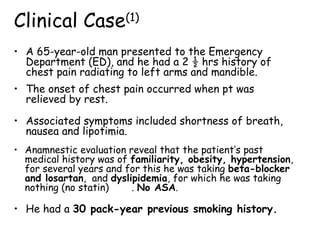
1. • A 65-year-old man presented to the Emergency
Department (ED), and he had a 2 ½ hrs history of
chest pain radiating to left arms and mandible.
Clinical Case(1)
• Anamnestic evaluation reveal that the patient’s past
medical history was of familiarity, obesity, hypertension,
for several years and for this he was taking beta-blocker
and losartan, and dyslipidemia, for which he was taking
nothing (no statin) . No ASA.
• He had a 30 pack-year previous smoking history.
• The onset of chest pain occurred when pt was
relieved by rest.
• Associated symptoms included shortness of breath,
nausea and lipotimia.
2. Clinical Case(1)
• Personal history reveal a previous AMI in ’91, treated
(HSR) with PCA on LAD artery and Posterior Inter-
ventricular right (PIV) artery whereas a pathologic
Obtuse Marginal Artery (OMA) was not treated
because complicated (no documentation).
• Pt reveal a subsequent coronary angiography evalutation in ’98
that confirm the patologic stenosis of OMA (no documentation)
with subsequent further indication to stress-test evalutation.
• Moreover, according with his cardiologist, no further stress test
evaluation was performed until now (9 yrs ago).
4. Clinical Case(1)
- R wave ≥0.04s in V1 or V2
- ST segment depr. V1 V3
- R/S ratio ≥1 in V1 and V2.
EKG (Emergency Departement)
R
R
R
S
S
• BP was 150/80 mmHg, and
HR was 85/min, whereas
the rest of the exam was
unremarkable.
5. Clinical Case(1)
• First Line Therapy
- Oxigen
- Nitrate (iv)
- ASA 500 mg (iv)
- Eparin (bolus + cont.
infusion iv)
• No changes in symptom
• An ECHO evaluation
reveal Total Ackinesia of
apex and posterior LV wall
R
R
R
S
S
Intensive CCU
6. • Turn ECG upside down and
look at it from the back.
• Changes in V1 and V2 which
might be over-looked at
first glance, will be seen as
abnormal Q waves, ST
elevation and increased T
wave inversion(2)
.
• R>0.04s and R≥S V1, showed
a high specificity (>99%)
and a high positive
predictive value (91%).
• R≥0.04s and R≥S V2, showed
95% of specificity, and 73%
positive predictive value.
Clinical Case(1)
Intensive CCU
7. • Turn ECG upside down and
look at it from the back.
• Changes in V1 and V2 which
might be over-looked at
first glance, will be seen as
abnormal Q waves, ST
elevation and increased T
wave inversion(2)
.
• R>0.04s and R≥S V1, showed
a high specificity (>99%)
and a high positive
predictive value (91%).
• R≥0.04s and R≥S V2, showed
95% of specificity, and 73%
positive predictive value.
Clinical Case(1)
Intensive CCU
8. • Turn ECG up-side down and look at it from the
back.
• Changes in V1 and V2 which might be over-looked
at first glance, will be seen as abnormal Q
waves, ST elevation and increased T wave
inversion(2)
.
• R>0.04s and R≥S V1, showed a high specificity
(>99%) and a high positive predictive value (91%).
• R≥0.04s and R≥S V2, showed 95% of specificity,
and 73% positive predictive value.
Clinical Case(1)
• Tall R waves in anterior leads (V1, V2 and V3)
are the electrical equivalent of Q waves in
the posterior leads (V7, V8, V9) (1,2)
10. Clinical Case(1-2)
• Tall R waves in
anterior leads (V1, V2
and V3) are the
electrical equivalent
of Q waves in the
posterior leads (V7,
V8, V9) (1,2)
• Turn ECG upside
down and look at
it from the back.
12. Clinical Case(1)
• No absolute or relative
CI were present to
fibrinolitic therapy, and
then was administrated
with immediately
disappeared of symptom
Intensive CCU
R
R
R
S
S
• 10 hrs after fibrinolisis,
pt develop cerebral
symptom with evidence at
TC scan of parenchimal
hemorragic expansion
13. Clinical Case(1)
Considerations
• Posterior wall of LV is typically supplied by the left
Cx coronary artery(1)
and is a challenging area for
identifying acute ischemia and AMI.
• During transmural AMI, the characteristic ST
-segment elevations seen in other areas of the
heart are not seen in I-PMI on standard 12-lead
EKG (1,2)
• Conventional ECG, even with correct placement of the
electrodes, may miss a true I-PMI
14. • Recently a consensus report from the ACC that was
endorsed by the American College of Emergency
Physicians (16)
use the presence of tall and prominent
R waves (typically defined as an R/S ratio≥1) in V1
and V2 to define posterior MI with ST horizontal or
down-sloping depression and carachteristic
symptoms .
• Different techniques have been developed to identify I-PMI,
including the use of posterior leads V7, V8 and V9
(3–10, 11, 12)
Clinical Case(1)
15. Clinical Case(1)
• It has been suggested that
tall R waves in anterior
leads (V1, V2 and V3) are
simply the electrical
equivalent of Q waves in
the posterior leads (V7, V8
and V9)(1,2)
• If acute I-PMI was
suspected in the ED, these
pt should be considered for
thrombolytic therapy or –
if possible – to immediate
interventional Cath. Lab. (3–6)
16. • This pt presented with suggestive symptom, previous
AMI and PCA procedure, several risk factor and EKG
analisis of tall R waves in V1,V2,V3 an down-sloping ST
depression in V1 V4 and a R/S ratio≥1 in V1 and V2
Clinical Case(1)
• No EKG signs reveal a previous MI
• ECHO analysis reveal ackinesia of apex and posterior
LV wall
• No Absolute or Relative CI were present for
fibrinolitic therapy
• Symptoms immediately disappeared after rTPA
17. • The true incidence of I-PMI is unknown but has been
reported between 0-12% (6,7,9,18)
of AMI when posterior
leads V7 through V9 were obtained.
• If this were the case, these two findings would
always occur simultaneously on a 15-lead ECG (a 12-
lead ECG + posterior leads).
Clinical Case(1)
18.
19. References(1)
1. Topol EJ, Van De Werf FJ. Acute myocardial infarction: Early
diagnosis and management. In: EJ Topol, ed. Textbook of
Cardiovascular Medicine. Philadelphia: Lippincott Williams and
Wilkins, 2002: 385-419.
2. Alexander RW, Pratt CM, Ryan TJ, Roberts R. ST-segment
elevation myocardial infarction: clinical presentation, diagnostic
evaluation, and medical management. In: Fuster V, Alexander
RW, ORourke RA, eds. Hursts The Heart. New York: McGraw
Hill, 2004: 1277-349.
3. Oraii S, Maleki M, Tavakolian AA, et al. Prevalence and outcome
of ST-segment elevation in posterior electrocardiographic leads
during acute myocardial infarction. J Electrocardiol 1999; 32:275-8.
4. Schamroth L. Posterior wall myocardial infarction. In: The 12-lead
Electrocardiogram, Book 1 (of 2). Boston: Blackwell, 1989:176-80.
20. References(1)
5. Bough EW, Boden WE, Korr KS, Gandsman EJ. Left ventricular
asynergy in electrocardiographic “posterior” myocardial infarction.
J Am Coll Cardiol 1984; 4:209-15.
6. Agarwal JB, Khaw K, Aurignac F, LoCurto A. Importance of
posterior chest leads in patients with suspected myocardial
infarction, but nondiagnostic, routine 12-lead electrocardiogram.
Am J Cardiol 1999; 83:323-6.
7. Huey BL, Beller GA, Kaiser DL, Gibson RS. A comprehensive
analysis of myocardial infarction due to left circum.ex artery
occlusion: comparison with infarction due to right coronary artery
and left anterior descending artery occlusion. J Am Coll Cardiol
1988; 12:1156-66.
8. Chaitman BR. Posterior myocardial infarction revisited. J Am
Coll Cardiol 1988; 12:167-8.
21. References(1)
9. Wang SF, Drew BJ. New electrocardiographic criteria for posterior
wall acute myocardial ischemia validated by a PTCA model of AMI.
Am J Cardiol 2001; 87:970-4.
10. Madird WL, Sanmarco ME, Gaarder TG, Selvester RH. Circum.ex
occlusion and posterior MI, diagnostic criteria and automated ECG
analysis programs. In: Bailey JJ, ed. Computerized Interpretation of the
ECG. XI. Proceedings of the Engineering Foundation Conferences. New
York: Engineering Foundation, 1986: 37-44.
11. Casas RE, Marriott HJL, Glancy DL. Value of leads V7-V9 in
diagnosing posterior wall AMI and other causes of tall R waves in V1-V2.
Am J Cardiol 1997; 80:508-9.
12. OKeefe JH, Sayed-Taha K, Gibson W, et al. Do patients with
left circum.ex coronary artery-related AMI without ST-segment
elevation bene.t from reperfusion therapy?Am J Card. 1995; 75:718-20.
22. • A 72-year-old woman complained of intermittent
chest pain for 3dys, which became severe and
continuous 4hs before presentation.
Clinical Case(2)
• Her cardiovascular risk factors were NIDDM for 30
yrs, hypertension for ten yrs and dyslipidaemia.
• BP was 148/74 mmHg, and HR was 82/min, whereas
the rest of the examination was unremarkable.
24. • Normal SR, tall R waves in V1 V3
• R wave >0.04s in V1, V2 and V3
• ST segment depression in V1-V4.
• Lateral and inferior leads did not
demonstrate T elevation.
• The EKG done in the ER; what
is the diagnosis?
Clinical Case(2)
25. • Coronary angiography showed an
occlusion of the proximal LCxCA
• Distal L-ADA 80% stenosis, a
dominant RCA with a 60-70%
stenosis in the postero-lateral
branch.
• PCA to the proximal LCxCA was
performed and balloon angioplasty
was also done to the obtuse
marginal branch of the LCxCA.
• A good final result with TIMI3
flow was obtained with resolution
of chest pain.
Clinical Case(2)
27. • I-PMI occurs in the posterior or postero-basal LV wall,
is rare and usually associated with an inf. or lat. MI(1,2)
.
DISCUSSION
Clinical Case(2)
• Incidence has been estimated at 3-4% of all AMI(3)
.
• ECG of posterior MI as described by Schamroth(4)
are:
- R wave ≥0.04s in V1 or V2
- Upright T waves in contiguous right
precordial leads
- ST segment depression in V1 V3
• R/S ratio ≥1 in leads V1 and V2.
• As MI evolves ST segment depression
decreases and the upright T amplitude
increases.
28. - R wave ≥0.04s in V1 or V2
- Upright T waves in contiguous right
precordial leads
- ST segment depression in V1 V3
• R/S ratio ≥1 in leads V1 and V2.
• As MI evolves ST segment
depression decreases and the
upright T amplitude increases.
EKG criteria
Clinical Case(2)
29. • Turn ECG upside down and
look at it from the back.
• Changes in V1 and V2 which
might be over-looked at first
glance, will be seen as abnormal
Q waves, ST elevation and
increased T wave inversion(2)
.
• R>0.04s and R≥S V1, showed a
high specificity (>99%) and a
high positive predictive value
(91%).
• R≥0.04s and R≥S V2, showed
95% of specificity, and 73%
positive predictive value.
Clinical Case(2)
30. • I-PMI has been found to be always due to LCX
occlusion(6)
.
• In another study, an abnormal R wave in V1 had a 96%
specificity for LCxCA vs RCA-related infarction, but
a sensitivity of only 21%(7)
.
• In addition, all patients with LCxCA-related MI and
abnormal R wave in lead V1, had multivessel disease(7)
• In spite of these, true posterior MIs are usually well-
tolerated(1)
.
• Conventional ECG, even with correct placement of the
electrodes, may miss a true I-PMI
Clinical Case(2)
31. • The use of additional chest leads on the posterior
thorax between the angle of the scapula and the
vertebral column, at the level of the 5th
intercostal
space (leads V7-9), will increase the sensitivity
through detection of Q waves(8)
.
• Some have questioned whether the conventional 1mm
ST elevations in the posterior leads were appropriate
and it has been found that the currently-used
criterion of 1mm to detect ischaemia is inadequate to
demonstrate ST segment elevation in the posterior
leads during LCxCA occlusion.
Clinical Case(2)
32. Possible mimics of ECG changes in a posterior
MI include other causes of tall R waves in V1:
– Right ventricular hypertrophy
- Right bundle branch block
- Wolf-Parkinson-White syndrome
- Normal variants
- Ischaemia of the anterior wall of the LV also
produces ST segment depression in leads V1-3 and
this must be differentiated from posterior MI.
Isolated PMI
Clinical Case(2)
33. • A 53-year-old man presented to the ED, and he had a
4 ½ hrs history of chest pain radiating to both arms.
• The onset of chest pain occurred while walking and
was not relieved by rest.
• Associated symptoms included shortness of breath,
nausea and diaphoresis. The patient’s only past
medical history was of hypertension, for which he was
taking losartan, and no other medications.
• He had a 30 pack-year smoking history.
Clinical Case(3)
34. • BP 177/102mmHg, HR 72b/m
RR 20 breaths/min, T. 36,5°C.
• Prompted by the ST depres-
sion and typical presentation
for MI, a reading for post.
leads V7 through V9 was
immediately obtained and
demonstrated ST elevation
• Standard management
including M.O.N.A. and heparin
• ED ECG showed downsloping
ST depression mostly
prominent in leads V1 V4
and tall R wave in V3 (not in
V1/V2)
Clinical Case(3)
35. • A cardiology consultation was obtained, and the pt
was taken promptly to the Cath. Lab, whereas
Troponin I and other laboratory values were pending
at that time.
• Cath. revealed total occlusion of the LCA, a normal
LDA, a normal left main artery, a 60% proximal lesion
of a small branch of the LCA and luminal irregularities
in LDA.
• Pt underwent uncomplicated stenting of the LCA, and
a subsequent Echo evaluation identified moderate to
severe posterior wall hypokinesis and was otherwise
unremarkable.
Clinical Case(3)
36. • Pt’s initial troponin I level was 1.4 ng/mL (range 0.0–
1.2 ng/mL) with a peak of 172 ng/mL 7 hours after
presentation. Five hours after presentation a single
CK measurement was obtained and was 3730 U/L with
a CK-MB of 191 ng/mL (relative index 5.1%).
• The only complication identified during hospitalization
was a single uncomplicated episode of hematemesis.
• The pt otherwise did well and was discharged home on
hospital day 4.
Clinical Case(3)
37. • No EKG ST elevat nor tall R
V1 or V2
• EKG reveal ST depression
in V1 and V2 and an up-right
T wave in V2
• ST elev. in leads V8 and V9.
• True incidence is unknown but has reported
between 0% to 12% (6,7,9,13,18)
when posterior
leads V7 through V9 is obtained
• Identif. pts with I-PMI with
standard EKG could be
challenging(1,2,10,12–14)
due to the
location of the AMI(1,2,14)
Clinical Case(3)
38. Clinical Case(3)
Introduction
• Posterior wall of LV is typically supplied by the left Cx
coronary artery (1) and is a challenging area of the heart
in which to identify acute ischemia and MI.
• During transmural AMI, the characteristic ST-segment
elevations seen in other areas of the heart are not seen
in isolated posterior myocardial infarctions (IPMI) on
standard 12-lead EKG (1,2)
• If A-IPMI were identified promptly in the emergency
department (ED), these patients could be considered
for thrombolytic therapy or immediate interventional
Cath. Lab. (3–6)
39. • Recently a consensus report from the ACC that was
endorsed by the American College of Emergency
Physicians (16)
use the presence of tall R waves
(typically defined as an R/S ratio ≥ 1 in V1 and V2 to
define posterior MI with ST depression and
carachteristic symptoms .
• Few techniques have been developed to identify IPMI,
including the use of posterior leads V7, V8 and V9 (3–10)
body surface mapping (11,12)
and the development of
specific EKG criteria other than ST elevation (13-15)
• We present a case of an acute I-PMI in which the ECG
lacked tall R waves in leads V1 and V2 and the use of
posterior leads identified ST-segment elevation that
changed pt management resulting in the pt going
promptly to the Cath. Lab.
Clinical Case(3)
40. • Current diagnostic criteria for acute I-PMI include:
horizontal STsegment depression, tall R waves, and
prominent upright T waves in V1, V2 and/or V3 (1,2)
Discussion
• In this case we had down-sloping ST segments and
lacked tall R waves in V1 and V2, an up-right T wave in
V2 and a tall R wave in V3 were present. It has been
suggested that tall R waves in anterior leads (V1, V2
and V3) are simply the electrical equivalent of Q
waves in the posterior leads (V7, V8 and V9)(1,2)
• If this were the case, these two findings would
always occur simultaneously on a 15-lead ECG (a 12-
lead ECG + posterior leads).
• Correlation between anterior R waves, posterior Q waves, and
the time course of their development in I-PMI needs further
study.