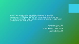
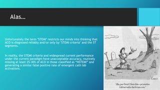
The trouble with STEMI
Sonja Maria
Senior academic, researcher & lecturer
The current guideline-recommended paradigm of acute MI
management (“STEMI vs. NSTEMI”) is irreversibly flawed, and has
prevented meaningful progress in the science of emergent reperfusion
therapy over the past 25 years
Pendell Meyers, MD
Scott Weingart, MD, FCCM
Stephen Smith, MD
STEMI
• ST Elevation Myocardial Infarction
NSTEMI
• Non ST Elevation Myocardial Infarction
ACO
• Acute Coronary Occlusion (ACO) or near occlusion, with insufficient collateral circulation, whose
myocardium is at imminent risk of irreversible infarction without immediate reperfusion therapy
OMI
• Occlusion Myocardial Ischaemia and/or Infarction
Firstly, lets define some
(un)familiar terms
Another passing fad… or
revolutionary?
STEMI - Became a fashionable term in early 2000’s
Replaced the ‘q wave’ terminology (Q-wave vs. Non-Q-wave)
Ambulance service guidelines introduce STEMI infarct protocols
≈ 2010 onwards
Becoming a ‘KPI focus’ for healthcare systems as society
recognises the expense of treatment costs in survivors1
Push to shorten needle to balloon times within paramedicine.
Alas…
Unfortunately the term "STEMI" restricts our minds into thinking that
ACO is diagnosed reliably and/or only by "STEMI criteria" and the ST
segments.
In reality, the STEMI criteria and widespread current performance
under the current paradigm have unacceptable accuracy, routinely
missing at least 25-30% of ACO in those classified as “NSTEMI” and
generating a similar false positive rate of emergent cath lab
activations.
The biggest problems
Overestimating
STEMI
1
Undertreating
NSTEMI
2
Not looking for
STEMI equivalents
3
Myth 1: NSTEMI
patients are
probably
healthier
STEMI (n=17287) NSTEMI
(n=28912)
Prior Myocardial
infarction
19.1% 29.2%
Prior Heart Failure 6.6% 16.1%
Prior PCI 30% 28.2%
Prior CABG 10.2% 25%
Prior stroke 7.4% 10.7%
Peripheral artery
disease
8.8% 15.6%
Hypertension 73.7% 82.5%
Diabetes Mellitis 25.6% 37.1%
Dyslipidemia 56.7% 68.7%
Current/recent smoke 19.9% 15.5%
Vora, A. N., Wang, T. Y., Hellkamp, A. S., Thomas, L., Henry, T. D., Goyal, A., & Roe, M. T. (2016). Differences in Short- and Long-Term Outcomes Among Older Patients With ST-Elevation
Versus Non-ST-Elevation Myocardial Infarction With Angiographically Proven Coronary Artery Disease. Circ Cardiovasc Qual Outcomes, 9(5), 513-522. doi:10.1161/circoutcomes.115.002312
Myth 2 – NSTEMI
and STEMI are
really different, so
we should manage
them differently
Answer: By definition, STEMI and NSTEMI
are only different with respect to the
reflection of acute myocardial ischaemia
and necrosis on the ECG. The pathogenesis
may actually be the same.
Bode, C., & Zirlik, A. (2007). STEMI and NSTEMI: the dangerous brothers. European heart journal, 28(12), 1403-1404. doi:10.1093/eurheartj/ehm159
Myth 3 –
Focus on the
STEMI patient
Answer: NSTEMI patients appear to be undertreated
with respect to reperfusion and also after discharge
from hospital. The similar prognosis of NSTEMI and
STEMI patients should lead to a more aggressive in-
hospital and secondary prevention treatment of both
groups, particularly the NSTEMI population
Bode, C., & Zirlik, A. (2007). STEMI and NSTEMI: the dangerous brothers. European heart journal, 28(12), 1403-1404. doi:10.1093/eurheartj/ehm159
The most Important
fact : STEMIs lie..
The big 5 mimickers
• Pericarditis
• Left Ventricular Hypertrophy
• Left Bundle Branch Block
• Ventricular rhythms
• Benign Early Repolarisation
Can you tell the difference?
STEMI
equivalents
a quick
snapshot
• Brugada syndrome
• Wellens A and B
• De Winters T wave
• aVR – how can we use it?
Brugada
Syndrome
Is a genetic disorder (affect 1 in 2000 people
worldwide)1
8 – 10 times more common in men (testosterone may
account for the difference)
Most commonly a mutation of the gene SCN5A
This gene provides instructions for making a sodium channel, which
normally transports positively charged sodium atoms (ions) into heart
muscle cells. Plays a critical role in maintaining the heart's normal
rhythm. Mutations in the SCN5A gene alter the structure or function
of the channel, which reduces the flow of sodium ions into cells. A
disruption in ion transport alters the way the heart beats, leading to
the abnormal heart rhythm characteristic of Brugada syndrome.
1. https://ghr.nlm.nih.gov/condition/brugada-syndrome#statistics
Brugada Syndrome - ECG
ST-segment elevation in right
precordial ECG leads and
associated with sudden cardiac
death in young adults. The
ECG manifestations are often
concealed but can be
unmasked by sodium channel
blockers and fever.
Other conduction defects can
include first-degree AV block,
intraventricular conduction
delay, right bundle branch
block, and sick sinus
syndrome.
Brugada Syndrome -
ECG (typical)
A) Normal electrocardiogram of the
precordial leads V1-3,
B) Changes in Brugada syndrome
3 Forms and their changes on the ECG
Type 1 has a coved type ST elevation with at least 2 mm (0.2
mV) j point elevation and a gradually descending ST
segment followed by a negative Twave.
Type 2 has a saddle-back pattern with a least 2 mm J-point
elevation and at least 1 mm ST elevation with a positive or
biphasic T-wave. Type 2 pattern can occasionally be seen in
healthy subjects.
Type 3 has either a coved (type 1 like) or a saddle-back (type
2 like) pattern, with less than 2 mm J-point elevation and less
than 1 mm ST elevation. Type 3 pattern is not rare in healthy
subjects.
Postema, P. G., Wolpert, C., Amin, A. S., Probst, V., Borggrefe, M., Roden, D. M., … Wilde, A. A. (2009). Drugs and Brugada syndrome patients: review of the literature, recommendations,
and an up-to-date website (www.brugadadrugs.org). Heart rhythm, 6(9), 1335–1341. doi:10.1016/j.hrthm.2009.07.002
Wellens
Syndrome
• Wellens syndrome describes a pattern of ECG
changes that represent a pre-infarction state of
coronary artery disease.
• Wellens syndrome results from a temporary
obstruction of the LAD coronary artery. Usually,
this is caused by the rupture of an atherosclerotic
plaque leading to LAD occlusion, with subsequent
clot lysis or other disruption of the occlusion
before complete myocardial infarction has taken
place
• The exact mechanism of the ECG changes of
Wellens syndrome is not known, but some theorise
that coronary artery spasm and stunned
myocardium cause it. Others postulate that it is
caused by repetitive transmural ischaemia-
reperfusion leading to myocardial oedema.
Wellens Syndrome - ECG
Wellens syndrome is not always
an acute process. It can develop
over days to weeks. The ECG
pattern often develops when
the patient is not experiencing
chest pain.
When the patient does
experience chest pain, the ST
segment and T-wave pattern
can appear to normalise into
hyperacute upright T waves (so-
called “pseudo-normalisation”)
or even develop into ST-
segment elevations.
Wellens – 2 types
Type A
• 25% of cases
• Biphasic pattern
• More likely to be misrecognised
non-specific
normal pattern
Type B
• 75% of cases
• Deep inverted symmetric T-Waves
The T waves evolve over time from a Type A to a Type B pattern
Wellens Syndrome
– ECG (Type A)
• Biphasic precordial T waves
with terminal negativity,
most prominent in V2-3.
• Minor precordial ST
elevation.
• Preserved R wave
progression (R wave in V3 >
3mm)
Wellens Syndrome
– ECG (Type B)
There are deep, symmetrical
T wave inversions throughout
the anterolateral leads (V1-6,
I, aVL).
De Winters T
wave
The de Winter ECG pattern is an anterior STEMI
equivalent that presents without obvious ST segment
elevation, but instead changes to the T wave.
First reported by de Winter in 2008.
Key features of De Winters
Key diagnostic
features include ST
depression and
peaked T waves in the
precordial leads.
The de Winter pattern
is seen in ~2% of acute
LAD occlusions and is
under-recognised by
clinicians.
Unfamiliarity with this
high-risk ECG pattern
lead to under
identification and
under treatment
Patients may be
instead misidentified
as NSTEMI
What does it look like?
Anything look odd ?
Cont’
• Tall, prominent, symmetric T
waves in the precordial leads
• Upsloping ST segment depression
>1mm at the J-point in the precordial
leads
• Absence of ST elevation in the
precordial leads
• ST segment elevation (0.5mm-
1mm) in aVR
• “Normal” STEMI morphology may
precede or follow the deWinter
pattern
Example - 20 minutes before ED arrival
Example – Arriving at ED
aVR – the forgotten lead
The lead aVR is a very
important lead in localisation
of Coronary Artery Disease.
Also can be used for
identification of arrhythmias
(PSVT), acute pericarditis,
tricyclic overdoses, tension
pneumothorax, dextrocardia,
pulmonary embolism and
malposition/technical errors
on taking an ECG,
aVR (the widow maker sign)
What are we doing about it?
National study on all
undergraduate teaching of
cardiology and STEMIs – what are
the learning objectives?
Use the study result to inform
curriculum changes
Consult with other academics
Inform Stakeholders
(Ambulance Services, Council of
Deans, NHMRC guidelines)
Continue our Research and stay on
top of changes.
Questions?
smaria@csu.edu.au

The trouble with STEMI

  • 1.
    The trouble withSTEMI Sonja Maria Senior academic, researcher & lecturer
  • 2.
    The current guideline-recommendedparadigm of acute MI management (“STEMI vs. NSTEMI”) is irreversibly flawed, and has prevented meaningful progress in the science of emergent reperfusion therapy over the past 25 years Pendell Meyers, MD Scott Weingart, MD, FCCM Stephen Smith, MD
  • 3.
    STEMI • ST ElevationMyocardial Infarction NSTEMI • Non ST Elevation Myocardial Infarction ACO • Acute Coronary Occlusion (ACO) or near occlusion, with insufficient collateral circulation, whose myocardium is at imminent risk of irreversible infarction without immediate reperfusion therapy OMI • Occlusion Myocardial Ischaemia and/or Infarction Firstly, lets define some (un)familiar terms
  • 4.
    Another passing fad…or revolutionary? STEMI - Became a fashionable term in early 2000’s Replaced the ‘q wave’ terminology (Q-wave vs. Non-Q-wave) Ambulance service guidelines introduce STEMI infarct protocols ≈ 2010 onwards Becoming a ‘KPI focus’ for healthcare systems as society recognises the expense of treatment costs in survivors1 Push to shorten needle to balloon times within paramedicine.
  • 5.
    Alas… Unfortunately the term"STEMI" restricts our minds into thinking that ACO is diagnosed reliably and/or only by "STEMI criteria" and the ST segments. In reality, the STEMI criteria and widespread current performance under the current paradigm have unacceptable accuracy, routinely missing at least 25-30% of ACO in those classified as “NSTEMI” and generating a similar false positive rate of emergent cath lab activations.
  • 6.
  • 7.
    Myth 1: NSTEMI patientsare probably healthier STEMI (n=17287) NSTEMI (n=28912) Prior Myocardial infarction 19.1% 29.2% Prior Heart Failure 6.6% 16.1% Prior PCI 30% 28.2% Prior CABG 10.2% 25% Prior stroke 7.4% 10.7% Peripheral artery disease 8.8% 15.6% Hypertension 73.7% 82.5% Diabetes Mellitis 25.6% 37.1% Dyslipidemia 56.7% 68.7% Current/recent smoke 19.9% 15.5% Vora, A. N., Wang, T. Y., Hellkamp, A. S., Thomas, L., Henry, T. D., Goyal, A., & Roe, M. T. (2016). Differences in Short- and Long-Term Outcomes Among Older Patients With ST-Elevation Versus Non-ST-Elevation Myocardial Infarction With Angiographically Proven Coronary Artery Disease. Circ Cardiovasc Qual Outcomes, 9(5), 513-522. doi:10.1161/circoutcomes.115.002312
  • 8.
    Myth 2 –NSTEMI and STEMI are really different, so we should manage them differently Answer: By definition, STEMI and NSTEMI are only different with respect to the reflection of acute myocardial ischaemia and necrosis on the ECG. The pathogenesis may actually be the same. Bode, C., & Zirlik, A. (2007). STEMI and NSTEMI: the dangerous brothers. European heart journal, 28(12), 1403-1404. doi:10.1093/eurheartj/ehm159
  • 9.
    Myth 3 – Focuson the STEMI patient Answer: NSTEMI patients appear to be undertreated with respect to reperfusion and also after discharge from hospital. The similar prognosis of NSTEMI and STEMI patients should lead to a more aggressive in- hospital and secondary prevention treatment of both groups, particularly the NSTEMI population Bode, C., & Zirlik, A. (2007). STEMI and NSTEMI: the dangerous brothers. European heart journal, 28(12), 1403-1404. doi:10.1093/eurheartj/ehm159
  • 10.
    The most Important fact: STEMIs lie.. The big 5 mimickers • Pericarditis • Left Ventricular Hypertrophy • Left Bundle Branch Block • Ventricular rhythms • Benign Early Repolarisation Can you tell the difference?
  • 11.
    STEMI equivalents a quick snapshot • Brugadasyndrome • Wellens A and B • De Winters T wave • aVR – how can we use it?
  • 12.
    Brugada Syndrome Is a geneticdisorder (affect 1 in 2000 people worldwide)1 8 – 10 times more common in men (testosterone may account for the difference) Most commonly a mutation of the gene SCN5A This gene provides instructions for making a sodium channel, which normally transports positively charged sodium atoms (ions) into heart muscle cells. Plays a critical role in maintaining the heart's normal rhythm. Mutations in the SCN5A gene alter the structure or function of the channel, which reduces the flow of sodium ions into cells. A disruption in ion transport alters the way the heart beats, leading to the abnormal heart rhythm characteristic of Brugada syndrome. 1. https://ghr.nlm.nih.gov/condition/brugada-syndrome#statistics
  • 13.
    Brugada Syndrome -ECG ST-segment elevation in right precordial ECG leads and associated with sudden cardiac death in young adults. The ECG manifestations are often concealed but can be unmasked by sodium channel blockers and fever. Other conduction defects can include first-degree AV block, intraventricular conduction delay, right bundle branch block, and sick sinus syndrome.
  • 14.
    Brugada Syndrome - ECG(typical) A) Normal electrocardiogram of the precordial leads V1-3, B) Changes in Brugada syndrome
  • 15.
    3 Forms andtheir changes on the ECG Type 1 has a coved type ST elevation with at least 2 mm (0.2 mV) j point elevation and a gradually descending ST segment followed by a negative Twave. Type 2 has a saddle-back pattern with a least 2 mm J-point elevation and at least 1 mm ST elevation with a positive or biphasic T-wave. Type 2 pattern can occasionally be seen in healthy subjects. Type 3 has either a coved (type 1 like) or a saddle-back (type 2 like) pattern, with less than 2 mm J-point elevation and less than 1 mm ST elevation. Type 3 pattern is not rare in healthy subjects. Postema, P. G., Wolpert, C., Amin, A. S., Probst, V., Borggrefe, M., Roden, D. M., … Wilde, A. A. (2009). Drugs and Brugada syndrome patients: review of the literature, recommendations, and an up-to-date website (www.brugadadrugs.org). Heart rhythm, 6(9), 1335–1341. doi:10.1016/j.hrthm.2009.07.002
  • 16.
    Wellens Syndrome • Wellens syndromedescribes a pattern of ECG changes that represent a pre-infarction state of coronary artery disease. • Wellens syndrome results from a temporary obstruction of the LAD coronary artery. Usually, this is caused by the rupture of an atherosclerotic plaque leading to LAD occlusion, with subsequent clot lysis or other disruption of the occlusion before complete myocardial infarction has taken place • The exact mechanism of the ECG changes of Wellens syndrome is not known, but some theorise that coronary artery spasm and stunned myocardium cause it. Others postulate that it is caused by repetitive transmural ischaemia- reperfusion leading to myocardial oedema.
  • 17.
    Wellens Syndrome -ECG Wellens syndrome is not always an acute process. It can develop over days to weeks. The ECG pattern often develops when the patient is not experiencing chest pain. When the patient does experience chest pain, the ST segment and T-wave pattern can appear to normalise into hyperacute upright T waves (so- called “pseudo-normalisation”) or even develop into ST- segment elevations.
  • 18.
    Wellens – 2types Type A • 25% of cases • Biphasic pattern • More likely to be misrecognised non-specific normal pattern Type B • 75% of cases • Deep inverted symmetric T-Waves The T waves evolve over time from a Type A to a Type B pattern
  • 19.
    Wellens Syndrome – ECG(Type A) • Biphasic precordial T waves with terminal negativity, most prominent in V2-3. • Minor precordial ST elevation. • Preserved R wave progression (R wave in V3 > 3mm)
  • 20.
    Wellens Syndrome – ECG(Type B) There are deep, symmetrical T wave inversions throughout the anterolateral leads (V1-6, I, aVL).
  • 21.
    De Winters T wave Thede Winter ECG pattern is an anterior STEMI equivalent that presents without obvious ST segment elevation, but instead changes to the T wave. First reported by de Winter in 2008.
  • 22.
    Key features ofDe Winters Key diagnostic features include ST depression and peaked T waves in the precordial leads. The de Winter pattern is seen in ~2% of acute LAD occlusions and is under-recognised by clinicians. Unfamiliarity with this high-risk ECG pattern lead to under identification and under treatment Patients may be instead misidentified as NSTEMI
  • 23.
    What does itlook like? Anything look odd ?
  • 24.
    Cont’ • Tall, prominent,symmetric T waves in the precordial leads • Upsloping ST segment depression >1mm at the J-point in the precordial leads • Absence of ST elevation in the precordial leads • ST segment elevation (0.5mm- 1mm) in aVR • “Normal” STEMI morphology may precede or follow the deWinter pattern
  • 25.
    Example - 20minutes before ED arrival
  • 26.
  • 27.
    aVR – theforgotten lead The lead aVR is a very important lead in localisation of Coronary Artery Disease. Also can be used for identification of arrhythmias (PSVT), acute pericarditis, tricyclic overdoses, tension pneumothorax, dextrocardia, pulmonary embolism and malposition/technical errors on taking an ECG,
  • 28.
    aVR (the widowmaker sign)
  • 29.
    What are wedoing about it? National study on all undergraduate teaching of cardiology and STEMIs – what are the learning objectives? Use the study result to inform curriculum changes Consult with other academics Inform Stakeholders (Ambulance Services, Council of Deans, NHMRC guidelines) Continue our Research and stay on top of changes.
  • 30.

Editor's Notes

  • #5 The treatment costs of CHD are currently estimated to represent about 26 % of the hospital budget for cardiovascular diseases. Owing to ageing of the population, however, treatment costs are expected to increase by 45 % by 2025
  • #8 Differences in Short- and Long-Term Outcomes Among Older Patients With ST-Elevation Verses Non–ST-Elevation Myocardial Infarction With Angiographically Proven Coronary Artery Disease http://circoutcomes.ahajournals.org.ezproxy.csu.edu.au/content/circcvoq/early/2016/09/06/CIRCOUTCOMES.115.002312.full.pdf Differences in Short- and Long-Term Outcomes Among Older Patients With ST-Elevation Verses Non–ST-Elevation Myocardial Infarction With Angiographically Proven Coronary Artery Disease
  • #9 Although this difference may be triggered by the size of the infarcted area, it may also be only the location of the infarct—an occluded circumflex artery does not project equally well on the ECG as does an occluded right coronary artery. Furthermore, specificity and sensitivity of ECG changes are influenced by several other factors including prior MI, bypass surgery, variation of coronary anatomy, bundle-branch block, and others. Are these differences meaningful—should they lead to different clinical approaches—as the guidelines currently recommend? (4) STEMI and NSTEMI: the dangerous brothers. Available from: https://www.researchgate.net/publication/6305848_STEMI_and_NSTEMI_the_dangerous_brothers [accessed Jul 16 2018].
  • #10  Montalescot et al. offer: NSTEMI patients appear to be undertreated with respect to reperfusion and also after discharge from hospital. The similar prognosis of NSTEMI and STEMI patients should lead to a more aggressive in-hospital and secondary prevention treatment of both groups, particularly the NSTEMI population (4) STEMI and NSTEMI: the dangerous brothers. Available from: https://www.researchgate.net/publication/6305848_STEMI_and_NSTEMI_the_dangerous_brothers [accessed Jul 16 2018].
  • #11 Add some stats in here about incidence
  • #13 1. https://ghr.nlm.nih.gov/condition/brugada-syndrome#statistics
  • #15  Brugada syndrome is characterized by cardiac conduction abnormalities (ST-segment abnormalities in leads V1-V3 on ECG and a high risk for ventricular arrhythmias) that can result in sudden death. Brugada syndrome presents primarily during adulthood although age at diagnosis may range from infancy to late adulthood. The mean age of sudden death is approximately 40 years. Clinical presentations may also include sudden infant death syndrome (SIDS; death of a child during the first year of life without an identifiable cause) and the sudden unexpected nocturnal death syndrome (SUNDS), a typical presentation in individuals from Southeast Asia.
  • #16 https://www.ncbi.nlm.nih.gov/pmc/articles/PMC2779019/ Three forms of the Brugada ECG pattern have been described[12] Type 1 has a coved type ST elevation with at least 2 mm (0.2 mV) J-point elevation and a gradually descending ST segment followed by a negative T-wave. Type 2 has a saddle-back pattern with a least 2 mm J-point elevation and at least 1 mm ST elevation with a positive or biphasic T-wave. Type 2 pattern can occasionally be seen in healthy subjects. Type 3 has either a coved (type 1 like) or a saddle-back (type 2 like) pattern, with less than 2 mm J-point elevation and less than 1 mm ST elevation. Type 3 pattern is not rare in healthy subjects. ECG pattern in Brugada syndrome. According to a recent consensus document, Type 1 ST segment elevation, either spontaneously present or induced with the sodium channel-blocker challenge test, is considered diagnostic. Type 2 and 3 may lead to suspicion, but provocation testing is required for diagnosis. The ECGs in the right and left panels are from the same patient before (right panel, type 3) and after (left panel, type 1) administration of Ajmaline.
  • #20 Biphasic precordial T waves with terminal negativity, most prominent in V2-3. Minor precordial ST elevation. Preserved R wave progression (R wave in V3 > 3mm)
  • #21 There are deep, symmetrical T wave inversions throughout the anterolateral leads (V1-6, I, aVL).
  • #24 So what looks odd about this?
  • #25 In patients presenting with chest pain, ST-segment depression at the J-point with upsloping ST-segments and tall, symmetrical T-waves in the precordial leads of the 12-lead ECG signifies proximal LAD artery occlusion. It is important for cardiologists and emergency care physicians to recognise this distinct ECG pattern, so they can triage such patients for immediate reperfusion therapy.– Verouden NJ 2009 Original reports of the de Winter pattern suggested that the ECG did not change or evolve until the culprit artery had been opened. Since then, cases have been reported where the deWinter pattern evolved from, or evolved to a “classic” anterior STEMI.
  • #26 Tall, prominent, symmetric T waves in the precordial leads Upsloping ST segment depression >1mm at the J-point in the precordial leads Absence of ST elevation in the precordial leads ST segment elevation (0.5mm-1mm) in aVR “Normal” STEMI morphology may precede or follow the deWinter pattern
  • #27 Tall, prominent, symmetric T waves in the precordial leads Upsloping ST segment depression >1mm at the J-point in the precordial leads Absence of ST elevation in the precordial leads ST segment elevation (0.5mm-1mm) in aVR “Normal” STEMI morphology may precede or follow the deWinter pattern
  • #28 The lead aVR is a very important lead in localisation of Coronary Artery Disease. In the presence of anterior ST elevation, ST elevation in lead aVR and V1 denotes proximal LAD obstruction where ST elevation is more in lead V1, than in aVR. In the presence of anterior ST depression, ST elevation in lead aVR indicates Left Main Coronary Artery (LMCA) Disease where ST elevation is more in aVR than in V1. In wide QRS tachycardia, tall R wave in aVR indicates Ventricular Tachycardia rather than SVT with aberrancy. In the presence of QS complexes in inferior leads, the lead aVR helps to differentiate between inferior wall MI (IWMI) and left anterior fascicular block (LAFB). Initial R in aVR is suggestive of IWMI and terminal R is suggestive of LAFB. In pericarditis, lead aVR is most often the only lead which shows reciprocal ST depression where as in Acute Infarction, usually a group of leads shows reciprocal depression. In the presence of persistent ST elevation in anterior chest leads, the R in aVR is suggestive of left ventricular aneurysm (Goldburger’s sign). In acute pulmonary embolism, ST elevation in lead aVR is a bad prognostic sign. In Tricyclic antidepressant toxicity, R in aVR more than 3 mm is an adverse prognostic sign. So in variety of conditions, the aVR is proved to be a valuable lead not only in diagnosis but also in predicting the prognosis.