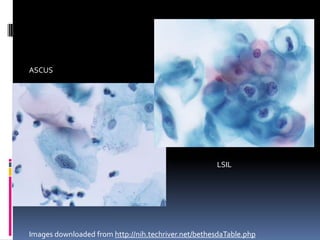
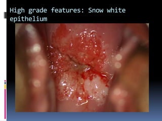
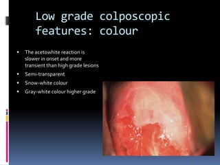
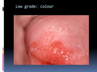
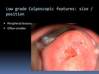
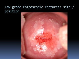
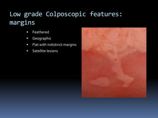
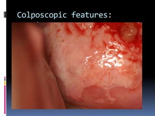
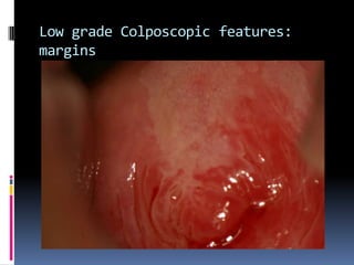
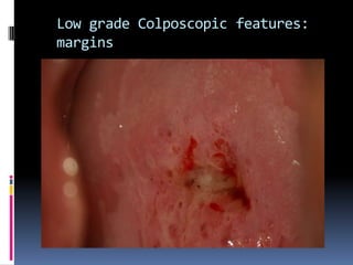
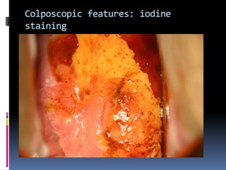
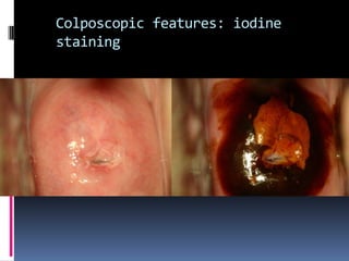
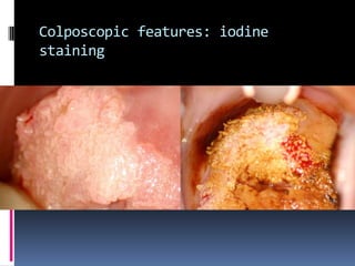
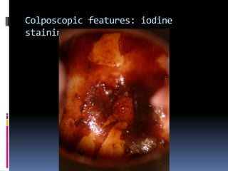
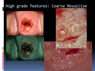
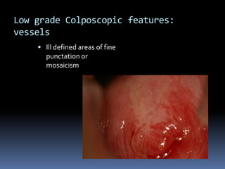
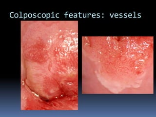
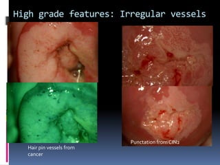
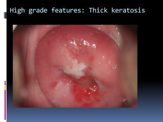
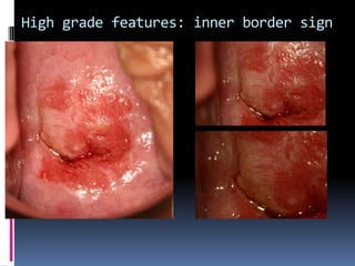
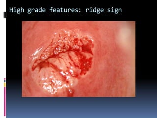
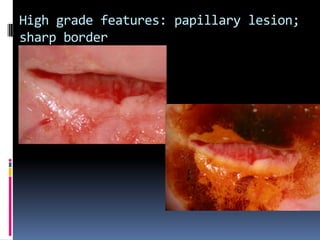
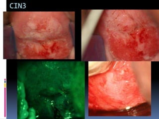
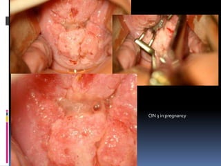
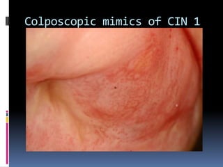
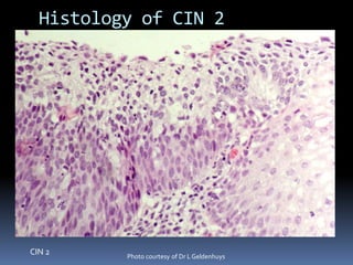
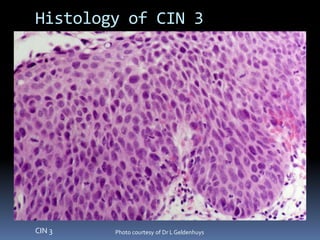
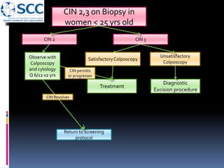
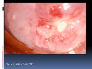
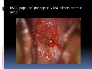
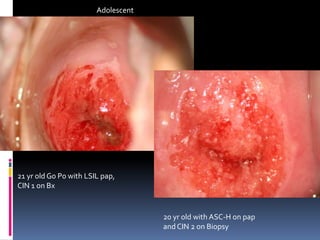
COLPOSCOPY OF CIN;
DIFFERENTIATING HIGH GRADE
FROM LOW GRADE LESIONS
James Bentley , Professor Department of Obstetrics and Gynecology,
Dalhousie University, Halifax NS, Canada
Normal
CIN 1
CIN 2
CIN 3
Cancer
Progression/Regression of CIN
Regress Persist Progress
to CIS
Progress
to
invasion
Months to
CIS from
baseline
CIN 1 57% 32% 11% 1% 58
CIN 2 43% 35% 22% 5% 38
CIN 3 32% <56% - >12% 12
OsterAG. Int J Gynecol Pathol 1993;12:86
Richart RM, Barron BA. A follow-up studyof patientswith cervical dysplasia.Am J Obstet
1969;105:386–393
Resolution of CIN 1 in adolescent is 90%
CIN 3/ CIS progression to cancer in 31% of cases
treated by Bx;
McCredie et al Lancet Oncology 2008 9(5) 425-34
ASCUS
LSIL
Images downloaded from http://nih.techriver.net/bethesdaTable.php
Results: hc2 +ve
Pap smear history Number of cases HybridCapture 2
+ve 1
95%Confidence
intervals
ASCUS/ASCUS 87 58 (67%) 56% to 76%
ASCUS/LSIL 33 23 (74%) 52% to 82%
LSIL/ASCUS 19 18 (95%) 73% to 100%
LSIL/LSIL 21 15 (71%) 49% to 86%
All cases 160 114 (72%) 64% to 78%
1 note 10 specimens had insufficient sample
•No significant difference between groups for hc2
Triage using repeat ASCUS or LSIL pap smears; is there any difference between categories? Bentley et al IFCPC
2008
Results: CIN2 + on Bx
Pap smear history Number of cases CIN 2 or greater 95%CI
ASCUS/ASCUS 87 23 (26%) 18% to 37%
ASCUS/LSIL 33 7 (21%) 10% to 38%
LSIL/ASCUS 19 2 (10.5%) 1.7% to 32%
LSIL/LSIL 21 3 (14%) 4% to 35%
All cases 160 35 (22%) 16% to 29%
•No significant difference between groups for histology
Triage using repeat ASCUS or LSIL pap smears; is there any difference between categories? Bentley et al IFCPC
2008
CIN2 and 3 after low grade
cytology
 ALTS trial:
 Progression to CIN2 or 3 in 13% of women referred
for the evaluation of LSIL or ASCUS HPV +ve smear
 NS Data:
 2ASCUS, 2 LSIL, or combination
 HR HPV +ve 72 %
 CIN2 or > 22%
ASC-H
Images downloaded from http://nih.techriver.net/bethesdaTable.php
CIN2 or > after ASC-H
 Significant pathology seen in the majority of
cases
 Barreth et al.:
 CIN2 or > in 70% of cases
 2.9% invasive disease
 1.7%AIS
HSIL
CIN2 or > after HSIL
 WrightASCCP:
 CIN2 or > 53%-66% with Biopsy
 90% if policy of immediate colposcopy
AGC cytology
Pathology finding1
CIN 1 7%
CIN 2 or 3 36%
Adenocarcinoma in situ 20%
Cervical Cancer 9%
Endometrial Pathology 29%
1Daniel A Int.J.Gynaecol.Obstet 2005; 91(3)238-242
2 Wright T Emerging Issues on HPV infections 2006 p
140-146
Cytology2 Any high-grade
lesion
High grade
glandular
AGC-NOS 9-14% 0-15%
AGC-N 27-96% 10-93%
ASC-H
Colposcopy
NoCIN
Manage as per SCC
guidelines
CIN1 or >
Colposcopy, cytology,
at 6 months x 2 (HPV
testing at 6 or 12
months ideally)
Return to screening
protocol
CIN 1 or >
No CIN
HPV +ve follow in
colposcopy clinic
HSIL
Colposcopy
(Bx, +/- ECC)
NoCIN 2, 3
Manage as per SCC
guidelines
CIN 2 or greater
Satisfactory
Colposcopy
Unsatisfactory
Colposcopy
Observe
with
Colposcopy
and cytology
Q 6/12 x2*
Return to screening
protocol
Diagnostic
Excision procedure
* Consider HPV testing
Cytology/histolo
gy review
disagreeagree
Colposcopic Approach
 Examine whole lower genital tract
 Use acetic acid liberally
 Beware the small lesion
 Take >1 biopsy
 Liberal use of ECC
 Always do ECC with unsatisfactory colposcopy
High grade features: Snow white
epithelium
Low grade colposcopic
features: colour
 The acetowhite reaction is
slower in onset and more
transient than high grade lesions
 Semi-transparent
 Snow-white colour
 Gray-white colour higher grade
Low grade: colour
Low grade Colposcopic features: size /
position
 Peripheral lesions
 Often smaller
Low grade Colposcopic features: size /
position
Low grade Colposcopic features:
margins
 Feathered
 Geographic
 Flat with indistinct margins
 Satellite lesions
Colposcopic features:
margins
Low grade Colposcopic features:
margins
Low grade Colposcopic features:
margins
Colposcopic features: iodine
staining
Colposcopic features: iodine
staining
Colposcopic features: iodine
staining
Colposcopic features: iodine
staining
High grade features: Coarse Mosaicism
Low grade Colposcopic features:
vessels
 Ill defined areas of fine
punctation or
mosaicism
Colposcopic features: vessels
High grade features: Irregular vessels
vascularity
Hair pin vessels from
cancer
Punctation from CIN2
High grade features: Thick keratosis
High grade features: inner border sign
High grade features: ridge sign
High grade features: papillary lesion;
sharp border
CIN3
CIN 3 in pregnancy
Colposcopic mimics of CIN 1
CIN 2 Photo courtesy of Dr LGeldenhuys
Histology of CIN 2
CIN 3 Photo courtesy of Dr LGeldenhuys
Histology of CIN 3
CIN 1 on Biopsy
or ECC
SatisfactoryColposcopy
Observe
with
Colposcopy
and cytology
Q 6/12 x2
Return to screening
protocol
Unsatisfactory
Colposcopy
Observe with
Colposcopy
and cytology
at 24 months2
Treatment1
1 consider ablative therapy for persistent CIN1
2 if cytology persists continue FU in colposcopy
Colposcopy and
cytology -ve
CIN persists or
progresses
Observe with
Colposcopy
and cytology
12 months
persisten
t
CIN 2,3 on
Biopsy
Return to screening
protocol
Diagnostic Excision
procedure
CIN 2,3
Treatment1
SatisfactoryColposcopy Unsatisfactory
Colposcopy
Follow-upat 6 and 12
months with colposcopy
and cytology
Follow-upat 6 months
with colposcopyand
cytologyand HPV2
OR
Treat per guidelines
CINNegative
1 LEEP or excision preferred for CIN 3
2 HPV testing for high risk HPV
CIN 2,3 on Biopsy in
women < 25 yrs old
CIN 2
Return to screening
protocol
Diagnostic
Excision procedure
CIN 3
Observe with
Colposcopy
and cytology
Q 6/12 x2 yrs
Treatment
SatisfactoryColposcopy
Unsatisfactory
Colposcopy
CIN persists
or progresses
CIN Resolves
CIN 3 with AIS (on final LEEP)
HSIL pap: colposcopic view after acetic
acid
21 yr old G0 P0 with LSIL pap,
CIN 1 on Bx
20 yr old with ASC-H on pap
andCIN 2 on Biopsy
Adolescent
Conclusion
 CIN 1 does not warrant therapy as most will
resolve spontaneously
 CIN 3 and CIN 2 are recognised cervical
cancer precursors
 They can be identified following both high
grade and low grade cytology
 The colposcopic features should allow
differentiation between CIN 1 and CIN 2/3

2 prof james bently differentiating high and low grade

  • 1.
    COLPOSCOPY OF CIN; DIFFERENTIATINGHIGH GRADE FROM LOW GRADE LESIONS James Bentley , Professor Department of Obstetrics and Gynecology, Dalhousie University, Halifax NS, Canada
  • 2.
  • 3.
    Progression/Regression of CIN RegressPersist Progress to CIS Progress to invasion Months to CIS from baseline CIN 1 57% 32% 11% 1% 58 CIN 2 43% 35% 22% 5% 38 CIN 3 32% <56% - >12% 12 OsterAG. Int J Gynecol Pathol 1993;12:86 Richart RM, Barron BA. A follow-up studyof patientswith cervical dysplasia.Am J Obstet 1969;105:386–393 Resolution of CIN 1 in adolescent is 90% CIN 3/ CIS progression to cancer in 31% of cases treated by Bx; McCredie et al Lancet Oncology 2008 9(5) 425-34
  • 4.
    ASCUS LSIL Images downloaded fromhttp://nih.techriver.net/bethesdaTable.php
  • 5.
    Results: hc2 +ve Papsmear history Number of cases HybridCapture 2 +ve 1 95%Confidence intervals ASCUS/ASCUS 87 58 (67%) 56% to 76% ASCUS/LSIL 33 23 (74%) 52% to 82% LSIL/ASCUS 19 18 (95%) 73% to 100% LSIL/LSIL 21 15 (71%) 49% to 86% All cases 160 114 (72%) 64% to 78% 1 note 10 specimens had insufficient sample •No significant difference between groups for hc2 Triage using repeat ASCUS or LSIL pap smears; is there any difference between categories? Bentley et al IFCPC 2008
  • 6.
    Results: CIN2 +on Bx Pap smear history Number of cases CIN 2 or greater 95%CI ASCUS/ASCUS 87 23 (26%) 18% to 37% ASCUS/LSIL 33 7 (21%) 10% to 38% LSIL/ASCUS 19 2 (10.5%) 1.7% to 32% LSIL/LSIL 21 3 (14%) 4% to 35% All cases 160 35 (22%) 16% to 29% •No significant difference between groups for histology Triage using repeat ASCUS or LSIL pap smears; is there any difference between categories? Bentley et al IFCPC 2008
  • 7.
    CIN2 and 3after low grade cytology  ALTS trial:  Progression to CIN2 or 3 in 13% of women referred for the evaluation of LSIL or ASCUS HPV +ve smear  NS Data:  2ASCUS, 2 LSIL, or combination  HR HPV +ve 72 %  CIN2 or > 22%
  • 8.
    ASC-H Images downloaded fromhttp://nih.techriver.net/bethesdaTable.php
  • 9.
    CIN2 or >after ASC-H  Significant pathology seen in the majority of cases  Barreth et al.:  CIN2 or > in 70% of cases  2.9% invasive disease  1.7%AIS
  • 10.
  • 11.
    CIN2 or >after HSIL  WrightASCCP:  CIN2 or > 53%-66% with Biopsy  90% if policy of immediate colposcopy
  • 12.
    AGC cytology Pathology finding1 CIN1 7% CIN 2 or 3 36% Adenocarcinoma in situ 20% Cervical Cancer 9% Endometrial Pathology 29% 1Daniel A Int.J.Gynaecol.Obstet 2005; 91(3)238-242 2 Wright T Emerging Issues on HPV infections 2006 p 140-146 Cytology2 Any high-grade lesion High grade glandular AGC-NOS 9-14% 0-15% AGC-N 27-96% 10-93%
  • 13.
    ASC-H Colposcopy NoCIN Manage as perSCC guidelines CIN1 or > Colposcopy, cytology, at 6 months x 2 (HPV testing at 6 or 12 months ideally) Return to screening protocol CIN 1 or > No CIN HPV +ve follow in colposcopy clinic
  • 14.
    HSIL Colposcopy (Bx, +/- ECC) NoCIN2, 3 Manage as per SCC guidelines CIN 2 or greater Satisfactory Colposcopy Unsatisfactory Colposcopy Observe with Colposcopy and cytology Q 6/12 x2* Return to screening protocol Diagnostic Excision procedure * Consider HPV testing Cytology/histolo gy review disagreeagree
  • 15.
    Colposcopic Approach  Examinewhole lower genital tract  Use acetic acid liberally  Beware the small lesion  Take >1 biopsy  Liberal use of ECC  Always do ECC with unsatisfactory colposcopy
  • 16.
    High grade features:Snow white epithelium
  • 17.
    Low grade colposcopic features:colour  The acetowhite reaction is slower in onset and more transient than high grade lesions  Semi-transparent  Snow-white colour  Gray-white colour higher grade
  • 18.
  • 19.
    Low grade Colposcopicfeatures: size / position  Peripheral lesions  Often smaller
  • 20.
    Low grade Colposcopicfeatures: size / position
  • 21.
    Low grade Colposcopicfeatures: margins  Feathered  Geographic  Flat with indistinct margins  Satellite lesions
  • 22.
  • 23.
    Low grade Colposcopicfeatures: margins
  • 24.
    Low grade Colposcopicfeatures: margins
  • 25.
  • 26.
  • 27.
  • 28.
  • 29.
    High grade features:Coarse Mosaicism
  • 30.
    Low grade Colposcopicfeatures: vessels  Ill defined areas of fine punctation or mosaicism
  • 31.
  • 32.
    High grade features:Irregular vessels vascularity Hair pin vessels from cancer Punctation from CIN2
  • 33.
    High grade features:Thick keratosis
  • 34.
    High grade features:inner border sign
  • 35.
  • 36.
    High grade features:papillary lesion; sharp border
  • 37.
  • 38.
    CIN 3 inpregnancy
  • 39.
  • 40.
    CIN 2 Photocourtesy of Dr LGeldenhuys Histology of CIN 2
  • 41.
    CIN 3 Photocourtesy of Dr LGeldenhuys Histology of CIN 3
  • 42.
    CIN 1 onBiopsy or ECC SatisfactoryColposcopy Observe with Colposcopy and cytology Q 6/12 x2 Return to screening protocol Unsatisfactory Colposcopy Observe with Colposcopy and cytology at 24 months2 Treatment1 1 consider ablative therapy for persistent CIN1 2 if cytology persists continue FU in colposcopy Colposcopy and cytology -ve CIN persists or progresses Observe with Colposcopy and cytology 12 months persisten t
  • 43.
    CIN 2,3 on Biopsy Returnto screening protocol Diagnostic Excision procedure CIN 2,3 Treatment1 SatisfactoryColposcopy Unsatisfactory Colposcopy Follow-upat 6 and 12 months with colposcopy and cytology Follow-upat 6 months with colposcopyand cytologyand HPV2 OR Treat per guidelines CINNegative 1 LEEP or excision preferred for CIN 3 2 HPV testing for high risk HPV
  • 44.
    CIN 2,3 onBiopsy in women < 25 yrs old CIN 2 Return to screening protocol Diagnostic Excision procedure CIN 3 Observe with Colposcopy and cytology Q 6/12 x2 yrs Treatment SatisfactoryColposcopy Unsatisfactory Colposcopy CIN persists or progresses CIN Resolves
  • 45.
    CIN 3 withAIS (on final LEEP)
  • 46.
    HSIL pap: colposcopicview after acetic acid
  • 47.
    21 yr oldG0 P0 with LSIL pap, CIN 1 on Bx 20 yr old with ASC-H on pap andCIN 2 on Biopsy Adolescent
  • 48.
    Conclusion  CIN 1does not warrant therapy as most will resolve spontaneously  CIN 3 and CIN 2 are recognised cervical cancer precursors  They can be identified following both high grade and low grade cytology  The colposcopic features should allow differentiation between CIN 1 and CIN 2/3