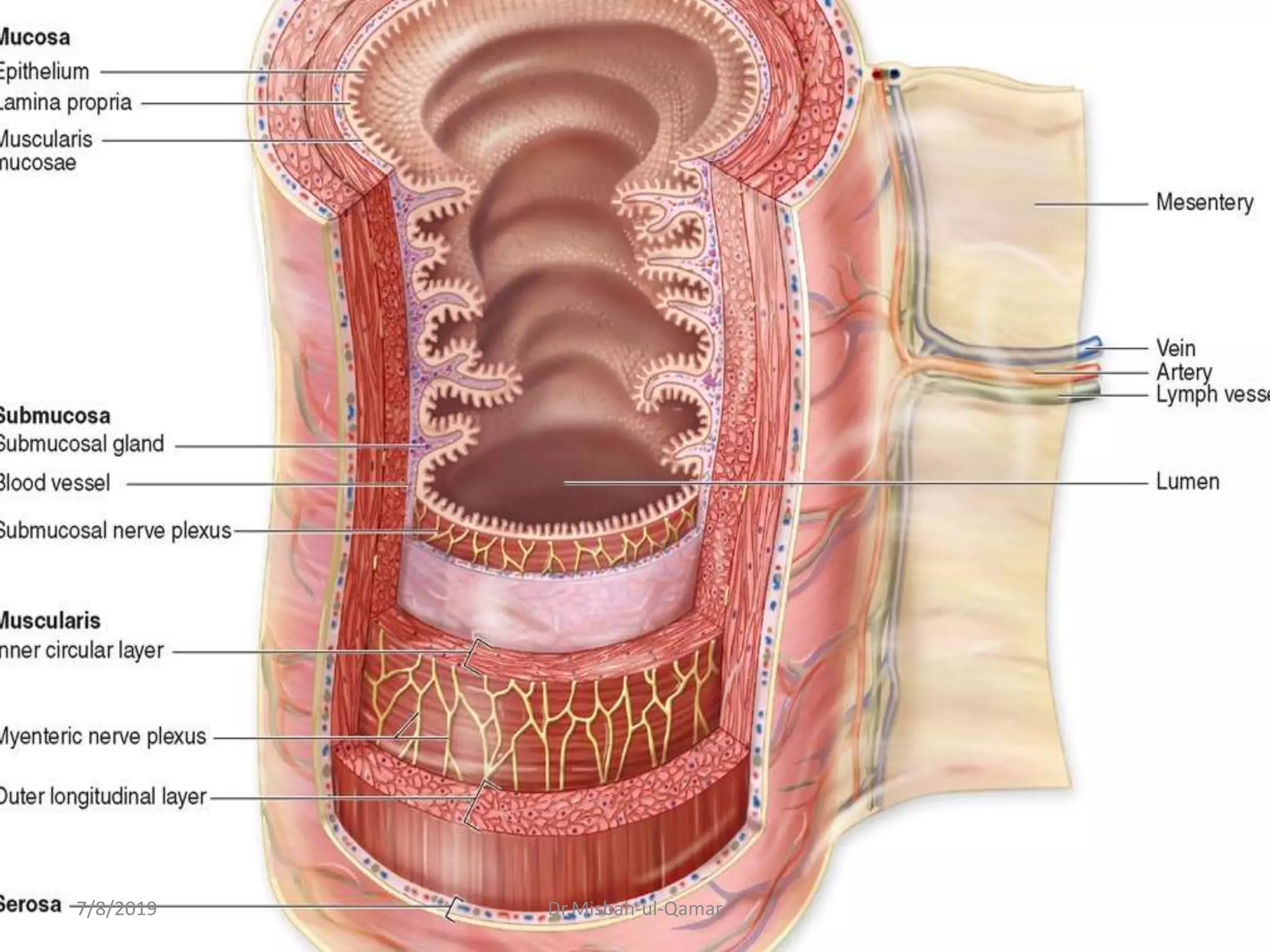
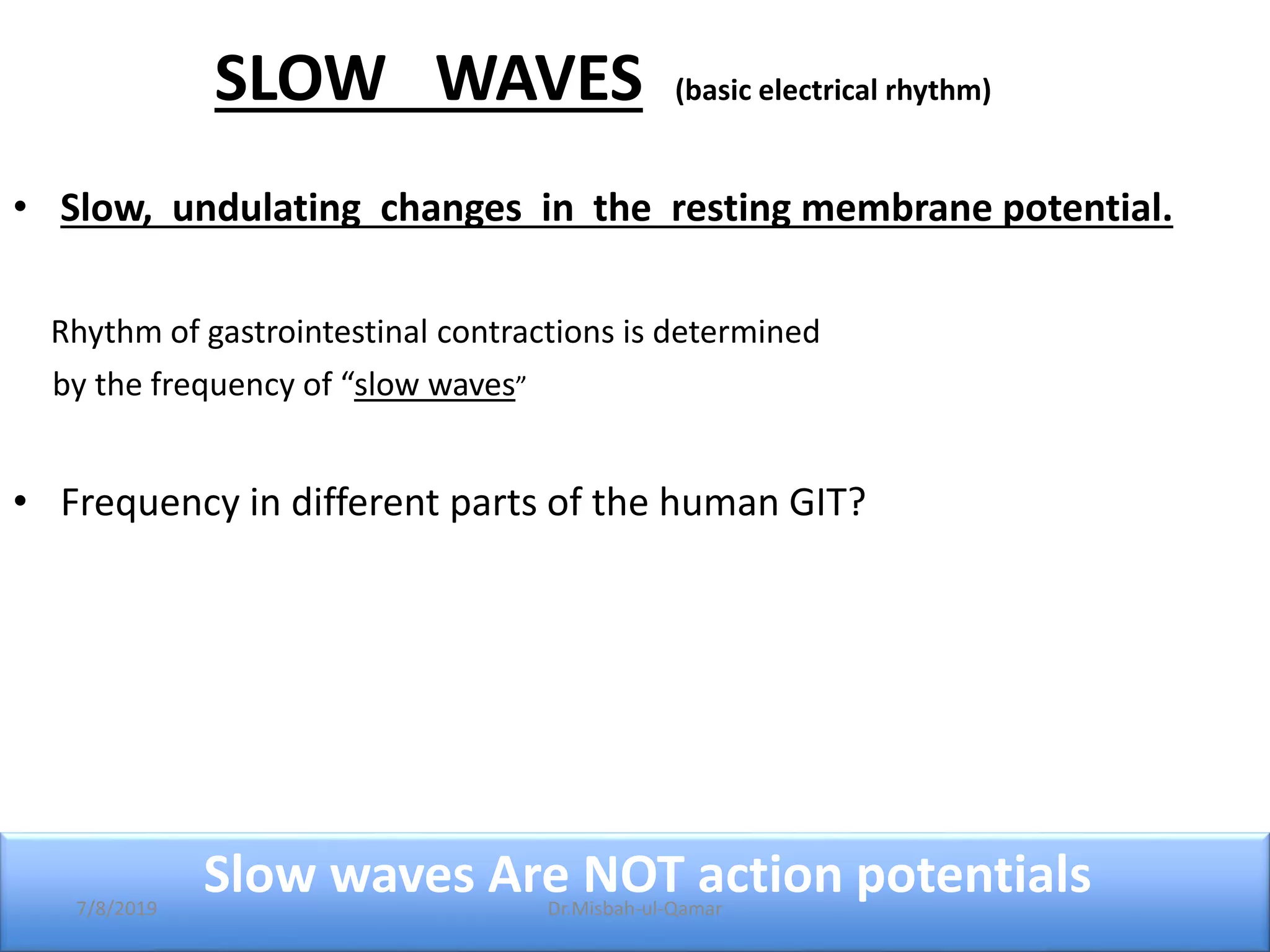
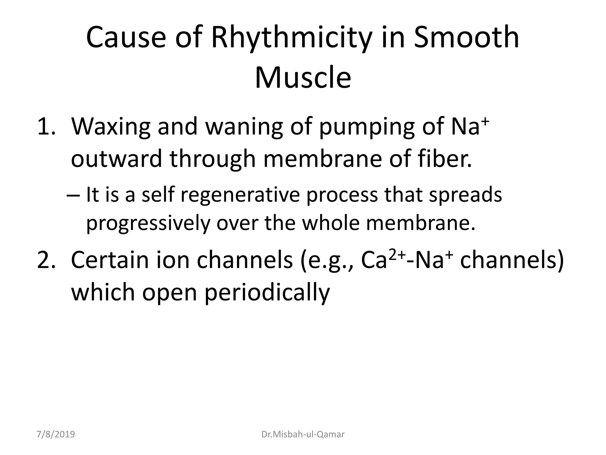
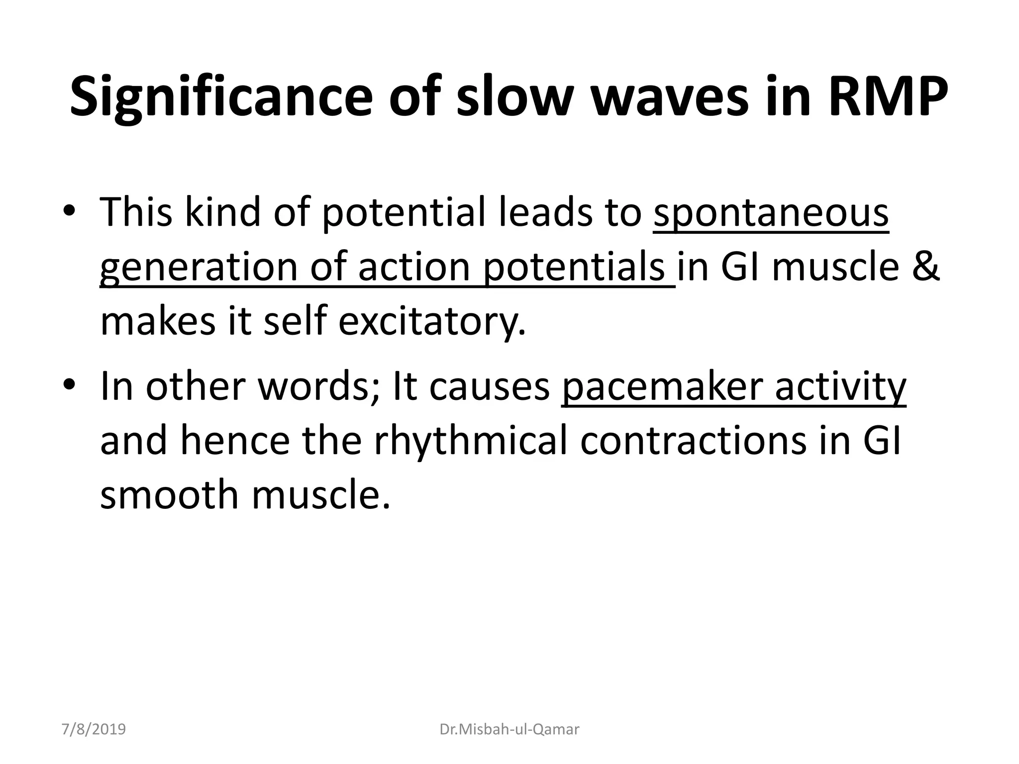
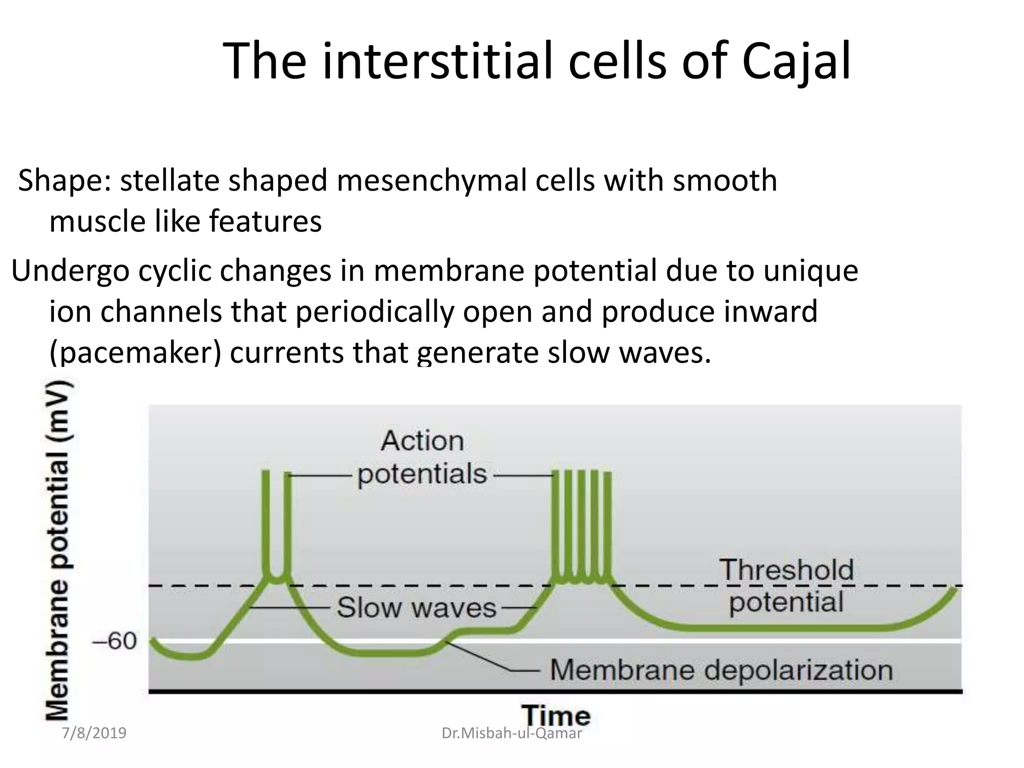
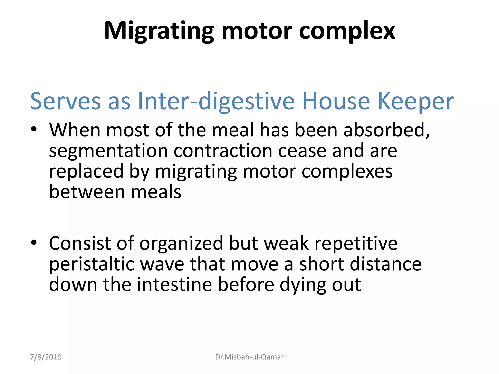
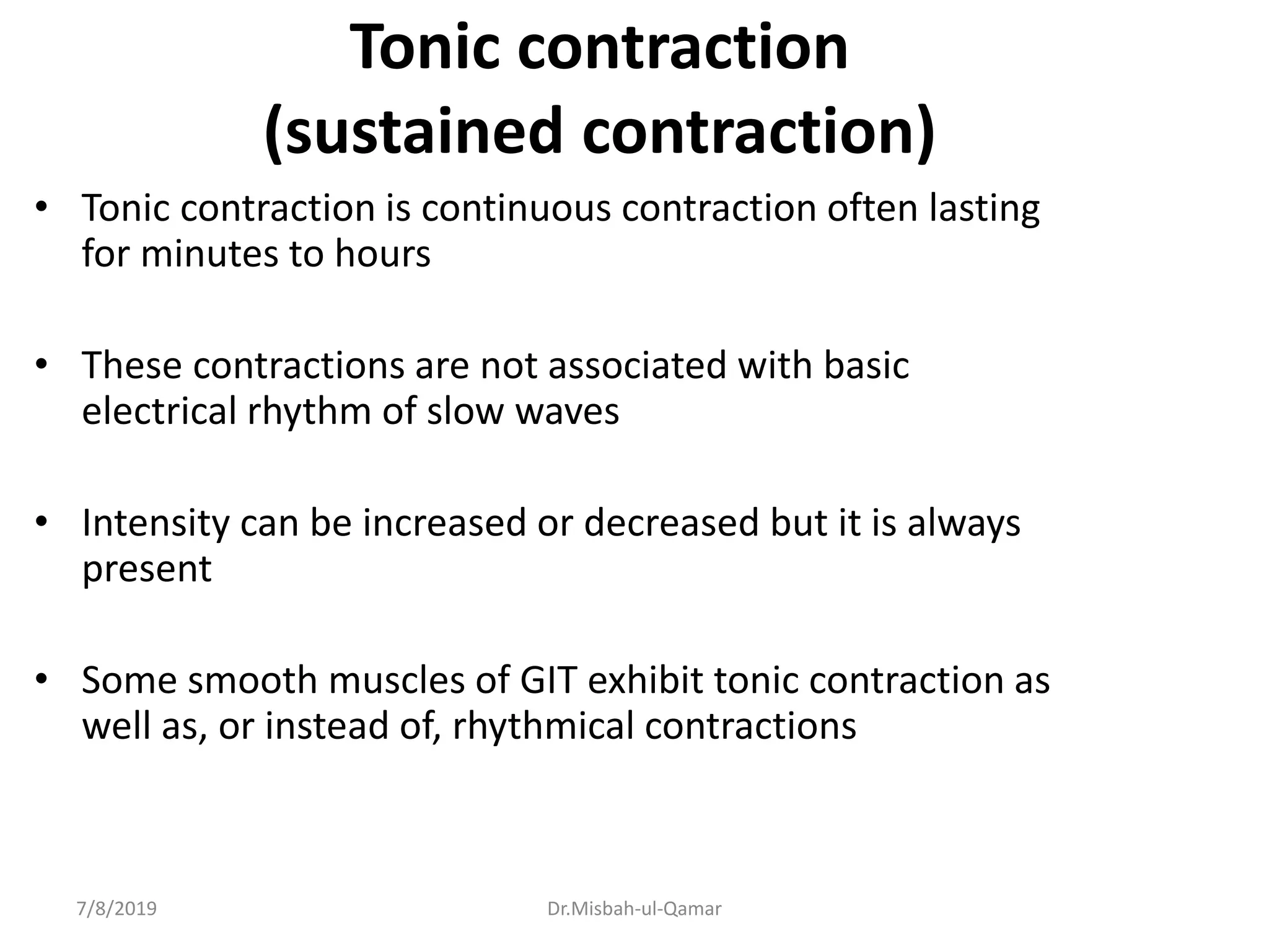
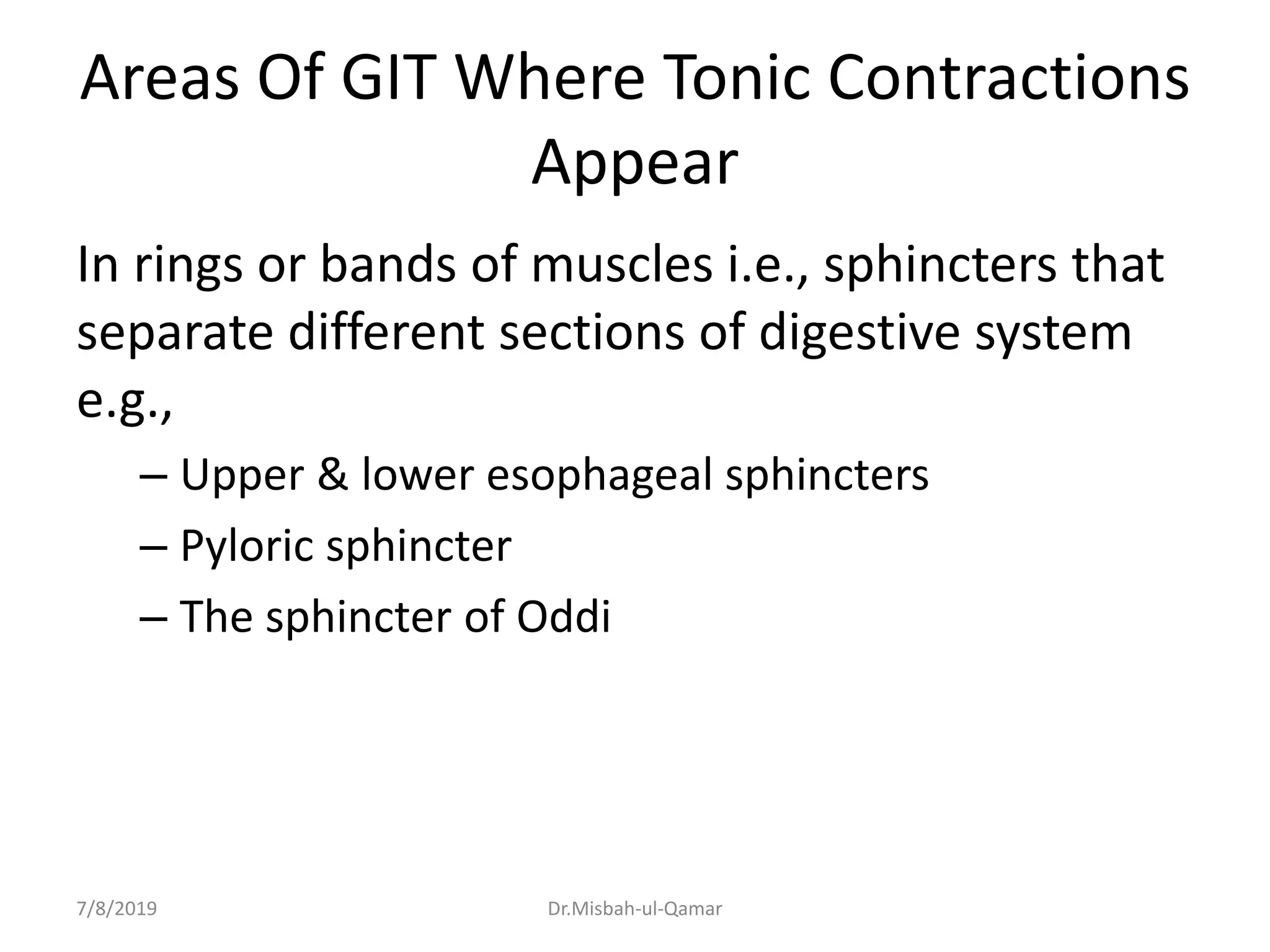
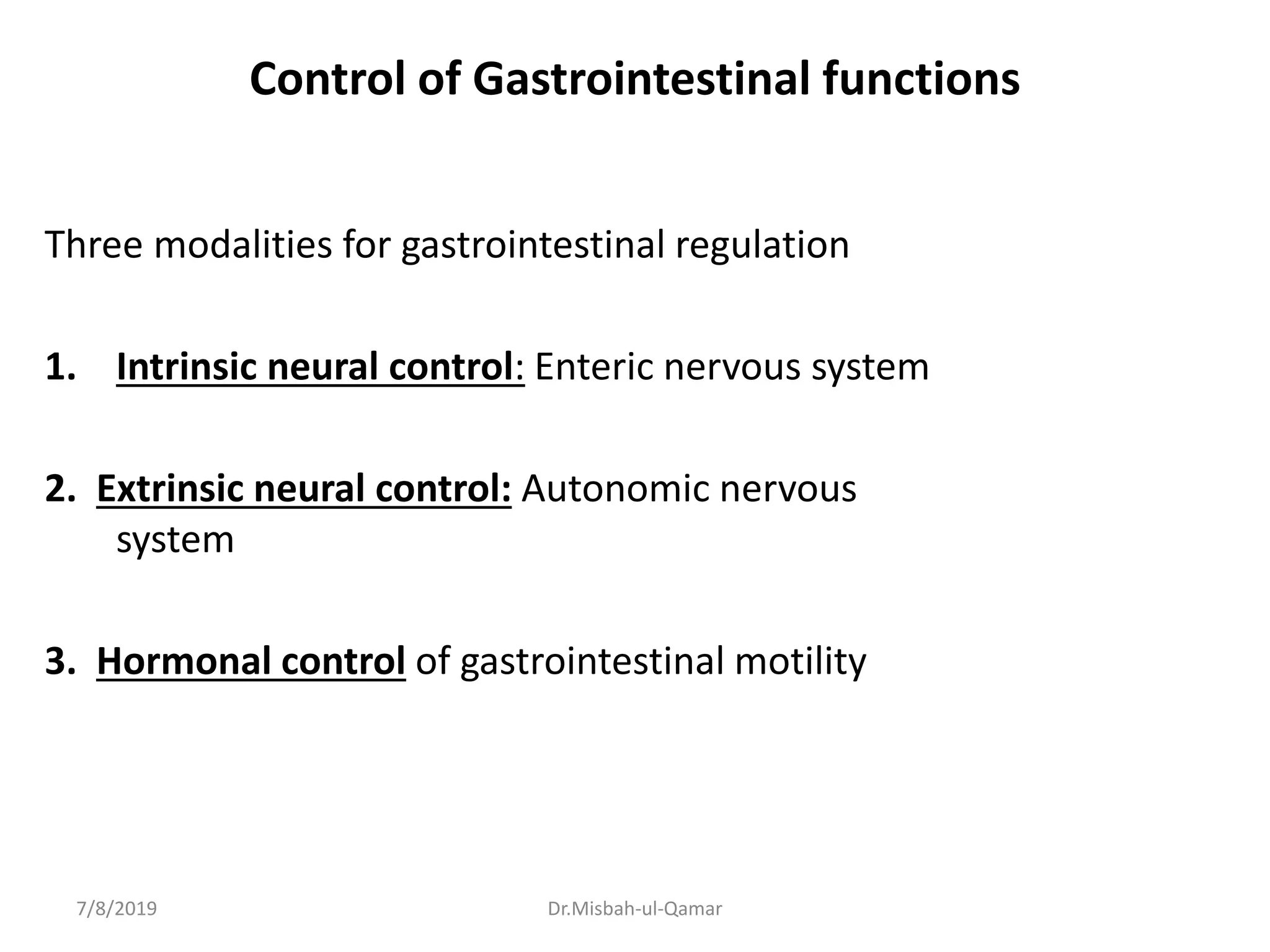
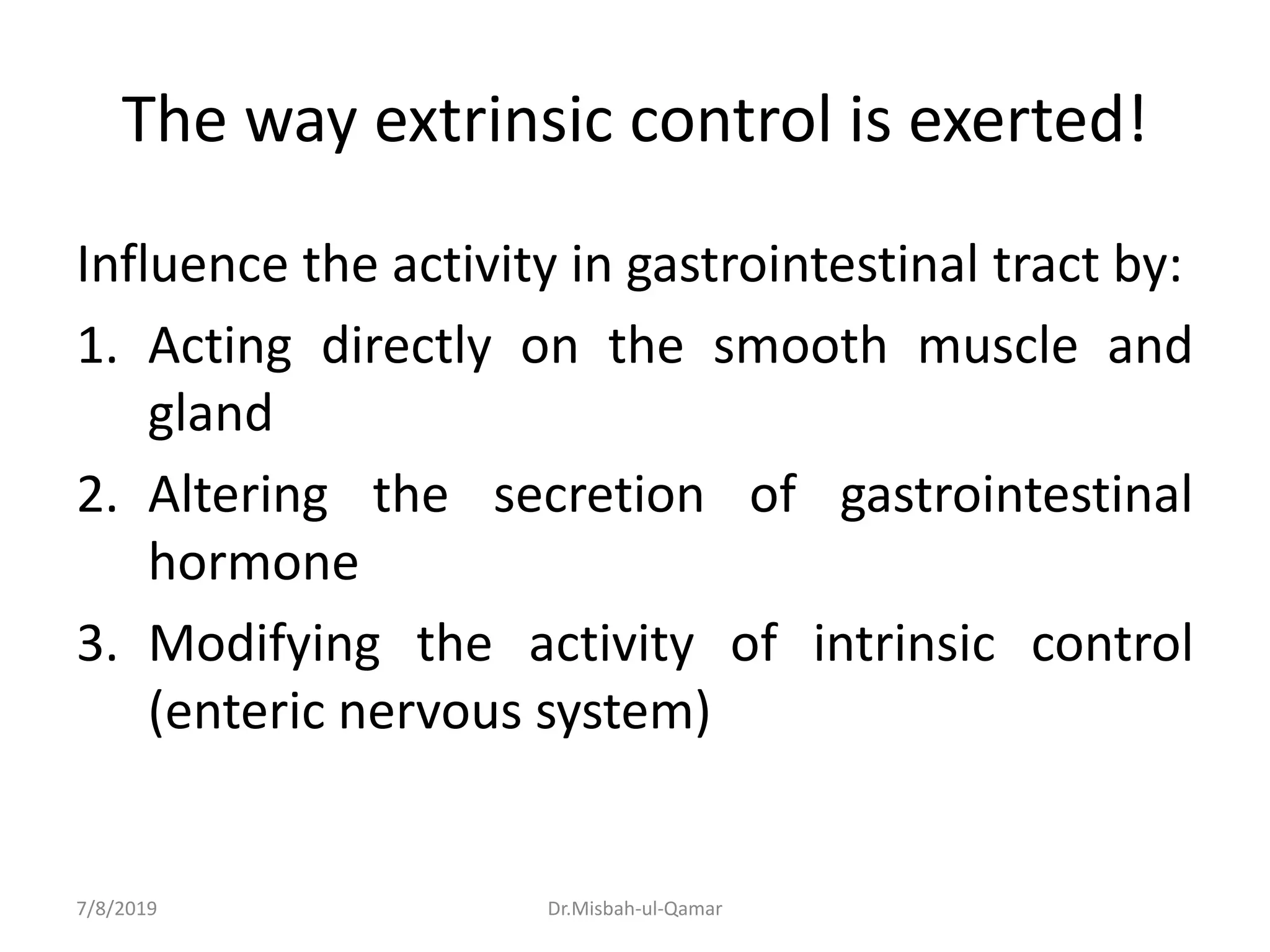
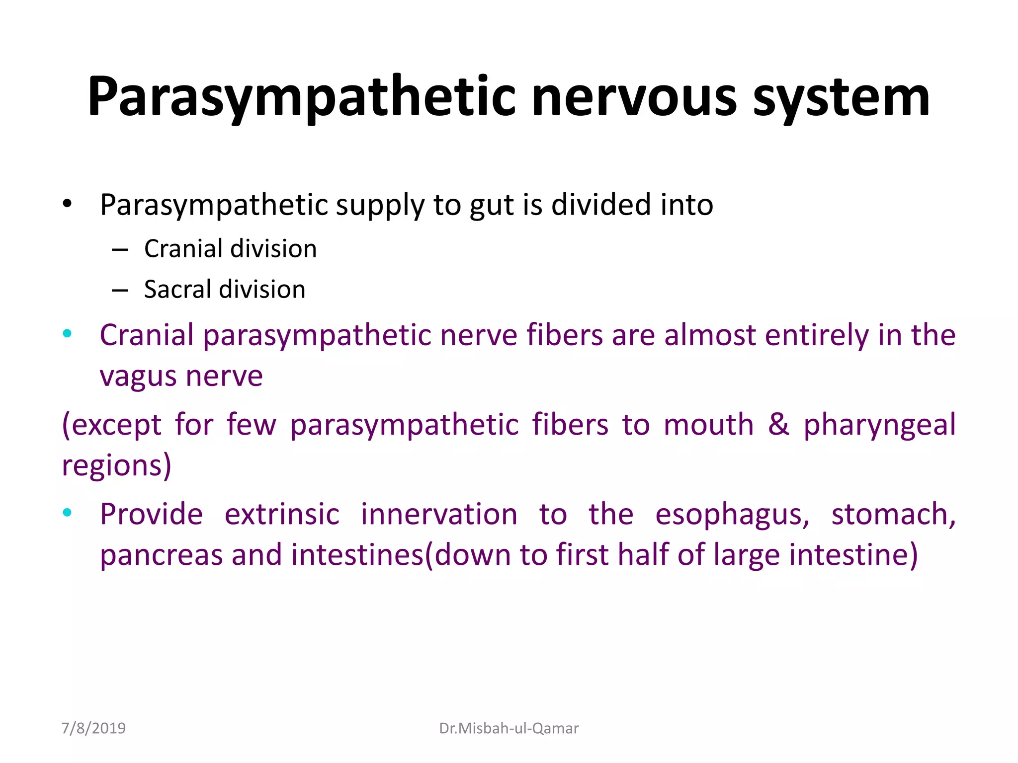
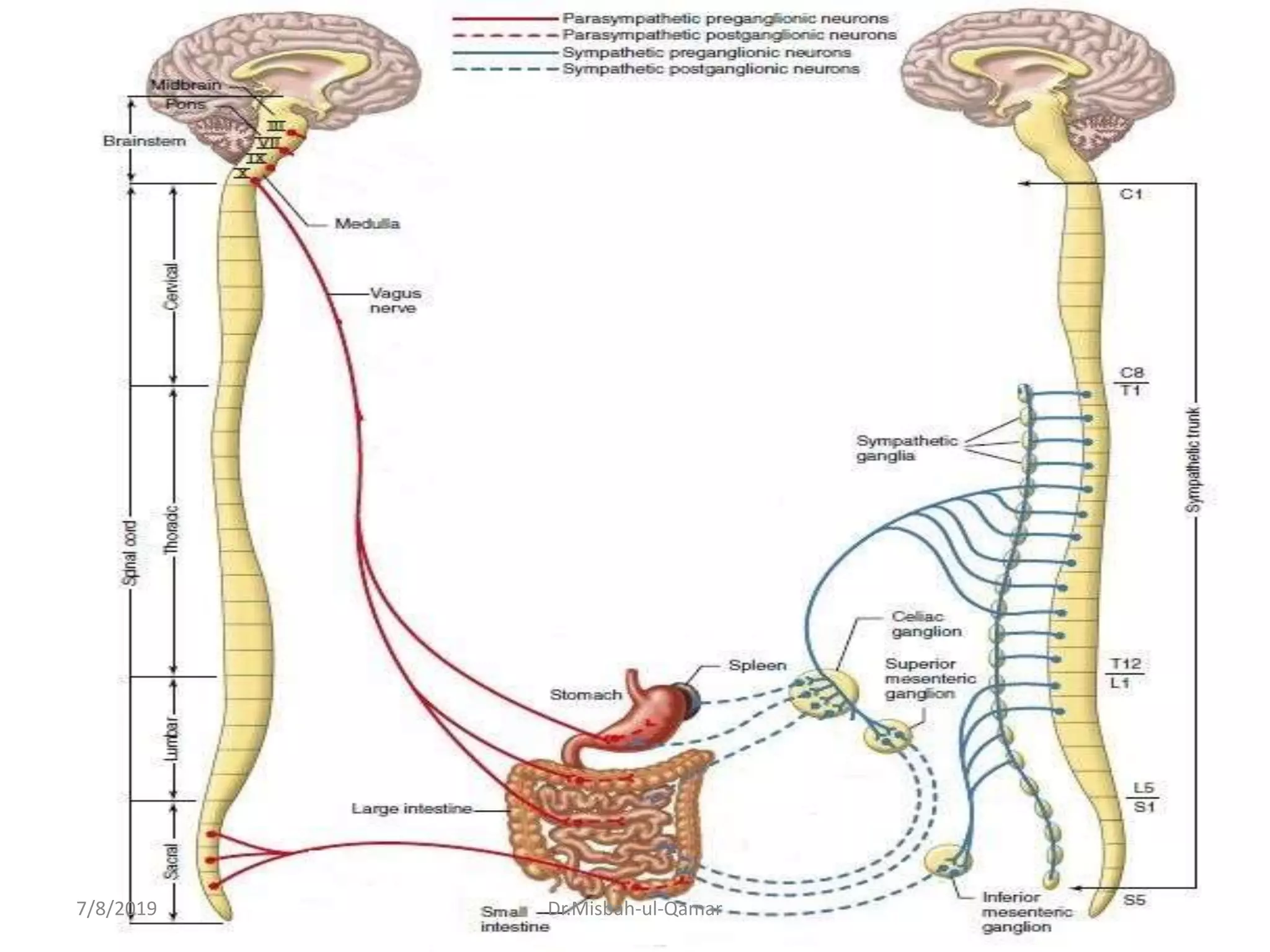
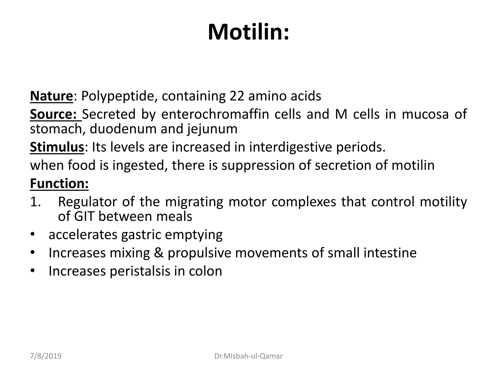
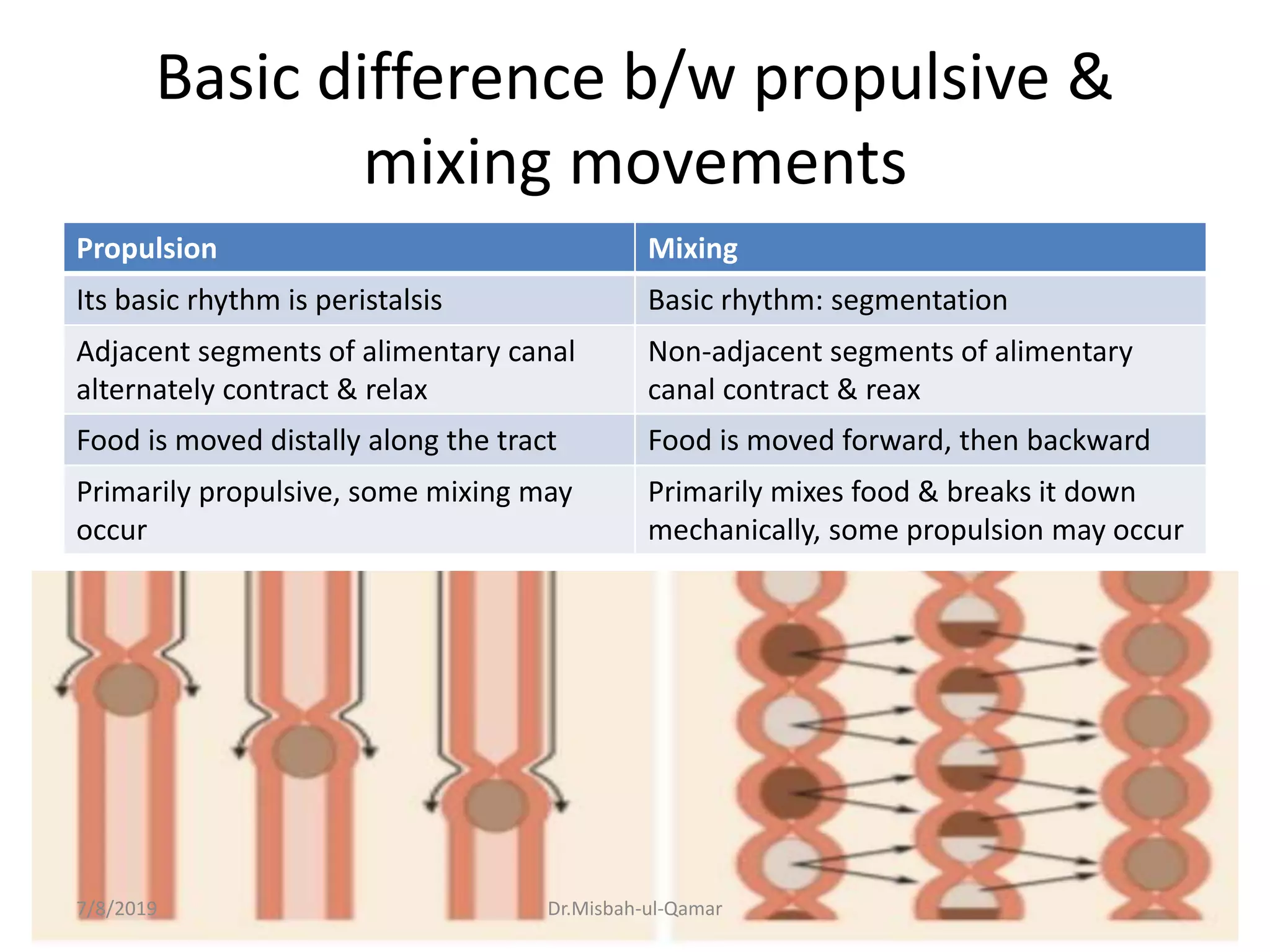
4. Transfer of nutrients from external to internal environment
7/8/2019 Dr.Misbah-ul-Qamar
5. Precise Need For Digestive System
• The basic chemical units of our food & our
body tissues are the same
• Why our food looks so different from the
tissues
7/8/2019 Dr.Misbah-ul-Qamar
6. This is how the digestive system renders 95% food available for body’s use
7/8/2019 Dr.Misbah-ul-Qamar
7. Sequence Of Nutrient Acquisition
• All inputs of nutrition are through the
gastrointestinal tract
• Excretory organs?
7/8/2019 Dr.Misbah-ul-Qamar
12. Motility (movement)
• It refers to the muscular contractions…
• Process of motility
• Tonic activity of GIT
– Tone of GIT
– Functions of tone
7/8/2019 Dr.Misbah-ul-Qamar
25. • Upper GIT
– Consists of structures that aid in the ingestion &
digestion of food
• Lower GIT
– Consists of small & large intestine
7/8/2019 Dr.Misbah-ul-Qamar
26. General principles of GI motility
Characteristics Of GI Wall,
Electrical Activity Of GI Smooth
Muscle.
7/8/2019 Dr.Misbah-ul-Qamar
27. Characteristics of GI wall
a. Layers of smooth muscle.
b. Syncytium.
7/8/2019 Dr.Misbah-ul-Qamar
28. General anatomy of gut wall
The digestive tract wall has four layers from
inside (lumen) to outside
1. Mucosa
2. Submucosa
3. Muscularis externa
4. Serosa (visceral peritoneum)
7/8/2019 Dr.Misbah-ul-Qamar
33. Epithelium Of Mucosa
– Stratified squamous (in mouth, esophagus & anus)
=tough
– Simple columnar in the rest of tract
• Secretes enzymes and absorbs nutrients
• Has specialized cells also
• Brush border & its functions
7/8/2019 Dr.Misbah-ul-Qamar
34. Lamina propria
• Thin layer of loose connective tissue
• Resident structures
7/8/2019 Dr.Misbah-ul-Qamar
35. Muscularis mucosae
• It forms only a thin layer, lying in deeper layers
of mucosa.
• Function: increasing absorption
7/8/2019 Dr.Misbah-ul-Qamar
37. Sub-mucosa
• It is composed of moderately dense areolar
connective tissue
• Regarded as a highly vascular layer
• Also contains a part of the submucosal plexus
of nerves
• In addition contains glands & lymphatic tissue
7/8/2019 Dr.Misbah-ul-Qamar
38. MUSCULARIS EXTERNA
• Skeletal muscle = voluntary control
– In mouth, pharynx, upper esophagus & anus
– Control over swallowing & defecation
• Smooth muscle = involuntary control
– Inner circular fibers & outer longitudinal fibers
– Mixes, crushes & propels food along by peristalsis
• This muscle layer also contains a part of nerve
plexus
7/8/2019 Dr.Misbah-ul-Qamar
39. Serosa
• It is an example of serous membrane which
secretes a slippery fluid
• This membrane covers all organs & walls of
cavities forms protective covering
• Consists of connective tissue covered with
simple squamous epithelium
7/8/2019 Dr.Misbah-ul-Qamar
40. Lets focus on GI smooth muscle
Gastrointestinal smooth muscles
• 200-500µm in length
• 2-10µm in diameter
7/8/2019 Dr.Misbah-ul-Qamar
41. Gastrointestinal Smooth Muscle as a
Syncytium.
• Longitudinal muscle layer
• Circular muscle layer
• Within each bundle, the muscle fibers are electrically connected with one another through
large numbers of Gap Junctions that allow electrical signals to travel readily from one fiber to
next.
• Each muscle layer functions as a Syncytium; that is, when an action potential is elicited
anywhere within the muscle mass, it generally travels in all directions in the muscle
• Smooth muscles are arranged in bundles as parallel fibers.
7/8/2019 Dr.Misbah-ul-Qamar
42. What is the function of a syncytium
• It allows coordinated contraction of muscles
along their entire length
• Function of syncytial smooth muscle in
intestine housekeeping
• It produces 2 types of contractions
– Tonic contractions---- to maintain organ dimension
against an imposed load (bolus of food)
– Forceful contractions---- these produce muscle
shortening to propel the bolus along GIT
7/8/2019 Dr.Misbah-ul-Qamar
43. Arrangement of bundles in each layer
• How each bundle is separate & connected as
well.
• Distance travelled by each AP
• Connection b/w circular & smooth muscle
layer also exists
7/8/2019 Dr.Misbah-ul-Qamar
44. GI MOTILITY ACHIEVED THROUGH
ELECTRICAL ACTIVITY OF SMOOTH
MUSCLE
The motility is brought about 2 types of contractions in GIT:
Phasic (rhythmic) & Tonic
7/8/2019 Dr.Misbah-ul-Qamar
45. Electrical activity of gastrointestinal
smooth muscles
• Normal resting membrane potential in the
smooth muscle fibers of the gut is between -
50 to-60mV
• The smooth muscles of gastrointestinal tract is
excited by continual slow electrical activity
7/8/2019 Dr.Misbah-ul-Qamar
46. Electrical Activity of Gastrointestinal Smooth Muscle
Two basic types of electrical waves:
(1) Slow waves. (Between -60 and -40 mv, i.e 15 mv)
(2) Spikes.
7/8/2019 Dr.Misbah-ul-Qamar
47. SLOW WAVES (basic electrical rhythm)
• Slow, undulating changes in the resting membrane potential.
Rhythm of gastrointestinal contractions is determined
by the frequency of “slow waves”
• Frequency in different parts of the human GIT?
Slow waves Are NOT action potentials7/8/2019 Dr.Misbah-ul-Qamar
48. Slow wave potential is different from
AP
Slow wave itself is not an AP but it can give
rise to AP.
Slow wave potentials fire the APs only when
they reach threshold.
Slow waves themselves cannot cause muscle
contractions unless they generate AP (or
depolarization of contraction threshold).
7/8/2019 Dr.Misbah-ul-Qamar
49. Cause of Rhythmicity in Smooth
Muscle
1. Waxing and waning of pumping of Na+
outward through membrane of fiber.
– It is a self regenerative process that spreads
progressively over the whole membrane.
2. Certain ion channels (e.g., Ca2+-Na+ channels)
which open periodically
7/8/2019 Dr.Misbah-ul-Qamar
50. Significance of slow waves in RMP
• This kind of potential leads to spontaneous
generation of action potentials in GI muscle &
makes it self excitatory.
• In other words; It causes pacemaker activity
and hence the rhythmical contractions in GI
smooth muscle.
7/8/2019 Dr.Misbah-ul-Qamar
52. Electrical Pacemakers For Smooth Muscle Cells (BER)
These specialized cells form a network with each other. They are interposed between
smooth muscle layers. They are connected with smooth muscle cells by gap junctions. So
that, electrical activity generated by these cells spread to adjacent smooth muscle cells.
7/8/2019 Dr.Misbah-ul-Qamar
53. The interstitial cells of Cajal
Shape: stellate shaped mesenchymal cells with smooth
muscle like features
Undergo cyclic changes in membrane potential due to unique
ion channels that periodically open and produce inward
(pacemaker) currents that generate slow waves.
.
7/8/2019 Dr.Misbah-ul-Qamar
54. Location of Cajal cells
– In stomach & small intestine, these cells are located in
outer circular muscle layer near myenteric plexus
– In colon, they are at sub-mucosal border of circular
muscle layer
• Rate of BER in different parts of gut?
– 4/min in stomach
– 12/min in duodenum
– 8/min in distal ileum
– In colon, BER rate rises from about 2/min at cecum to
about 6/min at sigmoid.
7/8/2019 Dr.Misbah-ul-Qamar
55. • Descending pattern of BER
– In stomach & small intestine, there is a
descending gradient in pacemaker frequency
– (pacemaker with highest frequency dominates)
• Significance of BER
– it coordinates peristaltic & other motor activity
– After vagotomy or transection of stomach wall,
peristalsis in stomach becomes irregular & chaotic
7/8/2019 Dr.Misbah-ul-Qamar
56. Slow waves usually Do Not cause muscle contraction.
Instead, they excite appearance of intermittent spike
potentials
The spike potentials excite the muscle contraction
7/8/2019 Dr.Misbah-ul-Qamar
57. Spike Potentials
• True action potential
• Occur when resting membrane potential/ peaks of slow
waves becomes more positive than – 40mV
7/8/2019 Dr.Misbah-ul-Qamar
58. Spike Potentials
• Frequency: 1-10 spikes/ sec (higher the slow wave
potential rises, greater the frequency of spike potentials)
• Duration : it is 10-40 times as long as AP in nerve. 10-20
msec (comparatively longer because of slowness of opening
& closing of Ca-Na channels)
• Depolarization: is caused by slow calcium-sodium
channel they allow especially large number of calcium ions
to enter along with smaller numbers of sodium ions
• Repolarization: is caused by opening of voltage gated
potassium channels
7/8/2019 Dr.Misbah-ul-Qamar
59. Spike Potentials
• Movement of large number of calcium into cell during
action potential causes muscle contraction.
Muscle fibers are connected with one another by gap
junctions allowing flow of ions from fiber to fiber
(Functional syncytium)
7/8/2019 Dr.Misbah-ul-Qamar
61. During fasting
• Between periods of digestion, the pattern of
electrical & motor activity in GI smooth
muscle become modified
• Cycles of motor activity (called MMC) migrate
aborally.
7/8/2019 Dr.Misbah-ul-Qamar
62. Migrating motor complex
Serves as Inter-digestive House Keeper
• When most of the meal has been absorbed,
segmentation contraction cease and are
replaced by migrating motor complexes
between meals
• Consist of organized but weak repetitive
peristaltic wave that move a short distance
down the intestine before dying out
7/8/2019 Dr.Misbah-ul-Qamar
63. MMC
• Begin in the stomach – to small intestine
(distal ileum)
• Migrate down @ 5cm/min, at interval of 90
min
• Phases of MMC
– Phase I: Basal phase quiescent period
– Phase II: Pre-burst phase period of irregular
electrical & mechanical activity
– Phase III: Burst phase burst of regular activity
7/8/2019 Dr.Misbah-ul-Qamar
64. Migrating motor complex
Function:
Sweep the remnants of
preceding meal,
secretions, mucosal debris
and bacteria forward
toward the colon
Prepares the gut for next
meal
7/8/2019 Dr.Misbah-ul-Qamar
65. Migrating myoelectric cycle
• Stimulated by hormone “motilin” circulating
levels of this hormone increase at intervals of
approx. 100-120 mins (coordinated with
contractile phases of MMC)
• Inhibited by ingestion of meal (suppression of
motilin release)
• Gastric secretion, bile flow, pancreatic
secretion & mucus discharge increase during
each MMC.
7/8/2019 Dr.Misbah-ul-Qamar
67. Changes in voltage of the RMP
• These changes occur in addition to slow waves
& spike potentials.
• Baseline voltage level of smooth muscle RMP
can also change
• Under normal conditions, RMP averages about
-56mV
• Multiple factors can change this level
7/8/2019 Dr.Misbah-ul-Qamar
68. Changes in voltage of the RMP (cont’d)
• When RMP becomes less negative it is
called depolarization of membrane muscle
becomes more excitable
• When RMP becomes more negative it is
called hyperpolarization of membrane
muscle becomes less excitable
7/8/2019 Dr.Misbah-ul-Qamar
69. Excitatory and Inhibitory
Factors
1) Depolarization:
-stretch
-acetylcholine
-parasympathetic stimulation
-specific GIT hormones
2) Hyperpolarization:
-epinephrine and norepinephrine
-sympathetic stimulation which mainly causes norepinephrine
secretion
7/8/2019 Dr.Misbah-ul-Qamar
71. Tonic contraction
(sustained contraction)
• Tonic contraction is continuous contraction often lasting
for minutes to hours
• These contractions are not associated with basic
electrical rhythm of slow waves
• Intensity can be increased or decreased but it is always
present
• Some smooth muscles of GIT exhibit tonic contraction as
well as, or instead of, rhythmical contractions
7/8/2019 Dr.Misbah-ul-Qamar
72. Causes of tonic contractions
1. Repetitive spike potentials
greater frequency greater degree of
contraction
2. Hormones/ certain other chemical factors
these bring about continuous partial
depolarization without causing action potential
3. Continuous entry of calcium into cell interior
brought about in ways not associated with
changes in membrane potential
7/8/2019 Dr.Misbah-ul-Qamar
74. Areas Of GIT Where Tonic Contractions
Appear
In rings or bands of muscles i.e., sphincters that
separate different sections of digestive system
e.g.,
– Upper & lower esophageal sphincters
– Pyloric sphincter
– The sphincter of Oddi
7/8/2019 Dr.Misbah-ul-Qamar
77. Control of Gastrointestinal functions
Three modalities for gastrointestinal regulation
1. Intrinsic neural control: Enteric nervous system
2. Extrinsic neural control: Autonomic nervous
system
3. Hormonal control of gastrointestinal motility
7/8/2019 Dr.Misbah-ul-Qamar
79. Extrinsic nervous control
It is exerted through autonomic nervous system
by its both divisions:
– Parasympathetic nervous system
– Sympathetic nervous system
• Connect to intrinsic system of control.
7/8/2019 Dr.Misbah-ul-Qamar
80. The way extrinsic control is exerted!
Influence the activity in gastrointestinal tract by:
1. Acting directly on the smooth muscle and
gland
2. Altering the secretion of gastrointestinal
hormone
3. Modifying the activity of intrinsic control
(enteric nervous system)
7/8/2019 Dr.Misbah-ul-Qamar
82. Parasympathetic nervous system
• Parasympathetic supply to gut is divided into
– Cranial division
– Sacral division
• Cranial parasympathetic nerve fibers are almost entirely in the
vagus nerve
(except for few parasympathetic fibers to mouth & pharyngeal
regions)
• Provide extrinsic innervation to the esophagus, stomach,
pancreas and intestines(down to first half of large intestine)
7/8/2019 Dr.Misbah-ul-Qamar
84. Parasympathetic nervous system
(cont’d)
• The sacral division of parasympathetic supply to
gut arises from S2,3,4 segments of spinal cord
• The parasympathetic nerve fibers pass through
pelvic nerves
• Innervate distal half of large intestine, all the way
to anus
• Sigmoid colon and rectum are better supplied
with parasympathetic fibers than other parts
(these fibers execute defecation reflex)
7/8/2019 Dr.Misbah-ul-Qamar
85. Summary of GI parasympathetics
a) Vagus nerve:
Esophagus upto proximal 2/3rd of transverse colon
b) Pelvic parasympathetic nerve: (S-2,3,4):
Distal 1/3 of transverse colon upto anus.
7/8/2019 Dr.Misbah-ul-Qamar
87. Parasympathetic nervous system
(cont’d)
• Fibers travelling in vagus & pelvic nerves are
preganglionic fibers
• Postganglionic neurons of GI parasympathetic
system are located in myenteric & submucosal
plexuses
7/8/2019 Dr.Misbah-ul-Qamar
89. Mode of action of para-sympathetics
Stimulation of parasympathetic nerves to gut
results in increased activity of the enteric
nervous system
– This results in increased motility and relaxation of
sphincters- increased motor activity
– The secretions from glands in gastrointestinal tract
is also increased- increased secretory activity
7/8/2019 Dr.Misbah-ul-Qamar
90. Summary of the effects exerted by
parasympathetic system
7/8/2019 Dr.Misbah-ul-Qamar
92. Sympathetic nerve supply to gut
Originate from T5 to L2 segments of spinal cord
Fibers leave spinal cord (splanchnic nerves)
Enter sympathetic chains lateral to spinal cord
To ganglia such as celiac and mesenteric ganglia
Post ganglionic fibers from here supply all the gut
7/8/2019 Dr.Misbah-ul-Qamar
94. Sympathetic nerve supply to gut
(cont’d)
• Neurotransmitter : Norepinephrine
• Stimulation of sympathetic nervous system inhibits the
activity of gastrointestinal tract
• Strong stimulation of sympathetics can inhibit motor movements of gut so
greatly that it can literally block movement of food
• Sympathetics innervate essentially all the GIT unlike parasympathetics
which are more extensive near oral cavity & anus
7/8/2019 Dr.Misbah-ul-Qamar
95. How sympathetic system exerts effects
on GIT
1. By direct effect of secreted norepinephrine
– this effect is slight
– it inhibits intestinal tract smooth muscles
– Exception is mucosal muscle which is excited
2. By an inhibitory effect of norepinephrine on
the neurons of entire enteric nervous system
– This effect has major role in its action on GIT
7/8/2019 Dr.Misbah-ul-Qamar
98. IT’S YOUR 2ND BRAIN!
That isn’t a butterfly in your stomach
7/8/2019 Dr.Misbah-ul-Qamar
99. intrinsic enteric nervous system
• Also called gut brain/ little brain/ second brain
• It is a highly developed control system
number of neurons: 100 million (nearly equal
to number in entire spinal cord)
7/8/2019 Dr.Misbah-ul-Qamar
100. Why it is called “brain of the gut”
• It can integrate sensory information & effect a
complex motor response independent of the
CNS.
• ENS releases a variety of neurotransmitters
just like CNS.
7/8/2019 Dr.Misbah-ul-Qamar
101. Enteric nervous system
• Begins in esophagus & extends to anus.
• The neurons can be excitatory or inhibitory
• Its activity can be modified by extrinsic nerves
• Sensory nerve endings which originate in GI
epithelium or gut wall also send afferent fibers to
enteric system these fibers are responsible for
local effects & also some reflexes
7/8/2019 Dr.Misbah-ul-Qamar
103. Plexuses of Enteric nervous system
Ganglionic & aganglionic plexuses
Ganglionated ones are mainly two
1. Inner plexus: Meissner’s/ Submucosal plexus for
secretion & local blood flow control
2. Outer plexus: Myenteric/ Aurbach plexuslie in
intermuscular plane b/w 2 layers of muscularis
propria for movement control mainly
*Aganglionic plexuses lie in other planes in gut wall
7/8/2019 Dr.Misbah-ul-Qamar
106. Both plexuses are connected to
autonomic system (extrinsic
control) & to each other
Both usually contain many
interstitial cells of Cajal also.
7/8/2019 Dr.Misbah-ul-Qamar
107. Ganglion of ENS
A ganglion is a compact body covered with a
collagen sheath.
It contains nerve cell bodies & processes
embedded in dense stroma of neurites &
Schwann cells.
Inter-ganglionic fascicles of nerve processes
connect adjacent ganglia.
Ganglion cells are variable in form (various
forms probably having different functions)
7/8/2019 Dr.Misbah-ul-Qamar
108. Sub-mucosal plexus
• Located in the sub-mucosal layer.
• Concerned mainly with controlling function within
the inner wall of each minute segment of intestine
• Many sensory signals originate from the GI
epithelium which are integrated in sub-mucosal
plexus to control local effects
Regional modifications
The intestine has a bi-layered sub-mucosal plexus
Esophagus lacks ganglia in sub-mucosal plexus
7/8/2019 Dr.Misbah-ul-Qamar
109. Submucosal plexus (cont’d)
Control local intestinal functions like:
1. Secretion
2. Blood Flow
3. Absorption
4. Local contraction of muscles in deeper parts
of mucosa
7/8/2019 Dr.Misbah-ul-Qamar
110. Myenteric / Auerbach’s plexus
Extends along the entire length of gastrointestinal tract
• It consists of a linear chain of many interconnecting neurons
• Neurons are present between circular and longitudinal layers
of smooth muscles
• Controls the muscle activity along the length of gut
• In organs where myenteric plexus is dense, it can be
subdivided into primary, 2ndary & tertiary plexuses (all in
same intermuscular plane)
7/8/2019 Dr.Misbah-ul-Qamar
112. Myenteric plexus
Can have excitatory and inhibitory effects by use of multiple
neurotransmitters.
• Excitatory neurotransmitters: Acetylcholine and Substance P
• Inhibitory neurotransmitters: NO and VIP
– (inhibitory signals are useful for inhibiting some of
intestinal sphincter muscles control movement of
food along successive segments)
• Pyloric sphincter (controls emptying of stomach)
• Sphincter of ileocecal valve (controls emptying
from small intestine into cecum)
7/8/2019 Dr.Misbah-ul-Qamar
113. Overall Effects of stimulation of Myenteric plexus
1. Increased tone of gut wall
2. Increased intensity of rhythmical contraction
3. Increased rate of rhythmical contractions
4. Increased velocity of conduction of excitatory
waves along the gut wall
7/8/2019 Dr.Misbah-ul-Qamar
114. Chagas disease
• Submucosal & myenteric plexus neuropathy can impair
motility.
• For example: A protozoan infestation of these plexus
neurons can lead to chagas disease which causes
distention & structural enlargements of esophagus &
colon.
• Regions with neuropathy can constrict but not relax
muscular layers the asymptomatic sections continue
to deliver food food is retained just proximal to
constricted area retention stretches these areas &,
over time, enlarges and contorts them.
7/8/2019 Dr.Misbah-ul-Qamar
115. Megacolon (Hirschsprung disease)
• Congenital abnormality of colonic motility
• Lack or deficiency of ganglion cells in myenteric plexus
and submucosal plexus in a segment of sigmoid colon-
aganglionic megacolon
• Cause of aganglionic area
– Failure of normal cranial to caudal migration of neural
crest cells during development
– Mutation in the gene of a receptor (endothelin B receptor)
which is required for normal migration of crest cells
• Characterized by anorexia, abdominal distention and
lassitude. Children with this disease defecate
infrequently; once every week
7/8/2019 Dr.Misbah-ul-Qamar
116. Megacolon
• Is diagnosed In infancy
• Defecation reflex and normal peristalsis fails to
occur in affected segment-rare bowel movement
occurring once every several days
• Allows tremendous quantities of fecal matter to
accumulate in colon-distended colon
• Symptoms can be completely relieved completely
if aganglionic segment is surgically removed and
the portion of colon above it anastomosed to the
rectum
7/8/2019 Dr.Misbah-ul-Qamar
121. GI sensory nervous system
• Many afferent sensory nerve fibers innervate
the gut
• Cell bodies of these afferent fibers are located
in
– Enteric nervous system or
– Dorsal root ganglia of spinal cord
7/8/2019 Dr.Misbah-ul-Qamar
122. GI sensory nervous system (cont’d)
• Sensory nerve endings originate in the GI
epithelium or gut wall
• Afferent signals are sent to both plexuses of
enteric system
• In addition to enteric system, afferents are sent
to
– Prevertebral ganglia of sympathetic system
– Spinal cord
– To brain stem (In vagus nerves) 80% of fibers in
vagus are afferent
7/8/2019 Dr.Misbah-ul-Qamar
123. Apprising enteric nervous system
• Enteric nervous system receive sensory inputs
from the receptors present in the epithelium
• Three main types of sensory receptors are
present in wall of digestive tract
1. Chemoreceptors
2. Mechanoreceptors
3. Osmoreceptors
4. Other sensory receptors are mesenteric
receptors, thermoreceptors & pain receptors
(nociceptors)
7/8/2019 Dr.Misbah-ul-Qamar
124. Sensations exerting through ENS
Stimulation of these receptors alter the activity
of gastrointestinal effector cells by:
• Eliciting neural reflexes
• Secretion of hormones
7/8/2019 Dr.Misbah-ul-Qamar
125. For example:
• Mechanoreceptors
• Chemoreceptors
These receptors respond to:
– stretch
– Osmolarity
– pH
– presence of substrate &
– end products of digestion
They initiate reflexes that:
– Activate or inhibit digestive glands
– Mix lumen contents & move them along
7/8/2019 Dr.Misbah-ul-Qamar
127. Input & response produced by GI
afferents
• Sensory fibers of these nerves can be
stimulated by
– Irritation of gut mucosa
– Excessive distension of gut
– Presence of specific chemical substances in gut
• Signals through these fibers can cause
excitation or inhibition of intestinal movement
or secretion
7/8/2019 Dr.Misbah-ul-Qamar
128. Most prompt function of GI sensory
system….. Reflex control
• They can elicit local reflexes within gut wall
itself
• They can cause reflexes that are relayed to gut
from either pre-vertebral ganglia or basal
regions of brain
7/8/2019 Dr.Misbah-ul-Qamar
130. Reflex control of GIT
• There are 3 types of reflexes which are
essential to gastrointestinal control
• These reflexes are supported by
– Anatomical arrangement of enteric nervous
system
– Connection of enteric nervous system with
sympathetic & parasympathetic systems
7/8/2019 Dr.Misbah-ul-Qamar
131. Types of GI reflexes
• Solely gut wall reflexes (local reflexes)
• Reflex from gut to ganglia to gut(short
reflexes)
• Reflex from gut to CNS to gut(long reflexes)
7/8/2019 Dr.Misbah-ul-Qamar
132. Gut wall reflexes
1) Reflexes that occur entirely within enteric
nervous system of GIT.
-Reflexes that control GI secretions,
-Peristalsis and mixing
-Also control local inhibitory effects
7/8/2019 Dr.Misbah-ul-Qamar
133. From gut to ganglia to gut
2) Reflexes from GIT to prevertebral sympathetic
ganglia and then back to GIT.
-these reflexes transmit signals long distance to other
areas of GIT
Examples
Gastro-colic reflex (signals from stomach to cause
evacuation of colon)
Entero-gastric reflex (signals from colon & small intestine
to inhibit stomach motility & secretion)
Colono-ileal reflex(from colon to inhibit emptying of ileal
contents into colon)
7/8/2019 Dr.Misbah-ul-Qamar
134. From gut to CNS to gut
3) Reflexes from GIT to spinal cord or brain stem and then back
to GIT.
-examples
1) Reflexes from stomach & duodenum to brainstem & back to
stomach (by way of vagus nerves)vagovagal reflex
2) Pain reflex cause general inhibition of entire GIT
3) Defecation reflex, it travels from colon & rectum to spinal cord
Signals from spinal cord produce powerful colonic, rectal &
abdominal contractions required for defecation
7/8/2019 Dr.Misbah-ul-Qamar
137. Enteric circuits could be independent
also
• PSNS and SNS nerves usually synapse with components of
ENS.
• ENS nerves are organized into myenteric & submucosal
plexuses.
• Many GI actions are regulated solely by neural circuits in
which a mechanoreceptor or chemoreceptor is stimulated
in mucosa and transmit the signal back to neurons in
submucosal plexus which stimulate other neurons in
submucosal & myenteric plexus regulation of endocrine
or secretory cells.
• Neurons of ENS are supported by enteric glial cells which
structurally & functionally resemble astrocytes in brain.
7/8/2019 Dr.Misbah-ul-Qamar
139. types of neurotransmitters secreted by enteric
neurons
From excitatory motor neurons
1. Acetylcholine
2. Substance P
From inhibitory motor neurons
1. ATP
2. Nitric oxide
3. VIP
From secreto-motor neurons
1. ACH
2. VIP
3. Histamine
Others
1. Norepinephrine
2. Serotonin
3. Dopamine
4. Cholecystokinin
5. Somatostatin
6. Leu-enkephalin, met-
enkephalin
7. Bombesin
7/8/2019 Dr.Misbah-ul-Qamar
140. Functions of neurotransmitters
secreted by enteric neurons
Through the extensive variety of
neurotransmitters and regulatory molecules ,
enteric system exerts multiple functions, e.g.,
• Acetylcholine most often excites GI activity
• Norepinephrine almost always inhibit GI
activity
• Epinephrine (secreted from adrenal medullae)
also inhibits GIT
7/8/2019 Dr.Misbah-ul-Qamar
141. • Specific functions of many of transmitters are not
well known
• Substance P contracts wall muscle & increases
salivary secretions.
• Enkephalins constrict circular muscle around
sphincters & decrease intestinal secretions.
• GRP (gastric-releasing peptide) acts on glands
only increases gastrin secretion.
• Neuropeptide Y relaxes wall muscle & decreases
intestinal secretions.
7/8/2019 Dr.Misbah-ul-Qamar
142. May act in hormonal fashion
7/8/2019 Dr.Misbah-ul-Qamar
143. Variety of actions achieved through
neurotransmitters
• In submucosal plexus, secretory neurons
primarily use VIP and ACh as
neurotransmitters, whereas sensory nerves
use substance P.
• In myenteric plexus, motor neurons use ACh
and nitric oxide, sensory neurons use
substance P, and the interneurons use ACh
and serotonin.
7/8/2019 Dr.Misbah-ul-Qamar
144. Pharmacological importance of certain
enteric neurotransmitters
• Certain enteric neurotransmitters are also
used elsewhere in the body.
• For example: A person on serotonin reuptake
inhibitors.
• These drugs alter serotonin levels regulating
GIT also.
• Patient may experience decreased GI motility
as a side effect
7/8/2019 Dr.Misbah-ul-Qamar
145. Neuro-muscular junction in GIT
It is the site where NTs are released from axons
of motor neurons to act on:
– Smooth muscle fibers
– Interstitial cells of Cajal
– Glands
– BVs
7/8/2019 Dr.Misbah-ul-Qamar
146. Structural significance
• These are simpler structures than motor end
plates of skeletal muscles.
• NTs are released from multiple varicosities of
motor axons which spread out along the axon.
• This is an adaptation for simultaneous
application of NT to a large number of muscle
fibers from a small number of motor axons.
7/8/2019 Dr.Misbah-ul-Qamar
147. Synaptic transmission in ENS is
modulated through pre & post-
synaptic actions
7/8/2019 Dr.Misbah-ul-Qamar
148. Synaptic transmission
• Slow EPSPs cause long lasting responses of
gut effectors during physiological stimuli
• Fast EPSPs cause rapid transfer of
information b/w elements of enteric
microcircuits
• Slow IPSPs e.g., shunting of blood by
sympathetic stimulation during exercise
7/8/2019 Dr.Misbah-ul-Qamar
149. Synaptic transmission (cont’d)
• Presynaptic inhibition: a mechanism for
selective shutdown of a microcircuit
• Presynaptic facilitation: selective
enhancement
7/8/2019 Dr.Misbah-ul-Qamar
151. Hormonal control
The gut hormones constitute a group of hormones secreted by
enteroendocrine cells in the stomach, pancreas & small intestine
that control various functions of digestive organs
Enteroendocrine cells do not form endocrine glands but are
spread throughout the digestive tract.
They exert their autocrine & paracrine actions also that integrate
all of GI function
7/8/2019 Dr.Misbah-ul-Qamar
152. hormones are released into portal circulation
They exert physiological actions on target cells
with specific receptors for the hormone
Effects of the hormones persist even after all
nervous connections have been severed
7/8/2019 Dr.Misbah-ul-Qamar
157. Gastrin
• Chemical Nature: Polypeptide hormone
secreted in two forms: G-34 and G-17
• Source: Produced by G cells in the antral
portion of the gastric mucosa
• Also secreted in duodenum & jejunum
7/8/2019 Dr.Misbah-ul-Qamar
158. Gastrin (cont’d)
Stimulant phases for secretion
– secreted from stomach during gastric phase of gastric
secretion
– from small intestine during intestinal phase
Stimulatory factors are
– Luminal: Presence of food in stomach peptide, amino
acids
– Stimulation of local nervous plexus in stomach & intestine
– Vasovagal reflex during gastric phase of gastric secretion
– Blood: Ca, Epinephrine
–
7/8/2019 Dr.Misbah-ul-Qamar
160. Feedback inhibition of gastrin
Release is inhibited by highly acidic pH (<2.0).
Acid in antrum inhibit gastrin secretion by 2
ways
• Direct action on G cell
• Stimulate release of somatostatin by D cell
In condition which parietal cells are damaged
(pernicious anemia) gastrin level is elevated.
7/8/2019 Dr.Misbah-ul-Qamar
161. Gastrin (cont’d)
• Actions:
– stimulates gastric glands to secrete gastric juice with more
pepsin & HCl
– Accelerates gastric motility
– Promotes growth of gastric mucosa
– Stimulates pancreatic juice secretion
MOA: it induces the insertion of K/H ATPase pumps into the apical
membrane of parietal cells ( which in turn increases H+ release)
• Abnormally elevated in following conditions:
– ZE syndrome
– gastrinoma
7/8/2019 Dr.Misbah-ul-Qamar
162. Zollinger-Ellison syndrome
• Gastrin is produced at excessive levels
• Often by a gastrinoma of the duodenum or the
pancreas
How it occurs
• In autoimmune gastritis, the immune system
attacks parietal cells hypochlorhydria
elevated gastrin level in attempt to
compensate eventually, all parietal cells lost &
loss of negative feedback on gastrin.
Gastrinoma is gastrin producing tumor, mostly benign
7/8/2019 Dr.Misbah-ul-Qamar
164. Pentagastrin
• Synthetic gastrin, composed of terminal four
amino acids of natural gastrin plus the amino
acid alanine.
• Has all physiological properties of natural
gastrin
7/8/2019 Dr.Misbah-ul-Qamar
165. Cholecystokinin/ Pancreozymin
Nature: Polypeptide in nature, contain 33 amino
acids
Source: Secreted by I cells of the mucosa of
upper small intestine( duodenum and jejunum)
A small quantity secreted in ileum also.
Stimuli: Secretion is stimulated by products of
protein and fat digestion and acid
7/8/2019 Dr.Misbah-ul-Qamar
166. Functions of CCK
1. Pancreatic juice secretion(
pancreatic enzyme and
bicarbonate)
2. Contraction of gall bladder and
relaxation of sphincter of oddi
3. Inhibits gastric motility &
emptying
4. Trophic effect on pancreas
• Increases motility of intestine
• Augments contraction of pyloric
sphincter
• As a whole it is responsible for
stimulating the digestion of fat &
protein.
• Also induces satiety by acting
through hypothalamus.
7/8/2019 Dr.Misbah-ul-Qamar
168. secretin
Nature: 27 amino-acid polypeptide
Its structure is similar to glucagon, VIP, GIP
Stored in an inactive form (prosecretin)
t1/2 : 5 min
Source: Secreted by S cells located in the mucosa of
upper small intestine (duodenum, jejunum and ileum)
Stimuli: Secretion is increased by products of protein
digestion, bile acid, fatty food & increased acidity in
duodenal content (pH< 4.5-5).
Inhibited by somatostatin.
7/8/2019 Dr.Misbah-ul-Qamar
169. Functions of secretin
1. Stimulate pepsin, pancreatic bicarbonate and biliary
bicarbonate secretion.
2. Also has trophic action of exocrine pancreas
3. Inhibits gastric acid secretion
• Inhibit motility of stomach
• Causes constriction of pyloric sphincter
• Enhances action of cholecystokinin on pancreatic
secretion
1. Inhibits gastric emptying by causing contraction of
pyloric sphincter
7/8/2019 Dr.Misbah-ul-Qamar
170. Combined action of secretin & CCK on
pancreas
7/8/2019 Dr.Misbah-ul-Qamar
171. Gastric Inhibitory Peptide ( GIP)
New name: Glucose dependent insulino-tropic polypeptide
Nature: a member of secretin family, peptide hormone
Source: Secreted by K cells in the mucosa of duodenum and
jejunum GIP receptors are found on beta cells of pancreas
Stimuli: Secretion is stimulated by glucose and fats in
duodenum, acid in stomach
Functions:
1. Main action: Stimulate insulin release
2. Inhibits gastric acid secretion by directly inhibiting parietal
cells or indirectly inhibit gastrin release via somatostatin
3. Mild effect in decreasing gastric motility
7/8/2019 Dr.Misbah-ul-Qamar
172. Vasoactive intestinal peptide (VIP)
• 28 amino-acid polypeptide
• Released in response to esophageal & gastric
distention, vagal stimulation, fatty acid &
ethanol in duodenum
• Amino acid & glucose do not affect VIP release
• t1/2: 2 min in circulation
7/8/2019 Dr.Misbah-ul-Qamar
173. Actions of VIP
• It seems to induce smooth muscle relaxation
(stomach, gallbladder), stimulate secretion of
water in pancreatic juice & bile cause
inhibition of gastric acid secretion &
absorption from intestinal lumen
• Also stimulate pepsinogen secretion from
chief cells
• Also found in heart & causes coronary
vasodilation through effect on CVS
7/8/2019 Dr.Misbah-ul-Qamar
174. Somatostatin
It basically is growth hormone inhibitory hormone
(GH-IH)
Nature: Peptide in nature
Source: it was first found in hypothalamus. In GIT it
is secreted by D cells in stomach, duodenum &
pancreatic islets
Presented in 2 forms
Somatostatin 14: from hypothalamus
Somatostatin 28: from GIT
7/8/2019 Dr.Misbah-ul-Qamar
175. Somatostatin (cont’d)
Stimuli: Its secretion is stimulated by acid in the
lumen
Also the presence of chyme with glucose & proteins
in stomach & small intestine
Functions:
1. It inhibits the secretion of gastrin, secretin, VIP,
GIP, motilin, insulin, glucagon & GH.
2. Also inhibits secretion of exocrine pancreas
7/8/2019 Dr.Misbah-ul-Qamar
176. Action Of Somatostatin On GIT
Results In
• Increased fluid absorption & decreased
secretion from intestine
• Decreased bile flow & gall bladder contraction
• Decreased gastric acid secretion & motility
• Decreased absorption of glucose, amino acids
& triglycerides
7/8/2019 Dr.Misbah-ul-Qamar
177. Motilin:
Nature: Polypeptide, containing 22 amino acids
Source: Secreted by enterochromaffin cells and M cells in mucosa of
stomach, duodenum and jejunum
Stimulus: Its levels are increased in interdigestive periods.
when food is ingested, there is suppression of secretion of motilin
Function:
1. Regulator of the migrating motor complexes that control motility
of GIT between meals
• accelerates gastric emptying
• Increases mixing & propulsive movements of small intestine
• Increases peristalsis in colon
7/8/2019 Dr.Misbah-ul-Qamar
179. Ghrelin
• Recently discovered peptide hormone
• Synthesized by epithelial cells in fundus of stomach. Mainly
secreted by oxyntic cells in the mucosa of stomach
• Stimulant for secretion:
– secretion increases during fasting
– Decreased when stomach full
• Actions:
– promotes secretion of growth hormone
– Induces appetite & food intake by acting via feeding center in
hypothalamus
– Stimulates gastric emptying
7/8/2019 Dr.Misbah-ul-Qamar
180. Peptide YY
• Fat is the major stimulant
• Inhibit GI motility
• Mainly fat causes its release from jejunum
7/8/2019 Dr.Misbah-ul-Qamar
181. Some other hormones which are
believed to act on GIT
Secreted by mucosa of GIT:
• Enkephalin
• Dynorphin
• Neurotensin
• Serotonin
• Urogastrone
• Enterocrinin
• Villikinin
• Gaunylin
• Bombesin
7/8/2019 Dr.Misbah-ul-Qamar
182. Actions of GI hormones
Actions Gastrin CCK Secre
tin
GIP
Acid secretion S I
Pancreatic HCO3 secretion S S
Pancreatic enzyme secretion S
Bile HCO3 S
Gallbladder contraction S
Gastric emptying I I
Gastric mucosal growth S
Pancreatic growth S S
Insulin secretion S
7/8/2019 Dr.Misbah-ul-Qamar
183. Paracrine control
• Other than hormones, GIT is affected by non-
neural signaling molecules which act in
paracrine fashion.
– Histamine (monoamine)
– Prostaglandins(eicosanoid)
– Somatostatin (peptide)
• Histamine is released in stomach, whereas
both prostaglandins & somatostatin are more
widespread in their release & actions.
7/8/2019 Dr.Misbah-ul-Qamar
185. How could NSAIDS affect GIT
• Prostaglandins are cyclooxygenase products
derived from arachidonic acid.
• Prostaglandins have an important role in
maintaining mucosal integrity.
• Cyclooxygenase inhibitors (i.e., aspirin & other
nonsteroidal anti-inflammatory drugs) can
cause stomach irritation.
7/8/2019 Dr.Misbah-ul-Qamar
186. General digestive phases
• Stomach and duodenal function can be divided
into 3 discrete phases:
• Cephalic phase
• Gastric phase
• Intestinal phase
• These phases allow for preparation, timing &
regulation feedback, e.g.,
– Cephalic is primarily feedforward regulation
– Gastric & intestinal phases are feedback mechanisms
7/8/2019 Dr.Misbah-ul-Qamar
187. Cephalic phase
• It is triggered by the thought of food or
conditions suggestive of previous food intake
(e.g., classical conditioning to eat after hearing a
dinner bell).
• Chemoreceptors & mechanoreceptors in the oral,
nasal cavities & throat that are stimulated by
tasting, chewing, swallowing & smelling food.
• Cephalic phase is primarily neural and causes ACh
& VIP release which stimulate secretion by
salivary glands, stomach, pancreas, & intestines.
7/8/2019 Dr.Misbah-ul-Qamar
188. • Gastric phase
• It begins when food & oral secretions enter the
stomach.
• It coincides with distention & stomach contents (amino
acids & peptides) and elicits neural, hormonal &
paracrine GI response.
Example of combination of signaling molecules : Gastric
acid secretion.
• It includes:
– ACh neural
– Gastrin hormonal
– Histamine paracrine
7/8/2019 Dr.Misbah-ul-Qamar
189. • Intestinal phase
• It begins when stomach contents enter the duodenum.
• It is linked to digested constituents of proteins & fats as
well as H+.
• It initiates primarily hormonal but also paracrine &
neural response.
• During this phase following are secreted
• CCK
• Gastrin
• Secretin
• GIP
7/8/2019 Dr.Misbah-ul-Qamar
190. Feeding tubes and intravenous feeding
• Feeding tubes are used to provide nutritional support in
certain conditions like:
– Patients with swallowing disorders
– Patients on mechanical ventilation
• Examples of feeding tubes are:
– NG tube (nasogastric)
– ND tube (nasoduodenal)
– PEG tube (percutaneous endoscopic gastrostomy)
• These deliver the nutrients past obstructed areas
thereby bypass majority of digestive phase initiation cues.
• This requires the feeding tube formula to be prepared in a
manner that will not require upper GI processing of food
7/8/2019 Dr.Misbah-ul-Qamar
191. • Nutrients infused intravenously bypass the
entire GI system so care must be taken to
include all required nutrients.
• Less kcals are necessary as 7% of energy
consumed by mouth is used to digest &
absorb nutrients.
7/8/2019 Dr.Misbah-ul-Qamar
192. Functional types of movements in the gastrointestinal tract
Muscular contractions mix and move forward the contents
of digestive tract
Mechanical activity of GIT can be classified as:
Fed state motility: in this state two types of movements
occur in digestive tract
1. Propulsive movement: Peristalsis (wave-like contractions)
2. Mixing movements: also called segmentation
Fasting state motility: Migrating motor complex (MMC)
7/8/2019 Dr.Misbah-ul-Qamar
193. Basic difference b/w propulsive &
mixing movements
Propulsion Mixing
Its basic rhythm is peristalsis Basic rhythm: segmentation
Adjacent segments of alimentary canal
alternately contract & relax
Non-adjacent segments of alimentary
canal contract & reax
Food is moved distally along the tract Food is moved forward, then backward
Primarily propulsive, some mixing may
occur
Primarily mixes food & breaks it down
mechanically, some propulsion may occur
7/8/2019 Dr.Misbah-ul-Qamar
194. Propulsive movements
• Is the basic movement of GIT that pushes the contents
forward through the digestive tract from esophagus to
rectum
• It occurs at an appropriate rate to accommodate
digestion & absorption of food in gut
• The propulsive movements are modified in different
parts of GIT for proper functioning
• The basic propulsive movement of GIT is peristalsis
7/8/2019 Dr.Misbah-ul-Qamar
195. What is peristalsis
It is the involuntary constriction & relaxation of
muscles of intestine, creating wave like
movements which push the contents forward
7/8/2019 Dr.Misbah-ul-Qamar
196. Peristaltic stimuli
• Distention of the gut
• Chemical irritation of gut epithelium
• Physical irritation
• Strong parasympathetic signal
7/8/2019 Dr.Misbah-ul-Qamar
197. Mechanism of peristalsis
• The usual stimulus for peristalsis is distension of gut
stretching of gut wall stimulation of enteric nervous
system
• Contractile ring appears in circular muscle 2-3cm
behind the point of distention around the gut and then
moves forward
• Segment of gut distal to contractile ring relaxes
• Any material in front of contractile ring moves forward
7/8/2019 Dr.Misbah-ul-Qamar
199. Control of peristalsis
Peristalsis is mediated by integrated activity of enteric
nervous system especially Myenteric plexus .
Peristalsis does not occur in the segment of GIT that has
congenital absence of myenteric plexus.
Also modified by extrinsic nervous system.
Increased by the action of gastrin, CCK, insulin, motilin &
serotonin while decreased by secretin & glucagon
It is greatly depressed or completely blocked by
anticholinergics (eg: atropine)
7/8/2019 Dr.Misbah-ul-Qamar
200. Other body tubes with peristaltic
activity
• Bile ducts
• Glandular ducts
• ureters
7/8/2019 Dr.Misbah-ul-Qamar
201. Peristaltic reflex
• complex pattern of appearance of contractile
ring on orad side of distended segment+
receptive relaxation
Receptive relaxation
– it is the relaxation of several centimeters of gut
wall ahead of distended part (toward anus)
– It allows the food to be propelled more easily
• It is called myenteric reflex also
7/8/2019 Dr.Misbah-ul-Qamar
202. Law of gut:
• Peristaltic reflex and anal direction of movement of
peristalsis is called law of gut
• Peristalsis can occur in either direction from
stimulated point but it normally dies out rapidly in
orad direction while continuing for considerable
distance( 5-10cm) towards anus
7/8/2019 Dr.Misbah-ul-Qamar
203. Mixing movement:
• These are local intermittent constrictive contractions occuring
every few centimeters in the gut wall
• These movements differ in different parts of alimentary tract
• Last for 5-30seconds
• The areas for appearance of these mixing constrictions keep
shuffling among different points of gut
• These movements are modified in different parts of GIT for proper
mixing
7/8/2019 Dr.Misbah-ul-Qamar
205. Functions of mixing movements
• Chop and shear the contents of GIT.
• Mix the food with digestive juices
• Facilitate the absorption by exposing all parts
of gastrointestinal contents to absorptive
surfaces
7/8/2019 Dr.Misbah-ul-Qamar
206. In some areas
• The peristaltic contractions cause most of the
mixing
• Example
• Specially true when forward progression of
intestinal contents is blocked by a shincter
• In this case, peristaltic wave can only churn
the contents instead of moving them forward
7/8/2019 Dr.Misbah-ul-Qamar
208. Gastrointestinal Blood Flow
• GI vessels are a part of splanchnic circulation
(visceral circulation).
• Splanchnic circulation consists of:
Portal circulatory system+ arterial blood flow into the liver
7/8/2019 Dr.Misbah-ul-Qamar
209. Unique feature of splanchnic
circulation
• Blood from mesenteric bed (from GIT &
pancreas) & spleen forms a major amount of
blood flowing to the liver
• This flow is called portal system
7/8/2019 Dr.Misbah-ul-Qamar
210. Design of gastrointestinal blood flow system
Blood from GIT, spleen, pancreas, liver
(portal vein)
Liver sinusoids
( hepatic vein)
Inferior Vena Cava
7/8/2019 Dr.Misbah-ul-Qamar
211. Significance of GI blood flow through
liver
• It prevents direct transport of potentially harmful
agents into body’s circulation
– Sinusoids of liver are lined by reticulo-endothelial cells
– These cells remove bacteria & other particulate
matter that might enter blood from GIT
• Chemical intermediary processing of certain
nutrients also occur in liver cells
– Both reticuloendothelial & hepatic cells absorb 7 store
nutrients (non-fat & water soluble) temporarily
7/8/2019 Dr.Misbah-ul-Qamar
212. Arterial blood supply to GIT is
achieved through
• Celiac artery to stomach
• Superior mesenteric arteries
• Inferior mesenteric arteries
7/8/2019 Dr.Misbah-ul-Qamar
216. Anatomy of the GI blood supply
• Arteries supply walls of small & large
intestines by forming an arching arterial
system
• What that means arteries give off circling
branches which then dig deeper
7/8/2019 Dr.Misbah-ul-Qamar
217. How arching system is formed
• Upon entering the walls, arteries branch & send
smaller arteries
• Smaller arteries circle around the gut in both
directions
• Tips of arteries meet on the side of gut wall
opposite to mesenteric attachment
• Smaller arteries from circling arteries penetrate
into intestinal wall & spread
1. Along muscle bundles
2. Into intestinal villi
3. Into submucosal vessels beneath epithelium
7/8/2019 Dr.Misbah-ul-Qamar
218. “Countercurrent”
Blood Flow in Villi
• It refers to the
specific pattern of
blood flow in villi of
intestine which could
vary in different
conditions.
7/8/2019 Dr.Misbah-ul-Qamar
219. “Countercurrent”
Blood Flow in Villi
• Arterial flow into villus is in opposite direction
to its venous out flow, So 80% O2 and other
nutrients diffuse directly from arterial end to
venous end without reaching the tip
• Under normal conditions it is not harmful
• Under diseased conditions like circulatory
shock ischemic death of villi may occur
7/8/2019 Dr.Misbah-ul-Qamar
220. Regulation of GI blood flow
• Intrinsic regulation
– Local metabolic control
– Local reflexes
– Locally produced vasoactive substances
• Extrinsic regulation
– Sympathetic innervation decrease in blood flow
– Circulatory vasoactive substances
(catecholamines)
– Systemic hemodynamic changes
7/8/2019 Dr.Misbah-ul-Qamar
221. Local metabolic control
• Responds by local vasodilator factors eg:
– Decreased oxygen
– Increased cellular metabolism
7/8/2019 Dr.Misbah-ul-Qamar
222. Other intrinsic factors
• Local reflexes occur as a response to the
presence of luminal contents
• Locally produced vasoactive substances
– Gastrin
– Secretin
– cholecystokinin
7/8/2019 Dr.Misbah-ul-Qamar
223. Nervous control of GI blood flow
• Parasympathetic stimulation: increased local
blood flow
• Sympathetic stimulation: decreased blood
flow due to intense vasoconstriction of
arterioles (but not for long time)
7/8/2019 Dr.Misbah-ul-Qamar
224. Importance of nervous depression of
GI blood flow
Sympathetic vasoconstriction takes place in gut in
conditions like
– Heavy exercise
– Circulatory shock
1. This vasoconstriction allows shutoff of GI blood
flow for short time when other parts of body
need extra blood flow
2. In dire situations (hemorrhagic shock)
Constriction of large volume GI veins displaces
large amount of blood into other parts of
circulation
7/8/2019 Dr.Misbah-ul-Qamar
225. Effect of gut activity on GI blood flow
Under normal conditions
• blood flow is directly proportional to level of local
activity
After a meal
• Motor, secretory & absorptive activity increases
• blood flow increases greatly at first
• 2-4 hours later, blood flow decreases back to
resting level
7/8/2019 Dr.Misbah-ul-Qamar
226. Causes of increased blood flow during
activity
1. Increased metabolic rate during activity decreased O2
concentration increased blood flow upto 50-100%
2. Vasodilator substances released from mucosa during digestive
process
Examples:
1. cholecystokinin,
2. VIP (vasoactive intestinal peptide),
3. gastrin
4. secretin
3. Mucosal vasodilation due to release of kinins from GI glands into
gut wall
Examples
1. kallidin
2. bradykinin
7/8/2019 Dr.Misbah-ul-Qamar
227. Summary of Main Factors increasing
GIT blood flow
Increased GIT activity
Vasodilators e.g., Cholecystokinin, VIP, Gastrin, Secretin, Kallidin, Bradykinin and Adenosin
Vasodilation due to decreased O2
Parasympathetic activation
7/8/2019 Dr.Misbah-ul-Qamar
228. Auto-regulatory escape
• Sympathetic stimulation causes intense
vasoconstriction and greatly decreased blood flow.
• After a few minutes of decreased blood flow local
metabolic vasodilator mechanisms elicited due to
ischemia cause re-vasodilation of vessels. This is called
autoregulatory escape
7/8/2019 Dr.Misbah-ul-Qamar