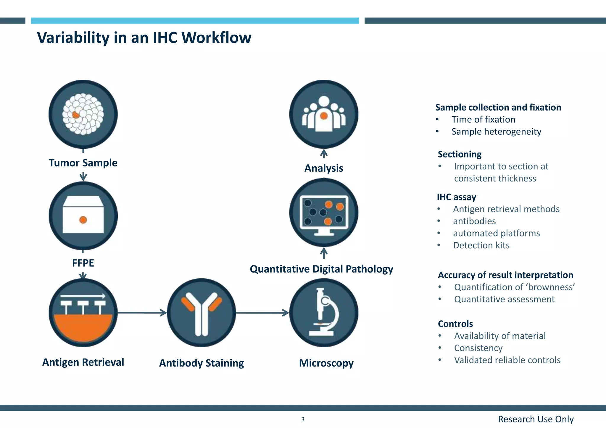
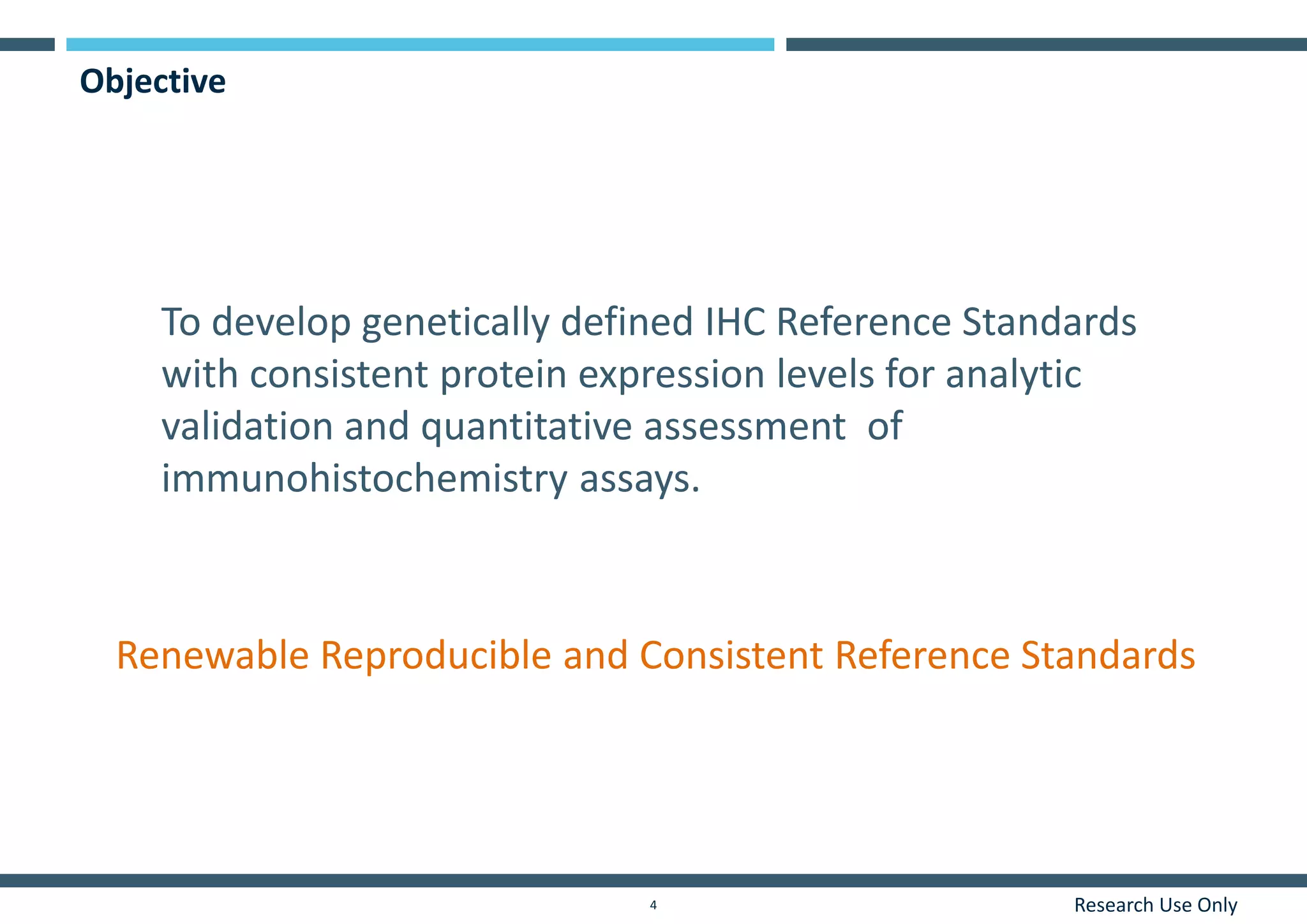
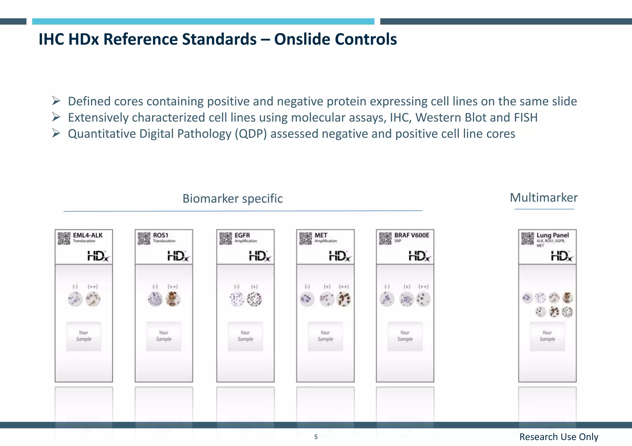
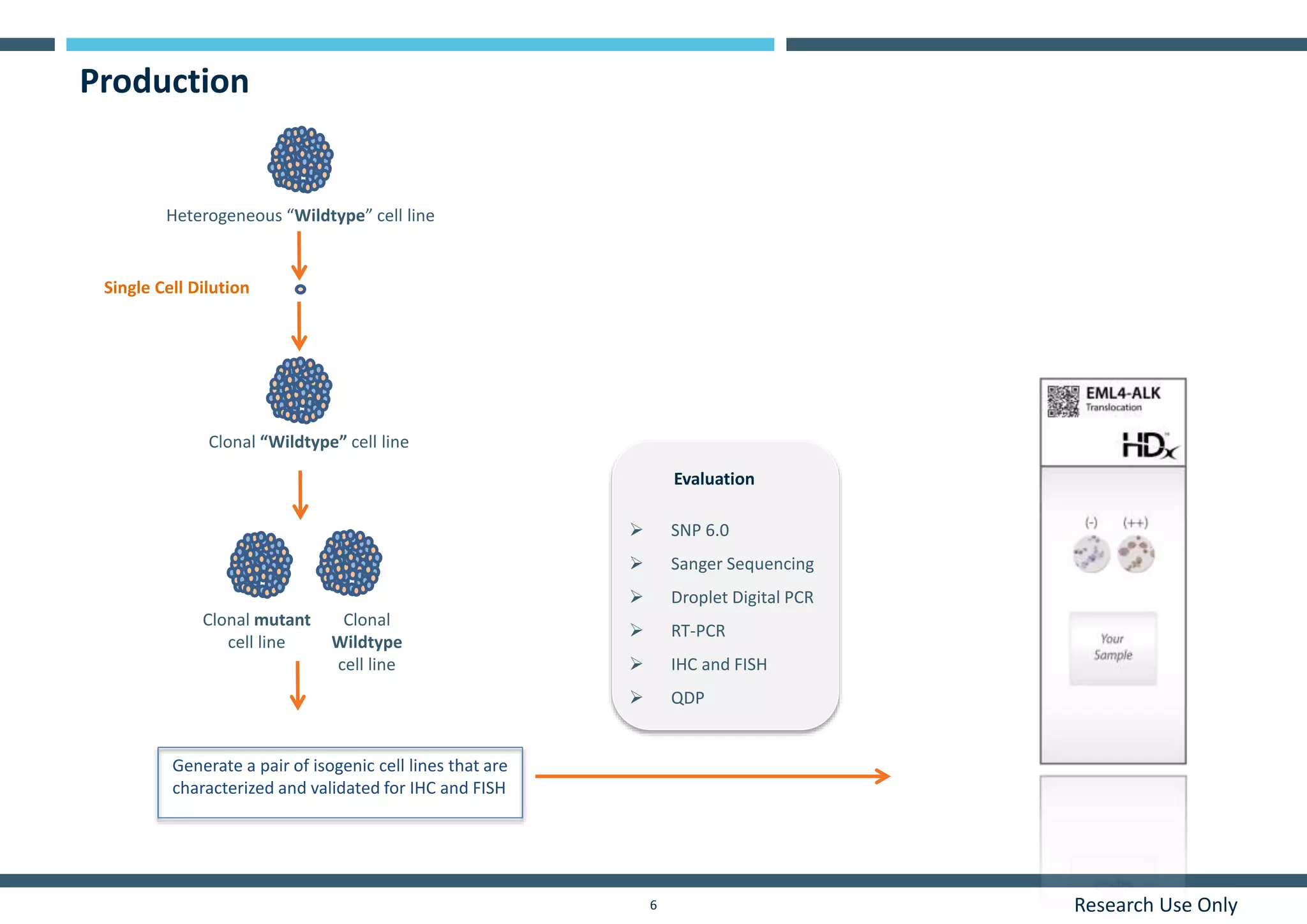
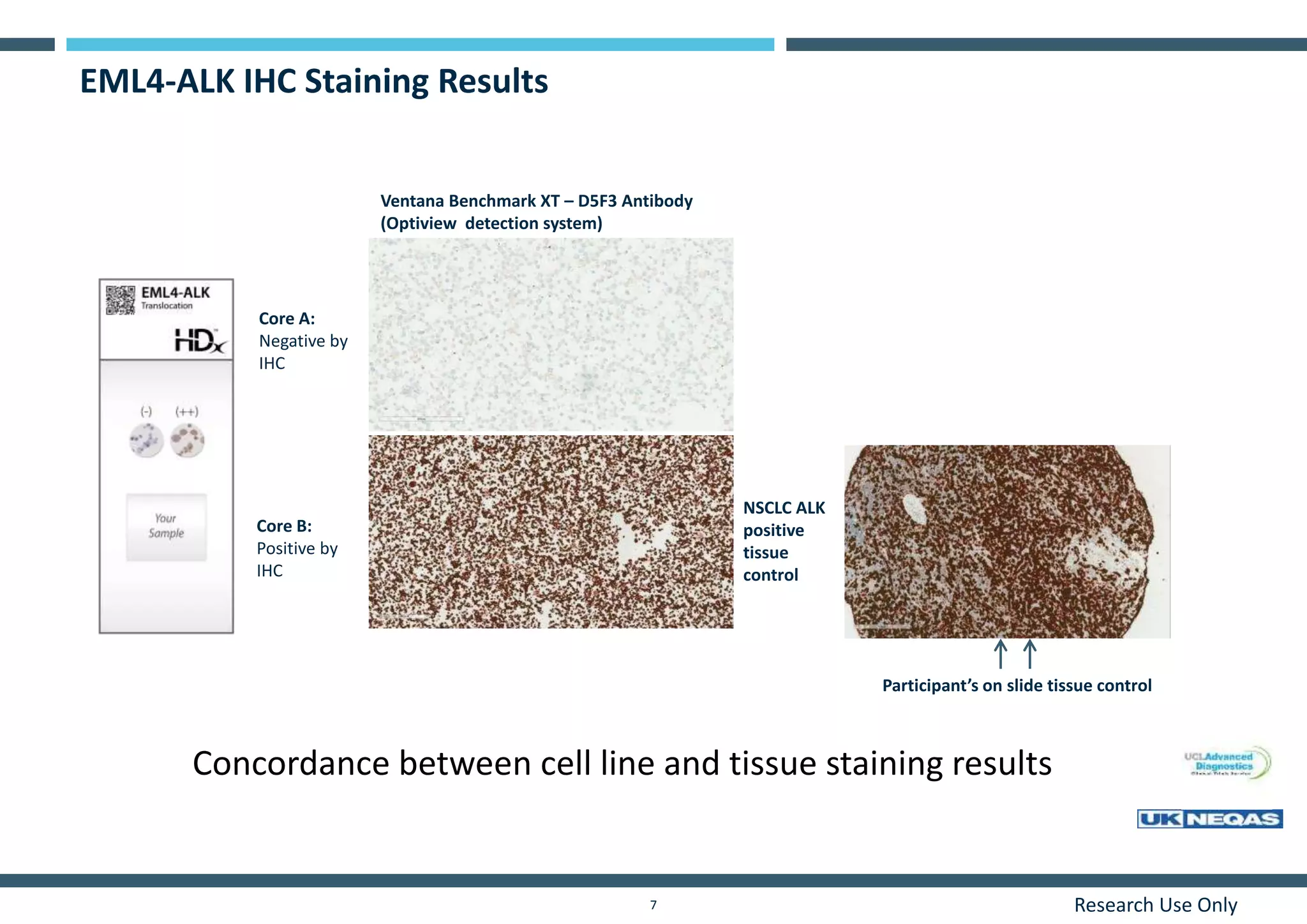
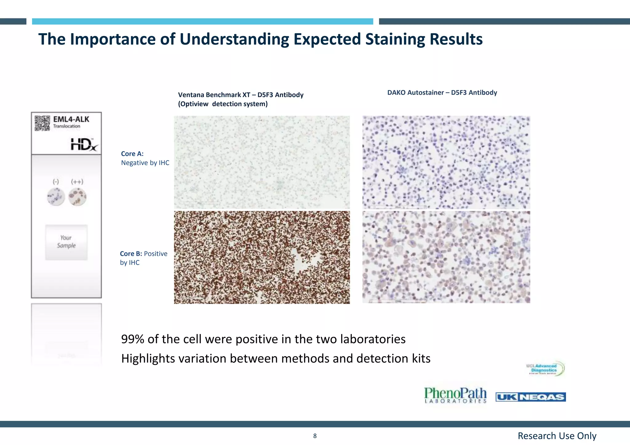
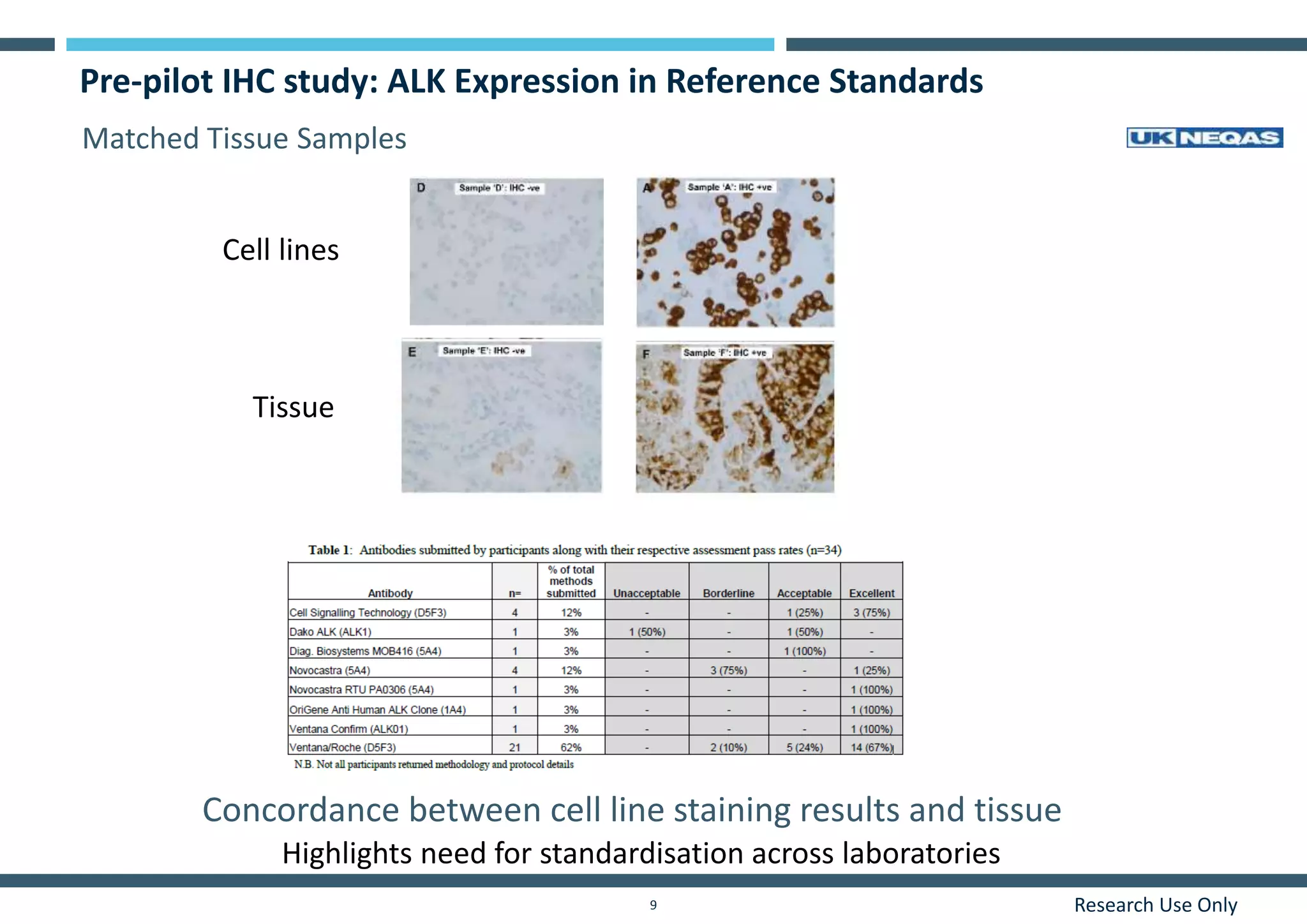
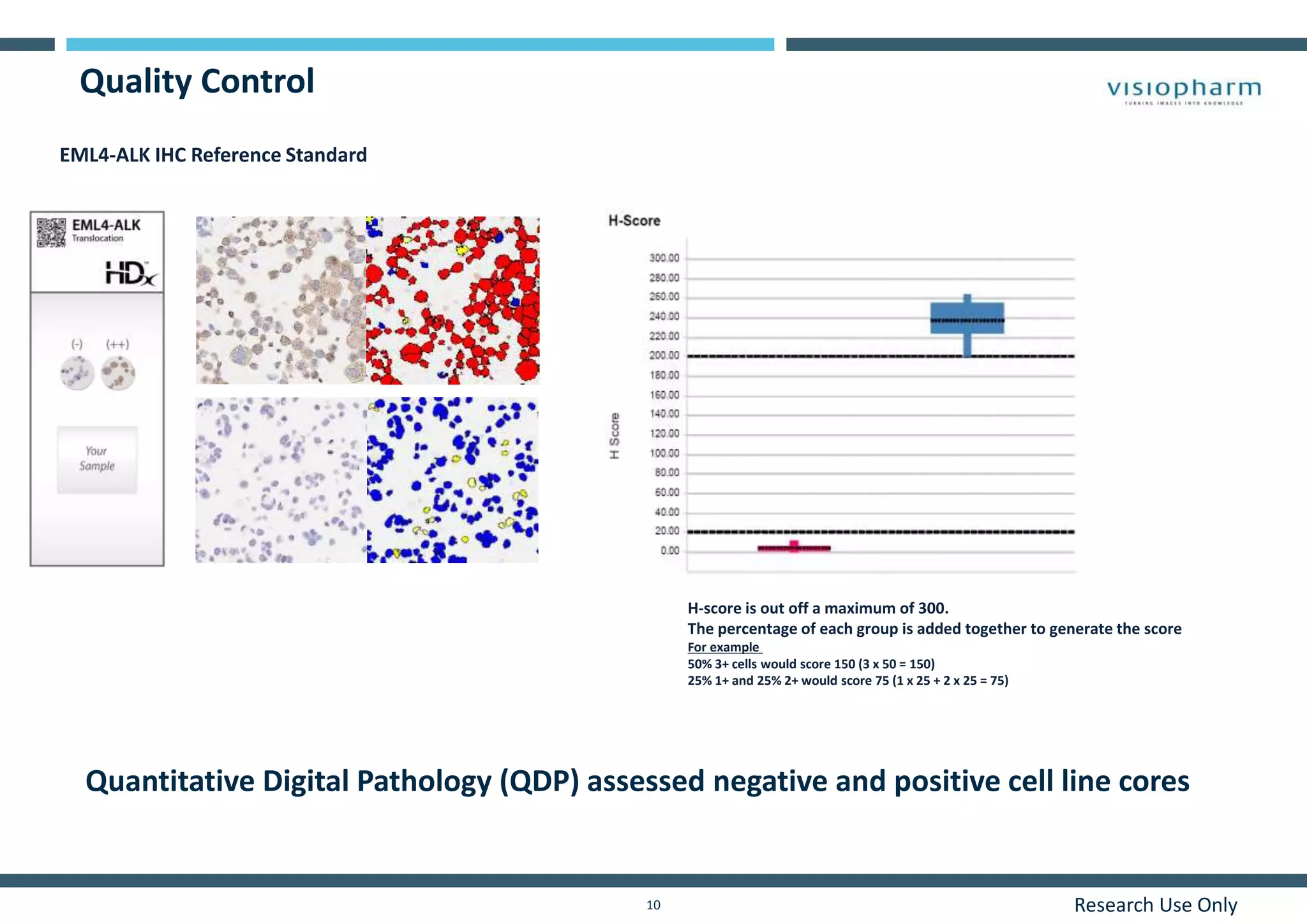
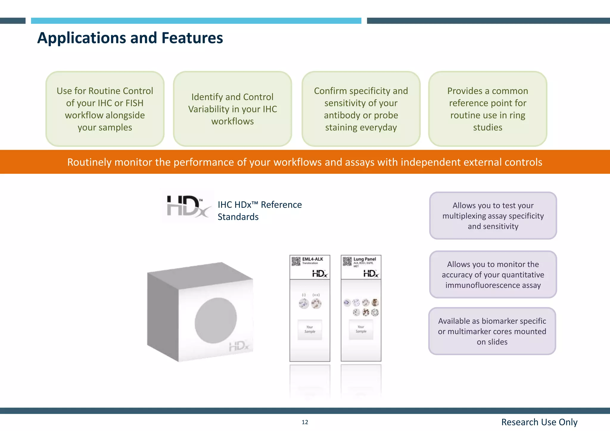
HORIZON DIAGNOSTICS
Research Use Only
Improving Immunohistochemistry
Standardization in your Laboratory
1
Dr Farah Patell-Socha and Dr Vicky Spivey
Research Use Only2
What is the impact of assay failure in
your laboratory and how do you
monitor for it?
Research Use Only3
Variability in an IHC Workflow
IHC assay
• Antigen retrieval methods
• antibodies
• automated platforms
• Detection kits
Sample collection and fixation
• Time of fixation
• Sample heterogeneity
Accuracy of result interpretation
• Quantification of ‘brownness’
• Quantitative assessment
Sectioning
• Important to section at
consistent thickness
Antigen Retrieval
FFPE
Antibody Staining Microscopy
Tumor Sample Analysis
Quantitative Digital Pathology
Controls
• Availability of material
• Consistency
• Validated reliable controls
Research Use Only4
Objective
To develop genetically defined IHC Reference Standards
with consistent protein expression levels for analytic
validation and quantitative assessment of
immunohistochemistry assays.
Renewable Reproducible and Consistent Reference Standards
Research Use Only5
IHC HDx Reference Standards – Onslide Controls
 Defined cores containing positive and negative protein expressing cell lines on the same slide
 Extensively characterized cell lines using molecular assays, IHC, Western Blot and FISH
 Quantitative Digital Pathology (QDP) assessed negative and positive cell line cores
(
-
)
Biomarker specific Multimarker
Research Use Only6
Production
Single Cell Dilution
Heterogeneous “Wildtype” cell line
Clonal “Wildtype” cell line
Generate a pair of isogenic cell lines that are
characterized and validated for IHC and FISH
Clonal mutant
cell line
Clonal
Wildtype
cell line
Evaluation
 SNP 6.0
 Sanger Sequencing
 Droplet Digital PCR
 RT-PCR
 IHC and FISH
 QDP
Research Use Only7
EML4-ALK IHC Staining Results
Core A:
Negative by
IHC
NSCLC ALK
positive
tissue
control
Core B:
Positive by
IHC
Participant’s on slide tissue control
Ventana Benchmark XT – D5F3 Antibody
(Optiview detection system)
Concordance between cell line and tissue staining results
Research Use Only8
The Importance of Understanding Expected Staining Results
Core A:
Negative by IHC
Core B: Positive
by IHC
DAKO Autostainer – D5F3 AntibodyVentana Benchmark XT – D5F3 Antibody
(Optiview detection system)
99% of the cell were positive in the two laboratories
Highlights variation between methods and detection kits
Research Use Only9
Pre-pilot IHC study: ALK Expression in Reference Standards
Cell lines
Tissue
Concordance between cell line staining results and tissue
Highlights need for standardisation across laboratories
Matched Tissue Samples
Research Use Only10
Quality Control
EML4-ALK IHC Reference Standard
H-score is out off a maximum of 300.
The percentage of each group is added together to generate the score
For example
50% 3+ cells would score 150 (3 x 50 = 150)
25% 1+ and 25% 2+ would score 75 (1 x 25 + 2 x 25 = 75)
Quantitative Digital Pathology (QDP) assessed negative and positive cell line cores
Research Use Only11
Key Points
 Characterized and genetically defined cell line standards
 Concordance between cell line and tissue staining results
 Highlights variation between methods and detection kits and highlights the need
for external controls
 Quantitative Digital Pathology (QDP) assessed negative and positive cell line cores
Research Use Only12
Applications and Features
Confirm specificity and
sensitivity of your
antibody or probe
staining everyday
Available as biomarker specific
or multimarker cores mounted
on slides
Routinely monitor the performance of your workflows and assays with independent external controls
Allows you to test your
multiplexing assay specificity
and sensitivity
Allows you to monitor the
accuracy of your quantitative
immunofluorescence assay
IHC HDx™ Reference
Standards
Use for Routine Control
of your IHC or FISH
workflow alongside
your samples
Identify and Control
Variability in your IHC
workflows
Provides a common
reference point for
routine use in ring
studies
Research Use Only13
Characterized consistent, reproducible and renewable
Reference Standards are ideal as External Controls
Is your assay
optimised?
What is the limit of
detection of your
workflow?
Are you interested in
achieving in
consistent results?
What is the impact of assay failure in
your laboratory and how do you
monitor for it?
Research Use Only14
Contact us!
Characterized consistent, reproducible and renewable
Reference Standards are ideal as External Controls
Characterized consistent, reproducible and renewable
Reference Standards are ideal as External Controls
Contact us:
f.patell-socha@horizondiscovery.com
v.spivey@horizondiscovery.com

Improving Immunohistochemistry Standardization in your Laboratory: Renewable Reproducible and Consistent Reference Standards

  • 1.
    HORIZON DIAGNOSTICS Research UseOnly Improving Immunohistochemistry Standardization in your Laboratory 1 Dr Farah Patell-Socha and Dr Vicky Spivey
  • 2.
    Research Use Only2 Whatis the impact of assay failure in your laboratory and how do you monitor for it?
  • 3.
    Research Use Only3 Variabilityin an IHC Workflow IHC assay • Antigen retrieval methods • antibodies • automated platforms • Detection kits Sample collection and fixation • Time of fixation • Sample heterogeneity Accuracy of result interpretation • Quantification of ‘brownness’ • Quantitative assessment Sectioning • Important to section at consistent thickness Antigen Retrieval FFPE Antibody Staining Microscopy Tumor Sample Analysis Quantitative Digital Pathology Controls • Availability of material • Consistency • Validated reliable controls
  • 4.
    Research Use Only4 Objective Todevelop genetically defined IHC Reference Standards with consistent protein expression levels for analytic validation and quantitative assessment of immunohistochemistry assays. Renewable Reproducible and Consistent Reference Standards
  • 5.
    Research Use Only5 IHCHDx Reference Standards – Onslide Controls  Defined cores containing positive and negative protein expressing cell lines on the same slide  Extensively characterized cell lines using molecular assays, IHC, Western Blot and FISH  Quantitative Digital Pathology (QDP) assessed negative and positive cell line cores ( - ) Biomarker specific Multimarker
  • 6.
    Research Use Only6 Production SingleCell Dilution Heterogeneous “Wildtype” cell line Clonal “Wildtype” cell line Generate a pair of isogenic cell lines that are characterized and validated for IHC and FISH Clonal mutant cell line Clonal Wildtype cell line Evaluation  SNP 6.0  Sanger Sequencing  Droplet Digital PCR  RT-PCR  IHC and FISH  QDP
  • 7.
    Research Use Only7 EML4-ALKIHC Staining Results Core A: Negative by IHC NSCLC ALK positive tissue control Core B: Positive by IHC Participant’s on slide tissue control Ventana Benchmark XT – D5F3 Antibody (Optiview detection system) Concordance between cell line and tissue staining results
  • 8.
    Research Use Only8 TheImportance of Understanding Expected Staining Results Core A: Negative by IHC Core B: Positive by IHC DAKO Autostainer – D5F3 AntibodyVentana Benchmark XT – D5F3 Antibody (Optiview detection system) 99% of the cell were positive in the two laboratories Highlights variation between methods and detection kits
  • 9.
    Research Use Only9 Pre-pilotIHC study: ALK Expression in Reference Standards Cell lines Tissue Concordance between cell line staining results and tissue Highlights need for standardisation across laboratories Matched Tissue Samples
  • 10.
    Research Use Only10 QualityControl EML4-ALK IHC Reference Standard H-score is out off a maximum of 300. The percentage of each group is added together to generate the score For example 50% 3+ cells would score 150 (3 x 50 = 150) 25% 1+ and 25% 2+ would score 75 (1 x 25 + 2 x 25 = 75) Quantitative Digital Pathology (QDP) assessed negative and positive cell line cores
  • 11.
    Research Use Only11 KeyPoints  Characterized and genetically defined cell line standards  Concordance between cell line and tissue staining results  Highlights variation between methods and detection kits and highlights the need for external controls  Quantitative Digital Pathology (QDP) assessed negative and positive cell line cores
  • 12.
    Research Use Only12 Applicationsand Features Confirm specificity and sensitivity of your antibody or probe staining everyday Available as biomarker specific or multimarker cores mounted on slides Routinely monitor the performance of your workflows and assays with independent external controls Allows you to test your multiplexing assay specificity and sensitivity Allows you to monitor the accuracy of your quantitative immunofluorescence assay IHC HDx™ Reference Standards Use for Routine Control of your IHC or FISH workflow alongside your samples Identify and Control Variability in your IHC workflows Provides a common reference point for routine use in ring studies
  • 13.
    Research Use Only13 Characterizedconsistent, reproducible and renewable Reference Standards are ideal as External Controls Is your assay optimised? What is the limit of detection of your workflow? Are you interested in achieving in consistent results? What is the impact of assay failure in your laboratory and how do you monitor for it?
  • 14.
    Research Use Only14 Contactus! Characterized consistent, reproducible and renewable Reference Standards are ideal as External Controls Characterized consistent, reproducible and renewable Reference Standards are ideal as External Controls Contact us: f.patell-socha@horizondiscovery.com v.spivey@horizondiscovery.com