2. What is the cell cycle?
All cells are derived from pre-existing cells
Cell cycle: defined as ordered sequences of events that
occurs in the cell in preparation of cell division.
The Cell cycle has two basic Function :
Copying cellular components and DNA duplication
Dividing the cell so that components are distributed evenly to
the daughter cells
The alternating “growth” and “division” activities of the
cell is called the “cell cycle”.
Cell division is an integral part of the cell cycle.
4. •In unicellular organisms, division of one cell
reproduces the entire organism (reproduction)
•Multicellular organisms depend on cell division for
• Growth (increase in numbers) and maintained
and repaired (adults renewal e.g. liver, skin).
Why the cell division occur?
Are all cells divide?
•NO some cell can’t divide e.g eye lens, nerve cell,
heart cells. Theses cells maintained and repaired by
replacing the intracellular component.
5. Types of Cell Reproduction
5
Asexual reproduction
involves a (smatic cell) single cell dividing to make 2 new,
identical daughter cells (eg.Mitosis)
Sexual reproduction
involves two cells (egg & sperm) joining to make a new
cell (zygote) that is NOT identical to the original cells (
e.g.Meiosis )
All diploid chromosomes is homologous except six
chromosome in male (y from father & x from mother)
Diploid (46
chr)
Haploid (23 chr)
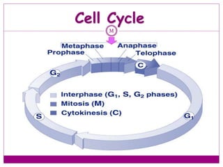
6. The “growth” activity corresponds to “Interphase”.
cell growth and copying of chromosomes in
preparation for cell division
The division activity corresponds to “M phase”.
What is the cell cycle basic function?
(mitosis andcytokinesis)
Interphase M Interphase M Interphase
G1 S G2 M G1 S G2 M G1 S G2
10. Interphase
G1 & G2 gap:
-The cell grows and mass protein and organelles are
duplicated.
-The cell monitor the internal and external
environment to ensure the condition suitable and
prepare for S and M.
-In unfavorable extracellular condition the cell delay
progression through G1.
-The cell may enter in G0 (resting state) it may stay in
until die or until the condition become favorable and
cell can row and divide.
11. Interphase
In yeast “Start” is at the end of G1; at this point the cell is
committed to DNA synthesis.
In mammals, this is called the “restriction point”. This point
late in G1 is a “checkpoint”; a cell will exit the cell cycle if
certain requirements to proceed to synthesis are not met.
After passing this point even the signal stimulating cell growth
removed the DNA will synthesis.
A second restriction point occurs in G2 before entry into
mitosis.
12. Interphase: G1
Events during G1
1 st stage of Cell growth after cell division
Preparation of chromosomes for replication
Duplication of cellular components (cytoplasm and organel)
Cell carries on its normal metabolic activities
G1 checkpoint (or restriction point); cell commits to division or
exits from cell cycle
13. S- phase
The instructions for making cell parts are
encoded in the DNA, so each new cell must
get a complete set of the DNA molecules
DNA must be copied or replicated before
cell division
Each new cell will then have an identical
copy of the DNA
13
Original DNA
strand
Two new, identical
DNA strands
14. Duplicated chromosomes
are called chromatids
& are held together by
the centromere
14Called Sister Chromatids
S- phase
15. DNA replication
Duplication of the centrosome
The centrosome is located near the nucleus of the cell and
contains the microtubule organizing center MTOC in animal
cells. It contains two centrioles surrounding by loose
collection of protein that migrate to the poles before cell
division and serve to organize the spindle.
S- phase
16. Interphase: G2
2 nd Cell growth stage
Occurs after DNA has been copied
All cell structures needed for division are made (e.g.
centrioles)
Both organelles & proteins are synthesized
Checkpoint (restriction point) for entry into M phase
19. PROPHASE
the first phase in mitosis and longest phase of mitosis.
THREE THINGS TO LOOK FOR:
1.chromosomes can be visible under light microscope as two
chromatids, in the shape of an “X” (chromatin condenses to
form chromosomes)
2.Nuclear envelope dissolves ( begin disagrregate)
3.Centrioles are present with some spindle fibers and the sister
chromatids are attached by their kinetochores to microtubules
from opposite poles
20. prophase
•kinetochore is a protein strucutre on
chromatide where microtubules will
bind to it in cell division to pull the
two sister chromatide a part
•each sister chromatid has its own
kinetochore (arise from centromer)
•sister chromatids become attached
by their kinetochores to microtubules
from opposite poles
•Used in cell division (mitosis &
miosis)
22. prometaphase
a system of
microtubules, called
the mitotic spindle,
organizes between the
two poles (opposite
ends) of the cell
each pole has a
microtubule organizing
center (MTOC)
in animals and some
other eukaryotes,
centrioles are found in
the MTOC
23. Late Prophase (prometaphase)
23
Nuclear membrane & nucleolus are broken
down
Chromosomes continue condensing & are
clearly visible
Spindle fibers called kinetochores attach to
the centromere of each chromosome
Spindle finishes forming between the poles of
the cell
25. METAPHASE
1.Chromosomes chromosomes are highly condensed line
up in the middle (meta phase plate)
2.Nuclear envelope is gone (no nucleus)
3.Spindle fibers (on opposite poles) are stretching
towards the chromosomes
4- the mitosis checkpoint appears to be here; progress
past metaphase is typically prevented until the
kinetochores are all attached to microtubules
28. Anaphase
Occurs rapidly
Sister chromatids are
pulled apart to
opposite poles of the
cell by kinetochore
fibers
the protein
tethers at the
centromere
between the
chromatids are
broken
each former sister
chromatid can
now be called a
chromosome
(daughter)
28
31. Telophase
31
Sister chromatids at opposite poles
Spindle disassembles
Nuclear envelope forms around each set of
sister chromatids and chromosome
condensed
Nucleolus reappears
CYTOKINESIS occurs (furrow)
Chromosomes reappear as chromatin
32. Mitosis: telophase
prophase is essentially
reversed
the mitotic spindle is
disintegrated
the chromosomes decondense
nuclear membranes reform
around the genetic material to
form two nuclei
each has an identical copy of
the genetic information
nucleoli reappear, and
interphase cellular functions
resume
*
35. Cell Division in Prokaryotes
Prokaryotes such as
bacteria divide into 2
identical cells by the
process of binary
fission
Single chromosome
makes a copy of
itself
Cell wall forms
between the
chromosomes dividing
the cell
35
Parent cell
2 identical daughter cells
Chromosome
doubles
Cell splits
36. What is the Cell Cycle?
Parent cells are diploid and make 2 daughter cells that
are also diploid with their own new nuclei.
Diploid means 2 of each chromosome: 2 (n)= 2
(23) = 46 chromosomes
It happens in all of your somatic(body) cells in order to
get the same DNA inside each cell. (your reproductive
cells do something different)
I P M A T C
I Peed on the MAT, see?
39. cytokinesis
Means division of the
cytoplasm
divides the cell into two
daughter cells
(cytoplasm, organelles
cytokinesis usually
begins in telophase and
ends shortly thereafter
41. Daughter Cells of Mitosis
41
Have the same number of chromosomes as
each other and as the parent cell from which
they were formed
Identical to each other, but smaller than
parent cell
Must grow in size to become mature cells (G1
of Interphase)
Ready to enter in new cell cycle
47. Name the Mitotic Stages:
47Interphase
Prophase
Metaphase
Anaphase
Telophase
Name this?
Name this?
48. Uncontrolled Mitosis
If mitosis is not
controlled, unlimited
cell division occurs
causing cancerous
tumors
Oncogenes are special
proteins that increase
the chance that a
normal cell develops
into a tumor cell
48
Cancer cells
53. Figure 12.16
G1 checkpoint
G1 G1
G0
(a) Cell receives a go-ahead
signal.
(b) Cell does not receive a
go-ahead signal.
54. Eukaryotic Cell Cycle
cyclins and cyclin-dependent protein kinases (Cdks)
cytokinins; growth factors; suppressors; cancer cells
55. Cell Cycle Checkpoints
The decision to proceed from one part of the cell cycle to
another depends on a variety of factors
Growth
DNA replication
DNA integrity
Cellular integrity
The mechanisms that the cell has to monitor these factors act at
“checkpoints”
Generally, the feedback from checkpoints is through negative
regulation—sending a signal to stop the progression of the cell cycle
rather than dialing back a positive signal.
Schematic of Cell Cycle Checkpoints
56. Cell Cycle Checkpoints
G1 (Restriction) Checkpoint
DNA Replication Checkpoint (end of G2)
Cell will not proceed with mitosis if DNA replication is not
complete
Cells with mutations in this checkpoint pathway or cultured
mammalian cells treated with caffeine will proceed through
mitosis with unreplicated DNA.
57. Cell Cycle Arrest
Cells have “checkpoints” where they “proof-read” DNA for damage
before continuing to cycle. This ensures faithful chromosome replication
and maintains genomic integrity.
Irradiation causes cells to arrest at these checkpoints
Cells tend to arrest at
• G1 - especially if they have wt p53. This may lead to apoptosis
• Intra S phase - initiation and elongation stages of DNA replication
are affected by p53 independent mechanisms
• G2 - most cells arrest here - allows chromatid repair prior to
segregation in M
• M phase - block in anaphase until all sister chromatids
have aligned properly on the spindle - Monitors spindle
integrity for cytokinesis
58. Cell Cycle Checkpoints
Growth checkpoints
In budding yeasts, division produces a small daughter cell and
a large mother cell. The daughter cell spends a longer time
growing in G1 before it can divide again. There is a minimum
size that must be reached before S phase can begin.
In animal cells, external growth factors play a major role in
providing signals about cell growth and differentiation and
regulating the cell cycle.
59. Cell Cycle Checkpoints
Spindle-attachment checkpoint
Before anaphase (separation of chromosomes) there is a
checkpoint to ensure the chromatids are correctly attached to
the mitotic spindle
The kinetochore (where the chromatids attach to the spindle)
is the structure that is monitored
60. Cell Cycle Checkpoints
Exiting Mitosis
Degradation of the M phase cyclin/cdk complex (aka MPF) is
required to proceed with the final activities of mitosis (spindle
disassembly and formation of the nuclear envelopes).
This degradation is accomplished by ubiquitinylation of the
complex.
The cdc20-APC complex is responsible for signaling the
degradation and exit from mitosis.