SlideShare a Scribd company logo
1 of 20
Starr et al., 2013. Chapter 8 and 11 p. 124-125 and 164-171, Edulink and
learning guide notes
 Chromosomes form when the chromatin network in the
nucleus of the cell, coil up, shortens and thickens.
 Each organism has a specific amount of chromosomes.
 All humans have 46 chromosomes.
 These chromosomes are arranged in identical pairs
called homologous chromosome pairs –
 Therefore humans have 23 homologous pairs of
chromosomes.
 These chromosomes is only visible during cell division
processes.
 One chromosome consist of 2 chromatids and one
centromere that attach the chromatids together.
 Each chromatid consist of genes which in turn consist of
DNA.
X andY chromosomes:
gonosomes/sex-chromosomes
Colored bands
represents different
genes
The cell cycle includes the following phases:
 Interphase (include G1- , S- [DNA synthesis]
and G2 phase)
 Mitosis
 Cytokinesis
 Growth
The result is 2 identical cells.
 Allow an organism to
grow.
 Repairs damaged
cells/tissue.
 Replace dead cells/tissue.
 Reproduction in some
simple organisms.
 In all somatic cells (include all body cells and
excludes the sex cells – sperm/egg cells)
 CONSIST OF A FEW PHASES:
 INTERPHASE
 PROPHASE
 METAPHASE
 ANAPHASE
 TELOPHASE
BioFlix: Mitosis
 Cell builds up enough energy for division
process.
 DNA replication occurs
 Cell look normal, like before division
 Nuclear envelope and nucleolus
disappear.
 Chromatin become more tightly
coiled, and condenses into
individual chromosomes.
Chromosomes arrange randomly
in the cell.
 Centrioli move to opposite poles,
with spindle fibers stretching
between them.
 The centrioli reached the opposite poles with
the spindle fibers in between.
 The chromosomes arrange randomly on the
equator, each single chromosome attaching
to a separate spindle fiber by means of the
centromere.
 The spindle fibers pull tight.
 The centromers attaching the chromatids of
the chromosomes split in half.
 Daughter chromosomes move to opposite
poles.
 Daughter chromosomes reach
poles.
 Nuclear envelope surrounds
chromosomes.
 Nucleolus reappear at each pole.
 Chromosomes become less
condense forming chromatin.
 Two identical nuclei has been
formed
 Invagination of the cytoplasm and plasma
membrane occurs. (Cleavage furrow forms in
animal cells and a cytoplasmic plate forms in
plant cells)
 Continues until the cell in divided into 2
separate cells. (Identical to one another and
to the original cell)
 Cancer is caused by the loss of cell cycle controls
in cancer cells.
 Cancer cells usually continue to divide well
beyond a single layer, forming a clump of
overlapping cells called a tumor.
 Malignant tumors invade surrounding tissues and
can metastasize exporting cancer cells to other
parts of the body, where they may form secondary
tumors.
 They do not exhibit anchorage dependence or
density dependent inhibition.
Ovarian cancer cells
dividing
 Divide into groups of 8.
 Design a research project for students using
the themeCancer.
 Complete the actual research project that
you have designed in detail as an example of
what you expect from a student.
Design
 Originality of the project 10
 Planning of the design 10
 Assessment you will be using 10
 Assessment criteria you will be using 10
 Design construction and set up. 10
 Clarity of task 10
 Cover page 10
 Index 10
 Content – scientific and true – no plagiarism 10
 Reference indicated in content 10
 Summary of references provided in Bibliography. 10
 Includes diagrams/ graphs/ tables/ statistics 10
 Includes own opinion and ideas. 10
 Neatness 10
 Overall impression (Did you do what the project required
you to do) 10
Total : 150
HAND IN 10 MAY 2013

More Related Content

What's hot (20)

Presentation1
Presentation1Presentation1
Presentation1
 
Mitosis
MitosisMitosis
Mitosis
 
Mitosis powerpoint
Mitosis powerpointMitosis powerpoint
Mitosis powerpoint
 
Mitosis pp presentation for gr 10 life sciences
Mitosis pp presentation for gr 10 life sciencesMitosis pp presentation for gr 10 life sciences
Mitosis pp presentation for gr 10 life sciences
 
Mitosis
MitosisMitosis
Mitosis
 
Cell division
Cell divisionCell division
Cell division
 
Presentation1
Presentation1Presentation1
Presentation1
 
Prof slides
Prof slidesProf slides
Prof slides
 
Introduction to mitosis
Introduction to mitosisIntroduction to mitosis
Introduction to mitosis
 
Mitosis
MitosisMitosis
Mitosis
 
Mitosis: An Interactive PowerPoint
Mitosis: An Interactive PowerPointMitosis: An Interactive PowerPoint
Mitosis: An Interactive PowerPoint
 
Mitosis meiosis powerpoint.ppsx
Mitosis meiosis powerpoint.ppsxMitosis meiosis powerpoint.ppsx
Mitosis meiosis powerpoint.ppsx
 
Cell cycle
Cell cycleCell cycle
Cell cycle
 
Cell division
Cell divisionCell division
Cell division
 
Cell division grade 10
Cell division grade 10Cell division grade 10
Cell division grade 10
 
Life Science Grade 10-Cell Division
Life Science Grade 10-Cell DivisionLife Science Grade 10-Cell Division
Life Science Grade 10-Cell Division
 
MITOSIS: LIFE SCIENCES FOR GRADE 10`S
MITOSIS: LIFE SCIENCES FOR GRADE 10`SMITOSIS: LIFE SCIENCES FOR GRADE 10`S
MITOSIS: LIFE SCIENCES FOR GRADE 10`S
 
Stages of mitosis final project itp
Stages of mitosis final project itpStages of mitosis final project itp
Stages of mitosis final project itp
 
Glossary DNA Cards
Glossary  DNA CardsGlossary  DNA Cards
Glossary DNA Cards
 
Chromosome rey b.
Chromosome   rey b.Chromosome   rey b.
Chromosome rey b.
 

Similar to Chromosomes and Cell Cycle Notes

Mitosis simphiwe
Mitosis simphiweMitosis simphiwe
Mitosis simphiweuj
 
Unit 3 mitosis2
Unit 3 mitosis2Unit 3 mitosis2
Unit 3 mitosis2oupas101
 
Cell division
Cell divisionCell division
Cell divisionbaasa
 
2.5 Cell Cycle and Mitosis PPT
2.5 Cell Cycle and Mitosis PPT2.5 Cell Cycle and Mitosis PPT
2.5 Cell Cycle and Mitosis PPTFernanda Silva
 
Chromosomal basis of heredity
Chromosomal basis of heredityChromosomal basis of heredity
Chromosomal basis of heredityBruno Mmassy
 
Chromosomes and cell cycle
Chromosomes and cell cycleChromosomes and cell cycle
Chromosomes and cell cyclejpochne
 
Mitosis and cell reproduction
Mitosis and cell reproductionMitosis and cell reproduction
Mitosis and cell reproductionPSPree
 
8 9 mitosis meiosis_and_genetics
8 9 mitosis meiosis_and_genetics8 9 mitosis meiosis_and_genetics
8 9 mitosis meiosis_and_geneticsJaden Francis
 
Unit 3 cell division
Unit 3 cell divisionUnit 3 cell division
Unit 3 cell divisionmoselane
 
Animal development lsc presentation
Animal development lsc presentationAnimal development lsc presentation
Animal development lsc presentationKoketso Pila
 

Similar to Chromosomes and Cell Cycle Notes (20)

Mitosis simphiwe
Mitosis simphiweMitosis simphiwe
Mitosis simphiwe
 
Unit 3 mitosis2
Unit 3 mitosis2Unit 3 mitosis2
Unit 3 mitosis2
 
5.1 Mitosis
5.1 Mitosis5.1 Mitosis
5.1 Mitosis
 
5.1 Mitosis
5.1 Mitosis5.1 Mitosis
5.1 Mitosis
 
Chapter8
Chapter8Chapter8
Chapter8
 
Cell division
Cell divisionCell division
Cell division
 
Mitosis
MitosisMitosis
Mitosis
 
Mitosis
MitosisMitosis
Mitosis
 
2.5 Cell Cycle and Mitosis PPT
2.5 Cell Cycle and Mitosis PPT2.5 Cell Cycle and Mitosis PPT
2.5 Cell Cycle and Mitosis PPT
 
Chromosomal basis of heredity
Chromosomal basis of heredityChromosomal basis of heredity
Chromosomal basis of heredity
 
Chromosomes and cell cycle
Chromosomes and cell cycleChromosomes and cell cycle
Chromosomes and cell cycle
 
Mitosis and cell reproduction
Mitosis and cell reproductionMitosis and cell reproduction
Mitosis and cell reproduction
 
Cell DIVISION/cycle
Cell DIVISION/cycleCell DIVISION/cycle
Cell DIVISION/cycle
 
8 9 mitosis meiosis_and_genetics
8 9 mitosis meiosis_and_genetics8 9 mitosis meiosis_and_genetics
8 9 mitosis meiosis_and_genetics
 
Cell DIVISION/cycle
Cell DIVISION/cycleCell DIVISION/cycle
Cell DIVISION/cycle
 
Meiosis Notes
Meiosis NotesMeiosis Notes
Meiosis Notes
 
Unit 3 cell division
Unit 3 cell divisionUnit 3 cell division
Unit 3 cell division
 
Mitosis
MitosisMitosis
Mitosis
 
Animal development lsc presentation
Animal development lsc presentationAnimal development lsc presentation
Animal development lsc presentation
 
Cell division 1
Cell division 1Cell division 1
Cell division 1
 

Chromosomes and Cell Cycle Notes

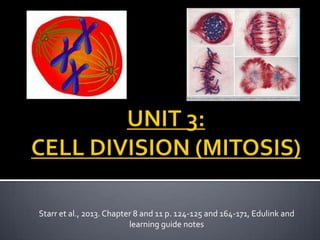
  • 1. Starr et al., 2013. Chapter 8 and 11 p. 124-125 and 164-171, Edulink and learning guide notes
  • 2.  Chromosomes form when the chromatin network in the nucleus of the cell, coil up, shortens and thickens.  Each organism has a specific amount of chromosomes.  All humans have 46 chromosomes.  These chromosomes are arranged in identical pairs called homologous chromosome pairs –  Therefore humans have 23 homologous pairs of chromosomes.  These chromosomes is only visible during cell division processes.  One chromosome consist of 2 chromatids and one centromere that attach the chromatids together.  Each chromatid consist of genes which in turn consist of DNA.
  • 3. X andY chromosomes: gonosomes/sex-chromosomes Colored bands represents different genes
  • 4. The cell cycle includes the following phases:  Interphase (include G1- , S- [DNA synthesis] and G2 phase)  Mitosis  Cytokinesis  Growth The result is 2 identical cells.
  • 5.  Allow an organism to grow.  Repairs damaged cells/tissue.  Replace dead cells/tissue.  Reproduction in some simple organisms.
  • 6.  In all somatic cells (include all body cells and excludes the sex cells – sperm/egg cells)
  • 7.  CONSIST OF A FEW PHASES:  INTERPHASE  PROPHASE  METAPHASE  ANAPHASE  TELOPHASE BioFlix: Mitosis
  • 8.  Cell builds up enough energy for division process.  DNA replication occurs  Cell look normal, like before division
  • 9.  Nuclear envelope and nucleolus disappear.  Chromatin become more tightly coiled, and condenses into individual chromosomes. Chromosomes arrange randomly in the cell.  Centrioli move to opposite poles, with spindle fibers stretching between them.
  • 10.  The centrioli reached the opposite poles with the spindle fibers in between.  The chromosomes arrange randomly on the equator, each single chromosome attaching to a separate spindle fiber by means of the centromere.
  • 11.  The spindle fibers pull tight.  The centromers attaching the chromatids of the chromosomes split in half.  Daughter chromosomes move to opposite poles.
  • 12.  Daughter chromosomes reach poles.  Nuclear envelope surrounds chromosomes.  Nucleolus reappear at each pole.  Chromosomes become less condense forming chromatin.  Two identical nuclei has been formed
  • 13.  Invagination of the cytoplasm and plasma membrane occurs. (Cleavage furrow forms in animal cells and a cytoplasmic plate forms in plant cells)  Continues until the cell in divided into 2 separate cells. (Identical to one another and to the original cell)
  • 14.
  • 15.  Cancer is caused by the loss of cell cycle controls in cancer cells.  Cancer cells usually continue to divide well beyond a single layer, forming a clump of overlapping cells called a tumor.  Malignant tumors invade surrounding tissues and can metastasize exporting cancer cells to other parts of the body, where they may form secondary tumors.  They do not exhibit anchorage dependence or density dependent inhibition.
  • 17.
  • 18.  Divide into groups of 8.  Design a research project for students using the themeCancer.  Complete the actual research project that you have designed in detail as an example of what you expect from a student.
  • 19. Design  Originality of the project 10  Planning of the design 10  Assessment you will be using 10  Assessment criteria you will be using 10  Design construction and set up. 10  Clarity of task 10
  • 20.  Cover page 10  Index 10  Content – scientific and true – no plagiarism 10  Reference indicated in content 10  Summary of references provided in Bibliography. 10  Includes diagrams/ graphs/ tables/ statistics 10  Includes own opinion and ideas. 10  Neatness 10  Overall impression (Did you do what the project required you to do) 10 Total : 150 HAND IN 10 MAY 2013