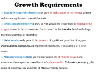
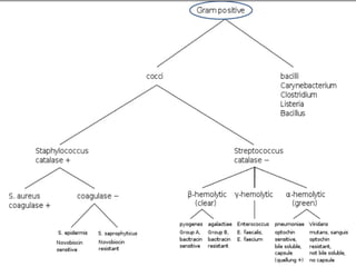
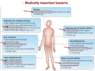
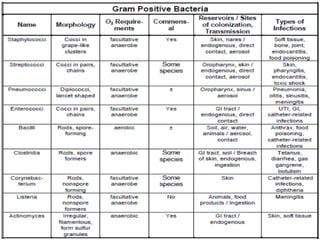
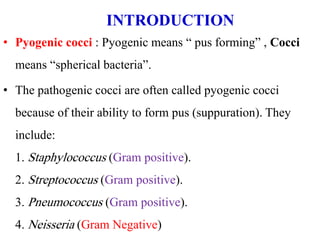
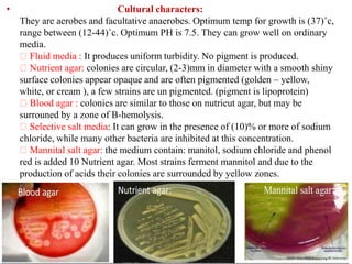
• The classification of bacteria serves a variety of different functions.
Because of this variety, bacteria may be grouped using many different
typing schemes.
• At present the typing schemes used by clinicians and clinical
microbiologists rely on phenotypic typing schemes. These schemes
utilize the bacterial morphology and staining properties of the organism,
as well as O2 growth requirements of the species combined with a
variety of biochemical tests.
• For clinicians the environmental reservoir of the organism, the vectors
and means of transmission of the pathogen are also of great importance.
Introduction
Classification of Bacteria
• On the Basis of Shape
• On the Basis of Staining
• On the Basis of Action
• On the Basis of Oxygen Requirement
• On the Basis of pH
• On the Basis of Temperature
• On the Basis of Structure
• On the Basis of Osmotic Pressure
Gram stain and bacterial morphology
Of all the different classification systems the, Gram stain has
withstood the test of time. Discovered by H.C. Gram in 1884 it
remains an important and useful technique to this day.
Gram positive bacteria stain blue-purple and Gram
negative bacteria stain red.
 The difference between the two groups is believed to be due to
a much larger peptidoglycan (cell wall) in Gram positives.
Some bacteria such as mycobacteria (the cause of
tuberculosis) are not reliably stained due to the large lipid
content of the peptidoglycan can called atypical bacteria.
Growth Requirements
Facultative anaerobic bacteria can grow in high oxygen or low oxygen content
and are among the more versatile bacteria.
strictly anaerobic bacteria grow only in conditions where there is minimal or no
oxygen present in the environment. Bacteria such as bacteroides found in the large
bowel are examples of anaerobes.
Strict aerobes only grow in the presence of significant quantities of oxygen.
Pseudomonas aeruginosa, an opportunistic pathogen, is an example of a strict
aerobe.
Microaerophilic bacteria grow under conditions of reduced oxygen and
sometimes also require increased levels of carbon dioxide. Neisseria species (e.g., the
cause of gonorrhea) are examples of Microaerophilic bacteria.
Serologic systems:
Selected antisera can be used to classify different bacterial species.
This may be based on either carbohydrate or protein antigens from
the bacterial cell wall or the capsular polysaccharide.
Environmental Reservoirs:
Environmental reservoirs are generally divided into those that are
endogenous (i.e., on or within the human body) and exogenous
(somewhere in the environment).
Genotypic systems:
Pattern or finger-print based techniques, sequence-based
techniques
Gram positive bacteria
• Have a large peptidoglycan structure.
• also capable of forming spores under stressful environmental
conditions such as when there is limited availability of carbon and
nitrogen. Spores therefore allow bacteria to survive exposure to extreme
conditions and can lead to reinfection (e.g., pseudomembranous colitis)
Distinguishing Features between Gram Positive and Negative Bacteria
Gram negative bacteria
• Have a small peptidoglycan layer but have an additional membrane, the
outer cytoplasmic membrane.
• This creates an additional permeability barrier and results in the need for
transport mechanisms across this membrane.
• A major component of the cytoplasmic membrane that is unique to Gram
negatives is endotoxin. Endotoxin has three components: the lipid A moiety,
the highly conserved core polysaccharide and the species specific O antigen
(also polysaccharide). In contrast with the secreted exotoxins, endotoxin is
cell associated but with bacterial division and death can be released. The
Lipid A moiety of endotoxin is responsible for sepsis and this may be fatal. It
is characterized clinically
by confusion, fever, drop in blood pressure and ultimately multi-organ failure.
• Propensity to cause disease, i.e., how likely are they to cause disease
Opportunistic pathogens – only cause disease in immunocompromised hosts
– AIDs patients
– Transplant patients on immunosuppressive drugs
– Cancer patients undergoing chemotherapy
– Patients who are already ill
– Opportunistic pathogens are often organisms that are typically normal flora.
Frank pathogens are always associated with disease
– Neisseria gonorrhoeae
– Shigella species
Facultative pathogens fall between the two extremes (opportunistic and frank)
and the majority of organisms that cause disease fall into this group
- Staphylococcus aureus and E. coli
Clinically significant bacteria
Virulence factors
Various factors involved in the host-parasite interaction determine
whether an organism will cause disease in the host:
– Virulence factors of the bacteria including:
• Capsules
• Pili
• IgA protease production
• Iron capturing ability
• Production of coagulase
• Production of toxins
• Ability to survive inside phagocytic cells.
Medically important bacteria
• Pyogenic cocci : Pyogenic means “ pus forming” , Cocci
means “spherical bacteria”.
• The pathogenic cocci are often called pyogenic cocci
because of their ability to form pus (suppuration). They
include:
1. Staphylococcus (Gram positive).
2. Streptococcus (Gram positive).
3. Pneumococcus (Gram positive).
4. Neisseria (Gram Negative)
INTRODUCTION
This microorganism is widely distributed in our
environment.
Some of them are members of the normal flora of humans,
Gram-positive , Lack spores and flagella, may have capsules.
Important human pathogens are like Staphylococcus aureus,
Staphylococcus epidermidis , Staphylococcus saprophyticus.
Genus Staphylococcus
• Resistance to environmental condition:
Staphylococcus aureus considered one of the hardest of all non
spore forming bacteria most strain are relatively heat stable, with
stand a temperature as high as 60°C for 30 min. also resist a high
concentration of salt (7.5% - 9% NaCl).
Staphylococci are spherical in shape usually arranged in grape
like clusters, grow rapidly on many types of ordinary bacterial
media ferment CHO and produced pigments varying from white to
deep yellow.
• Antigenic structure of
Staphylococcus aureus:
• Virulence factors
Peptidoglycan: a polysaccharide
polymer provides rigid exoskeleton of
the cell wall characterized by:
Stimulate the production of IL-1 and
opsonic antibodies.
The peptidoglycan has endotoxin
activity.
Teichoic acids:
They are polymers of glycerol or
ribitol phosphate. These polymers are
linked to the peptidoglycan and can be
antigenic.
Protein A :
Is a cell wall component of
Staphylococcus aureus strains
that binds to the FC portion of IgG
molecule.
• Extracellular substances: (enzymes and toxin)
Most extracellular substances produced by Staphylococcus aureus are
antigenic (stimulate the production of antibodies).
• These are:-
Catalase:
Is an enzyme that breaks hydrogen peroxide into O2 and H2O.
It’s useful in differentiation between members of genusStaphylococcus
aureus and genus Streptococcus.
Proteinase:
Is an enzyme which breaks down protein material.
Lipase: digest oils; enhances colonization on skin
Is an enzyme which breaks down lipid in the skin, lipoprotein in blood.
• Coagulase:
It’s an enzyme like protein; it clots citrated or oxalated human or rabbit
plasma in the presence of serum factors. produced by 97% of
human isolates; diagnostic.
This enzyme converts fibrinogen (soluble) into fibrin (insoluble).
Fibrin can be deposited on the surface of bacteria forming a wall
around the bacteria which has an important role in:
A. Protection of bacteria from phagocytosis.
B. Preventing the action of antibiotics.
• Hyaluronidase: digests connective tissue
Enzyme breaks down hyaluronic acid (is important substance in
binding tissue cells together therefore this enzyme helps the bacteria to
spread in the tissues. some time it is called (spreading factor)
• β - Lactamase:
break down β -lactam ring of penicillin.
• Staphylokinase: digests blood clots
 TOXINS
• Hemolysins (exotoxins): (α, β, γ) – lyse red blood cells
Include several toxins act on cell membrane of RBCs of Varity of
animal species.
• Leukocidin :lyses neutrophils and macrophages
Toxic substance causes degradation of WBC leading to cell death
• Exfoliative toxins: separates the epidermis from the dermis .
(scaled skin syndrome) this syndrome is common among small
children.
(it’s antigenic, stimulate production of antitoxins.
• Enterotoxins: induce gastrointestinal distress
Heat stable.
Resistant to the gut enzymes and acidity.
are the proteins that cause nausea, vomiting and diarrhea.
an important cause of food poisoning.
produced by Staphylococcus aureus growing in meat and dairy products.
• Toxic shock syndrome toxin 1 : (TSST-1):
Associated with toxic shock syndrome TSS.
Produced by some strains of Staphylococcus aureus in human this
toxin cause TSS which is a systemic infection characterized by high
fever, hypotension and shock with multisystem involvement.
• Staphylococci are variably sensitive to many antimicrobial drug
and resistance falls into several classes:
1. β- Lactamase production:
• This type of bacteria can be found in hospitals because above 90%
of hospitals strains of Staphylococcus aureus are β-lactamase
producer so antibiotics are useless when giving to the patients.
2. Resistance to naphcillin , methicillin and oxacillin
3. Tolerance: Indicates that Staphylococci are inhibited by the drug
but not killed (i.e. no growth, no multiplication and when removing the
drug the bacteria will grow and multiply).
Tolerance is due to the lack of the activation of autolytic enzymes in
the cell wall.
Pathogenicity of Staphylococcus aureus:
• Staphylococcus aureus
produces human infection by
its capability to invade and
spread into tissue and by
production of certain enzymes
and toxins.
• Disease spectrum: from
moderate to severe.
• Moderate infection:-
Range from localized to systemic
• Localized cutaneous infections – invade skin through wounds,
follicles, or glands
• folliculitis – superficial inflammation of hair follicle; usually
resolved with no complications but can progress
- furuncle – boil; inflammation of hair follicle or sebaceous gland
progresses into abscess or pustule
– carbuncle – larger and deeper lesion created by aggregation and
interconnection of a cluster of furuncles
– impetigo – bubble-like swellings that can break and peel away;
most common in newborns
• Sever infection:- (Bacteremia):
primary origin is bacteria from
another infected site or medical
devices; endocarditis possible
A. Osteomyelitis: Infection
reaches the bone by
hematogenous route. Infection is
established in the
metaphysis; abscess forms.
B. Pneumonia: In post operative
patients or fallowing viral
infection.
C. Arthritis.
D. Deep organ abscess as
brain or lung abscess.
• Diseases associated with toxin production
Staphylococcus food poisoning due to ingestion of
enterotoxin.
Scalded skin syndrome SSS (retter’s disease) associated
with exfolitive toxin.
Toxic shock syndrome toxin associated with TSST-1
• Other Staphylococci
Coagulase-negative staphylococcus (CNS) ; frequently involved
in nosocomial and opportunistic infections.
• Staphylococcus epidermidis
All humans carry this bacteria in the deep layer of skin its usually
associated with nosocomial infections and with foreign objects (i.e.
catheters, shunts, pacemaker wires, heart valves replacement). lives
on skin and mucous membranes; endocarditis, bacteremia, UTI
• S. hominis – lives around apocrine sweat glands
• S. capitis – live on scalp, face, external ear
• All 3 may cause wound infections by penetrating through broken
skin.
• Staphylococcus saprophyticus: infrequently lives on skin,
intestine, vagina; UTI
when become pathogenic, it causes upper and lower urinary tract
infection in young women.
• Identification of Staphylococcus in Samples
Frequently isolated from pus, tissue exudates, sputum, urine, and
blood Cultivation, catalase, biochemical testing, coagulase test
• Cultural characters:
They are aerobes and facultative anaerobes. Optimum temp for growth is (37)˚c,
range between (12-44)˚c. Optimum PH is 7.5. They can grow well on ordinary
media.
Fluid media : It produces uniform turbidity. No pigment is produced.
Nutrient agar: colonies are circular, (2-3)mm in diameter with a smooth shiny
surface colonies appear opaque and are often pigmented (golden – yellow,
white, or cream ), a few strains are un pigmented. (pigment is lipoprotein)
Blood agar : colonies are similar to those on nutrieut agar, but may be
surrouned by a zone of B-hemolysis.
Selective salt media: It can grow in the presence of (10)% or more of sodium
chloride, while many other bacteria are inhibited at this concentration.
Mannital salt agar: the medium contain: manitol, sodium chloride and phenol
red is added 10 Nutrient agar. Most strains ferment mannitol and due to the
production of acids their colonies are surrounded by yellow zones.
• Biochemical reactions:
It's ferment number of sugar producing acid and no gas (glucose,
lactose , sucrose, mannitol).
It's catalase +ve,
oxidase -ve
It liquefies gelatin
It is lipolytic in media containing egg yolk medium.
They also produce phosphatase.
Note:
Fermentation of mannitol carries diagnostic significance because most
strains of Staph. aureus ferment mannitol, while most strains of Staph.
epidermidis and Staph. saprophytics are mannitol negative.
• Clinical Concerns and Treatment
• 95% have penicillinase and are resistant to penicillin and ampicillin.
• MRSA – methicillin-resistant S. aureus – carry multiple resistance
• Abscesses have to be surgically perforated.
• Systemic infections require intensive lengthy therapy.
• Prevention of Staphylococcal Infections
Universal precautions by healthcare providers to prevent nosocomial
infections.
Hygiene and cleansing
1+2 bacteria 1.pdf

1+2 bacteria 1.pdf

  • 2.
    • The classificationof bacteria serves a variety of different functions. Because of this variety, bacteria may be grouped using many different typing schemes. • At present the typing schemes used by clinicians and clinical microbiologists rely on phenotypic typing schemes. These schemes utilize the bacterial morphology and staining properties of the organism, as well as O2 growth requirements of the species combined with a variety of biochemical tests. • For clinicians the environmental reservoir of the organism, the vectors and means of transmission of the pathogen are also of great importance. Introduction
  • 3.
    Classification of Bacteria •On the Basis of Shape • On the Basis of Staining • On the Basis of Action • On the Basis of Oxygen Requirement • On the Basis of pH • On the Basis of Temperature • On the Basis of Structure • On the Basis of Osmotic Pressure
  • 4.
    Gram stain andbacterial morphology Of all the different classification systems the, Gram stain has withstood the test of time. Discovered by H.C. Gram in 1884 it remains an important and useful technique to this day. Gram positive bacteria stain blue-purple and Gram negative bacteria stain red.  The difference between the two groups is believed to be due to a much larger peptidoglycan (cell wall) in Gram positives. Some bacteria such as mycobacteria (the cause of tuberculosis) are not reliably stained due to the large lipid content of the peptidoglycan can called atypical bacteria.
  • 5.
    Growth Requirements Facultative anaerobicbacteria can grow in high oxygen or low oxygen content and are among the more versatile bacteria. strictly anaerobic bacteria grow only in conditions where there is minimal or no oxygen present in the environment. Bacteria such as bacteroides found in the large bowel are examples of anaerobes. Strict aerobes only grow in the presence of significant quantities of oxygen. Pseudomonas aeruginosa, an opportunistic pathogen, is an example of a strict aerobe. Microaerophilic bacteria grow under conditions of reduced oxygen and sometimes also require increased levels of carbon dioxide. Neisseria species (e.g., the cause of gonorrhea) are examples of Microaerophilic bacteria.
  • 7.
    Serologic systems: Selected antiseracan be used to classify different bacterial species. This may be based on either carbohydrate or protein antigens from the bacterial cell wall or the capsular polysaccharide. Environmental Reservoirs: Environmental reservoirs are generally divided into those that are endogenous (i.e., on or within the human body) and exogenous (somewhere in the environment). Genotypic systems: Pattern or finger-print based techniques, sequence-based techniques
  • 8.
    Gram positive bacteria •Have a large peptidoglycan structure. • also capable of forming spores under stressful environmental conditions such as when there is limited availability of carbon and nitrogen. Spores therefore allow bacteria to survive exposure to extreme conditions and can lead to reinfection (e.g., pseudomembranous colitis) Distinguishing Features between Gram Positive and Negative Bacteria
  • 9.
    Gram negative bacteria •Have a small peptidoglycan layer but have an additional membrane, the outer cytoplasmic membrane. • This creates an additional permeability barrier and results in the need for transport mechanisms across this membrane. • A major component of the cytoplasmic membrane that is unique to Gram negatives is endotoxin. Endotoxin has three components: the lipid A moiety, the highly conserved core polysaccharide and the species specific O antigen (also polysaccharide). In contrast with the secreted exotoxins, endotoxin is cell associated but with bacterial division and death can be released. The Lipid A moiety of endotoxin is responsible for sepsis and this may be fatal. It is characterized clinically by confusion, fever, drop in blood pressure and ultimately multi-organ failure.
  • 11.
    • Propensity tocause disease, i.e., how likely are they to cause disease Opportunistic pathogens – only cause disease in immunocompromised hosts – AIDs patients – Transplant patients on immunosuppressive drugs – Cancer patients undergoing chemotherapy – Patients who are already ill – Opportunistic pathogens are often organisms that are typically normal flora. Frank pathogens are always associated with disease – Neisseria gonorrhoeae – Shigella species Facultative pathogens fall between the two extremes (opportunistic and frank) and the majority of organisms that cause disease fall into this group - Staphylococcus aureus and E. coli Clinically significant bacteria
  • 12.
    Virulence factors Various factorsinvolved in the host-parasite interaction determine whether an organism will cause disease in the host: – Virulence factors of the bacteria including: • Capsules • Pili • IgA protease production • Iron capturing ability • Production of coagulase • Production of toxins • Ability to survive inside phagocytic cells.
  • 13.
  • 16.
    • Pyogenic cocci: Pyogenic means “ pus forming” , Cocci means “spherical bacteria”. • The pathogenic cocci are often called pyogenic cocci because of their ability to form pus (suppuration). They include: 1. Staphylococcus (Gram positive). 2. Streptococcus (Gram positive). 3. Pneumococcus (Gram positive). 4. Neisseria (Gram Negative) INTRODUCTION
  • 18.
    This microorganism iswidely distributed in our environment. Some of them are members of the normal flora of humans, Gram-positive , Lack spores and flagella, may have capsules. Important human pathogens are like Staphylococcus aureus, Staphylococcus epidermidis , Staphylococcus saprophyticus. Genus Staphylococcus
  • 19.
    • Resistance toenvironmental condition: Staphylococcus aureus considered one of the hardest of all non spore forming bacteria most strain are relatively heat stable, with stand a temperature as high as 60°C for 30 min. also resist a high concentration of salt (7.5% - 9% NaCl). Staphylococci are spherical in shape usually arranged in grape like clusters, grow rapidly on many types of ordinary bacterial media ferment CHO and produced pigments varying from white to deep yellow.
  • 20.
    • Antigenic structureof Staphylococcus aureus: • Virulence factors Peptidoglycan: a polysaccharide polymer provides rigid exoskeleton of the cell wall characterized by: Stimulate the production of IL-1 and opsonic antibodies. The peptidoglycan has endotoxin activity. Teichoic acids: They are polymers of glycerol or ribitol phosphate. These polymers are linked to the peptidoglycan and can be antigenic. Protein A : Is a cell wall component of Staphylococcus aureus strains that binds to the FC portion of IgG molecule.
  • 21.
    • Extracellular substances:(enzymes and toxin) Most extracellular substances produced by Staphylococcus aureus are antigenic (stimulate the production of antibodies). • These are:- Catalase: Is an enzyme that breaks hydrogen peroxide into O2 and H2O. It’s useful in differentiation between members of genusStaphylococcus aureus and genus Streptococcus. Proteinase: Is an enzyme which breaks down protein material. Lipase: digest oils; enhances colonization on skin Is an enzyme which breaks down lipid in the skin, lipoprotein in blood.
  • 22.
    • Coagulase: It’s anenzyme like protein; it clots citrated or oxalated human or rabbit plasma in the presence of serum factors. produced by 97% of human isolates; diagnostic. This enzyme converts fibrinogen (soluble) into fibrin (insoluble). Fibrin can be deposited on the surface of bacteria forming a wall around the bacteria which has an important role in: A. Protection of bacteria from phagocytosis. B. Preventing the action of antibiotics. • Hyaluronidase: digests connective tissue Enzyme breaks down hyaluronic acid (is important substance in binding tissue cells together therefore this enzyme helps the bacteria to spread in the tissues. some time it is called (spreading factor) • β - Lactamase: break down β -lactam ring of penicillin. • Staphylokinase: digests blood clots
  • 23.
     TOXINS • Hemolysins(exotoxins): (α, β, γ) – lyse red blood cells Include several toxins act on cell membrane of RBCs of Varity of animal species. • Leukocidin :lyses neutrophils and macrophages Toxic substance causes degradation of WBC leading to cell death • Exfoliative toxins: separates the epidermis from the dermis . (scaled skin syndrome) this syndrome is common among small children. (it’s antigenic, stimulate production of antitoxins. • Enterotoxins: induce gastrointestinal distress Heat stable. Resistant to the gut enzymes and acidity. are the proteins that cause nausea, vomiting and diarrhea. an important cause of food poisoning. produced by Staphylococcus aureus growing in meat and dairy products.
  • 24.
    • Toxic shocksyndrome toxin 1 : (TSST-1): Associated with toxic shock syndrome TSS. Produced by some strains of Staphylococcus aureus in human this toxin cause TSS which is a systemic infection characterized by high fever, hypotension and shock with multisystem involvement. • Staphylococci are variably sensitive to many antimicrobial drug and resistance falls into several classes: 1. β- Lactamase production: • This type of bacteria can be found in hospitals because above 90% of hospitals strains of Staphylococcus aureus are β-lactamase producer so antibiotics are useless when giving to the patients. 2. Resistance to naphcillin , methicillin and oxacillin 3. Tolerance: Indicates that Staphylococci are inhibited by the drug but not killed (i.e. no growth, no multiplication and when removing the drug the bacteria will grow and multiply). Tolerance is due to the lack of the activation of autolytic enzymes in the cell wall.
  • 25.
    Pathogenicity of Staphylococcusaureus: • Staphylococcus aureus produces human infection by its capability to invade and spread into tissue and by production of certain enzymes and toxins. • Disease spectrum: from moderate to severe.
  • 26.
    • Moderate infection:- Rangefrom localized to systemic • Localized cutaneous infections – invade skin through wounds, follicles, or glands • folliculitis – superficial inflammation of hair follicle; usually resolved with no complications but can progress - furuncle – boil; inflammation of hair follicle or sebaceous gland progresses into abscess or pustule – carbuncle – larger and deeper lesion created by aggregation and interconnection of a cluster of furuncles – impetigo – bubble-like swellings that can break and peel away; most common in newborns
  • 27.
    • Sever infection:-(Bacteremia): primary origin is bacteria from another infected site or medical devices; endocarditis possible A. Osteomyelitis: Infection reaches the bone by hematogenous route. Infection is established in the metaphysis; abscess forms. B. Pneumonia: In post operative patients or fallowing viral infection. C. Arthritis. D. Deep organ abscess as brain or lung abscess.
  • 28.
    • Diseases associatedwith toxin production Staphylococcus food poisoning due to ingestion of enterotoxin. Scalded skin syndrome SSS (retter’s disease) associated with exfolitive toxin. Toxic shock syndrome toxin associated with TSST-1
  • 29.
    • Other Staphylococci Coagulase-negativestaphylococcus (CNS) ; frequently involved in nosocomial and opportunistic infections. • Staphylococcus epidermidis All humans carry this bacteria in the deep layer of skin its usually associated with nosocomial infections and with foreign objects (i.e. catheters, shunts, pacemaker wires, heart valves replacement). lives on skin and mucous membranes; endocarditis, bacteremia, UTI • S. hominis – lives around apocrine sweat glands • S. capitis – live on scalp, face, external ear • All 3 may cause wound infections by penetrating through broken skin. • Staphylococcus saprophyticus: infrequently lives on skin, intestine, vagina; UTI when become pathogenic, it causes upper and lower urinary tract infection in young women.
  • 30.
    • Identification ofStaphylococcus in Samples Frequently isolated from pus, tissue exudates, sputum, urine, and blood Cultivation, catalase, biochemical testing, coagulase test
  • 31.
    • Cultural characters: Theyare aerobes and facultative anaerobes. Optimum temp for growth is (37)˚c, range between (12-44)˚c. Optimum PH is 7.5. They can grow well on ordinary media. Fluid media : It produces uniform turbidity. No pigment is produced. Nutrient agar: colonies are circular, (2-3)mm in diameter with a smooth shiny surface colonies appear opaque and are often pigmented (golden – yellow, white, or cream ), a few strains are un pigmented. (pigment is lipoprotein) Blood agar : colonies are similar to those on nutrieut agar, but may be surrouned by a zone of B-hemolysis. Selective salt media: It can grow in the presence of (10)% or more of sodium chloride, while many other bacteria are inhibited at this concentration. Mannital salt agar: the medium contain: manitol, sodium chloride and phenol red is added 10 Nutrient agar. Most strains ferment mannitol and due to the production of acids their colonies are surrounded by yellow zones.
  • 32.
    • Biochemical reactions: It'sferment number of sugar producing acid and no gas (glucose, lactose , sucrose, mannitol). It's catalase +ve, oxidase -ve It liquefies gelatin It is lipolytic in media containing egg yolk medium. They also produce phosphatase. Note: Fermentation of mannitol carries diagnostic significance because most strains of Staph. aureus ferment mannitol, while most strains of Staph. epidermidis and Staph. saprophytics are mannitol negative.
  • 33.
    • Clinical Concernsand Treatment • 95% have penicillinase and are resistant to penicillin and ampicillin. • MRSA – methicillin-resistant S. aureus – carry multiple resistance • Abscesses have to be surgically perforated. • Systemic infections require intensive lengthy therapy. • Prevention of Staphylococcal Infections Universal precautions by healthcare providers to prevent nosocomial infections. Hygiene and cleansing