Download as PDF, PPTX


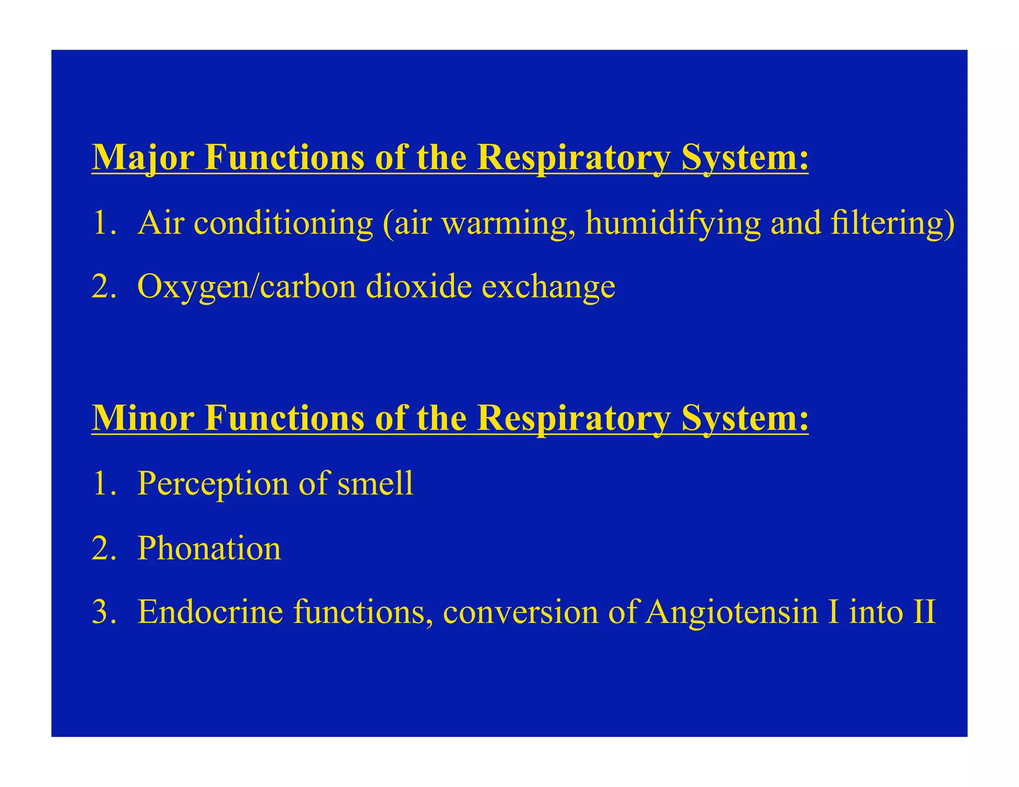
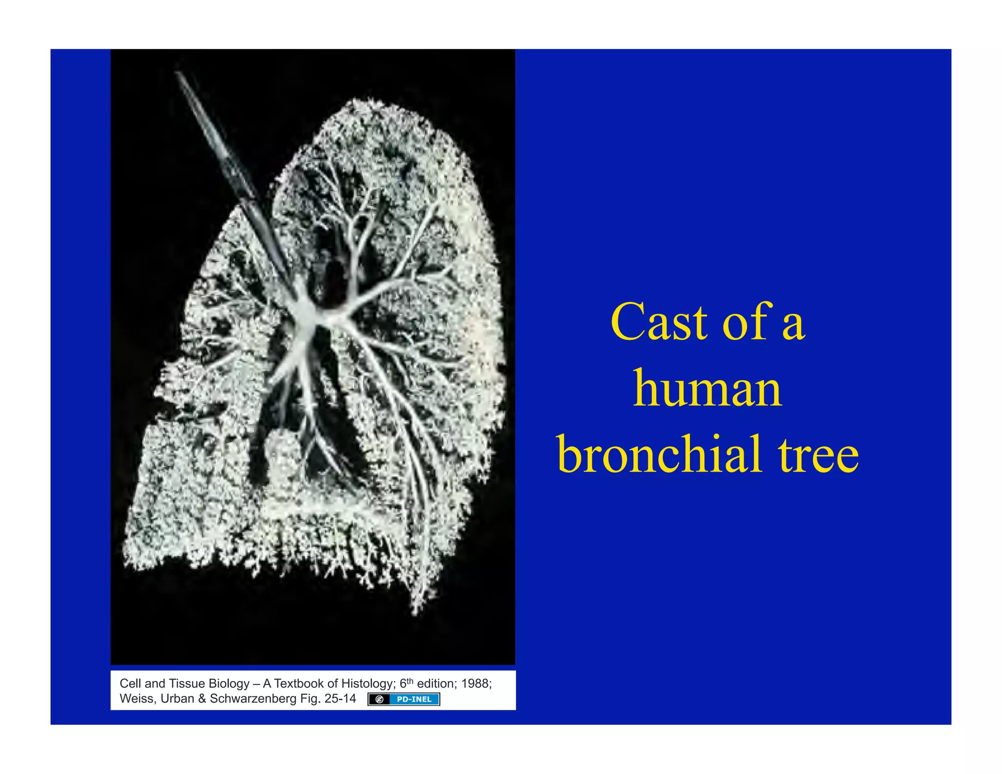

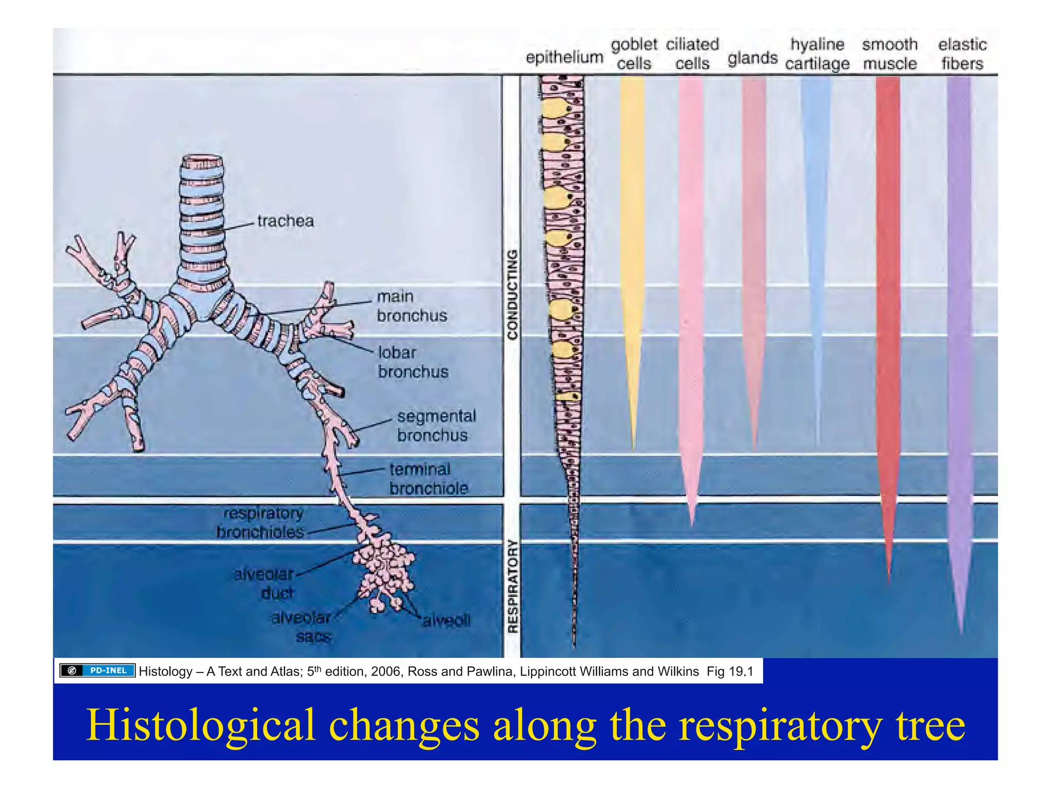


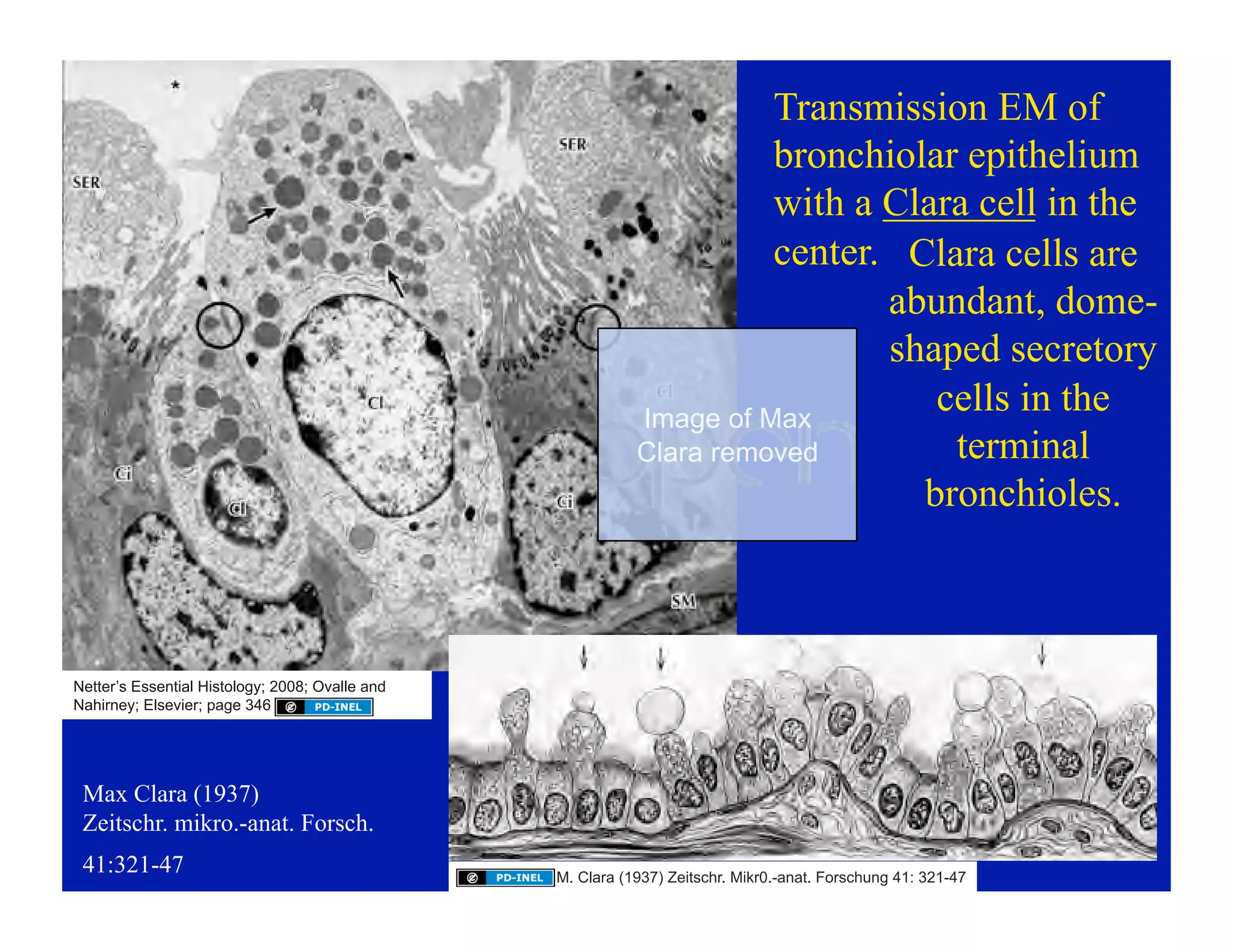


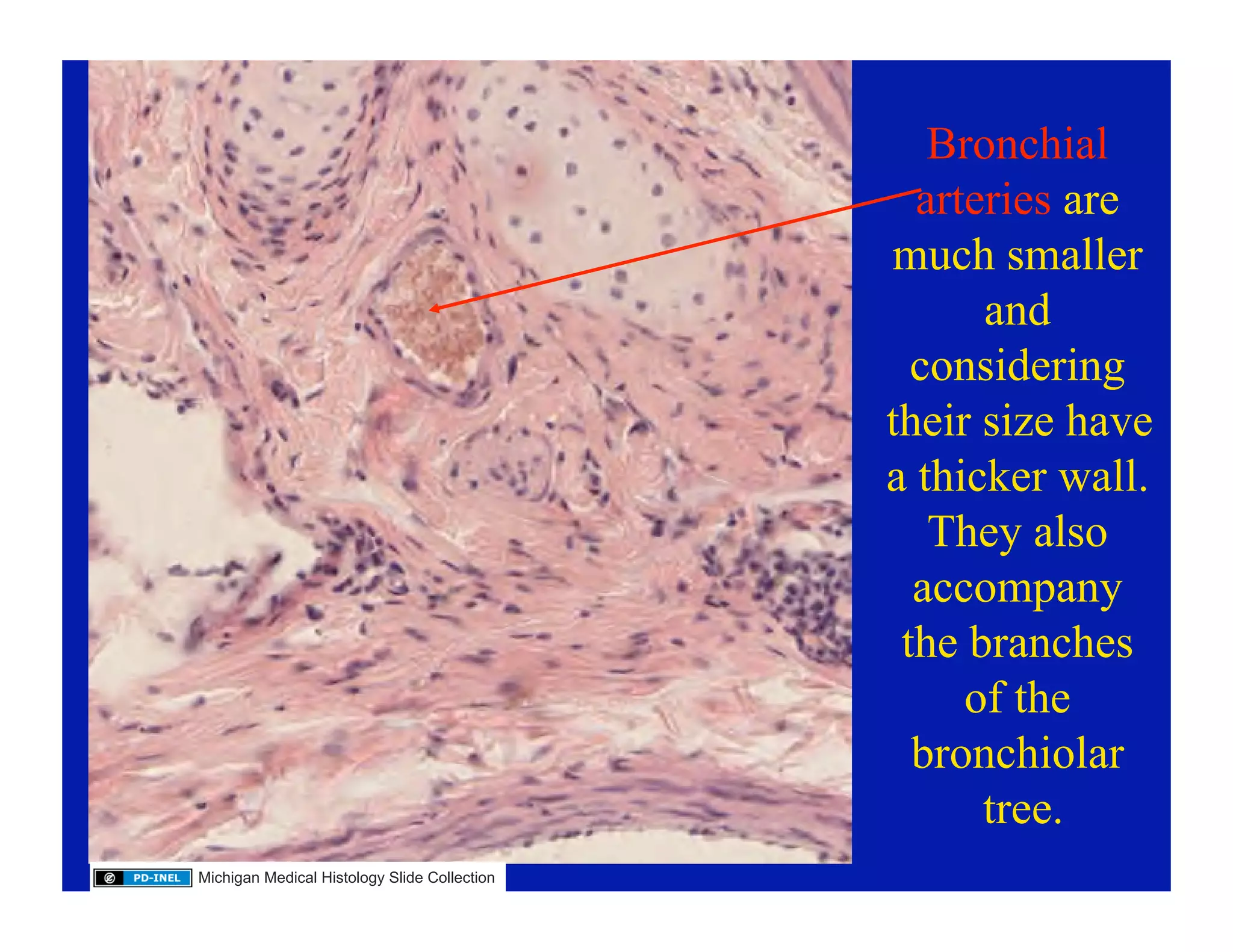


The document discusses the histology of the respiratory tract, detailing the structure and function of various components such as the nasal cavity, trachea, bronchi, and alveoli. It outlines the major and minor functions of the respiratory system, emphasizes the importance of cellular components like clara cells, and explains the organization of intrapulmonary blood circulation. The content is intended for educational purposes and includes specific licensing details regarding its use and sharing.