Downloaded 28 times
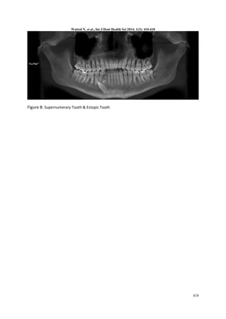
![Watted N. et al., Int J Dent Health Sci 2014; 1(3): 410-418
411
heredity factors and general diseases have
been suggested (1,3,4) .
The development of extra permanent
teeth can be classified as “heterotopic” –
teeth developing outside the alveolar
region or “normotopic”. The latter
includes teeth that develop in the alveolar
region and erupt in a relatively normal
orientation. Much variation in the
morphology of supernumerary teeth has
also been described. These teeth may be
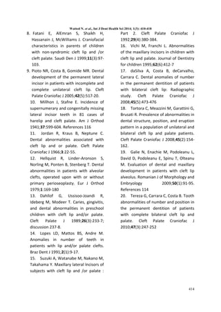
normal in shape and size,normal in shape
but reduced in size, of conical shape or,
lastly, abnormal in shape as well as
reduced in size(2,4) Single or multiple
supernumerary teeth can be unilateral or
bilateral and it has been shown that the
anterior maxilla and mandibular premolar
regions are most commonly affected .
Multiple supernumerary teeth most often
affect the mandibular premolar region (5).
In the primary dentition, the incidence is
said to be 0.3%-0.8% and in the
permanent dentition 1.5%-3.5% [4]. The
low prevalence of supernumerary teeth in
primary dentition is lower because it is
under reported [5] and it is often
overlooked, because the supernumerary
`teeth are often of normal shape
(supplemental type), erupt normally using
spaces in primary dentition, and appear to
be in proper alignment; and can be
mistaken for germination and fusion
anomalies [6] . There seems to be a racial
variation in the prevalence of
supernumeraries with a frequency higher
than 3% in Mongoloid races [7] . There is
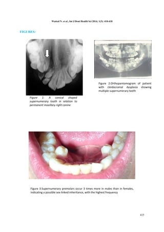
no significant sex distribution in primary
supernumerary teeth; however, males
have been shown to be affected more in
the permanent dentition than females.
Supernumerary teeth are estimated to
occur in the maxilla 8.2 to 10 times more
frequently than the mandible, and most
commonly affect the premaxilla[6,7,8] .
Effects of supernumerary teeth on the
developing dentition may vary. There may
be no effect and supernumerary teeth
may be discovered either following their
eruption or as a chance during
radiographic examination. Failure of
eruption and/or ectopic eruption of
adjacent permanent teeth is the most
frequently reported occurrence in almost
30 to 60 per cent of cases. Crowding may
occur when multiple supernumeraries are
present. Supernumerary teeth may also
cause root resorption, malformation of
adjacent teeth such as dilaceration,
diastema and loss of vitality of adjacent
teeth,
DISCUSSION:
Millhon & Stafne studied 108 cases of
hair lip and cleft palate to identify the
incidence of supernumerary teeth of the
lateral incisor teeth. They discovered that
the incidence of supernumerary teeth in
the cleft palate sample was high, and
related the cause to the splitting of the
lateral incisor tooth bud at the cleft area
(10 ) .
Jordan et al. researched dental
abnormalities with cleft lip and or palate.
They examined 10 human cleft lip and
palate fetuses. The other sample was
made up of maxillary and mandibular
dental casts of 192 individuals with CLP.](https://image.slidesharecdn.com/01032014-140710132333-phpapp01/85/Supernumerary-teeth-hyperdontia-2-320.jpg)

Supernumerary teeth, or extra teeth beyond the normal number, are a developmental dental anomaly that have been found to occur more frequently in patients with cleft lip and palate. This document reviews several studies that have examined the prevalence of supernumerary teeth in patients with cleft lip and/or palate, finding reported rates ranging from 11.7% to 29.2%. Multiple studies found the highest prevalence of supernumerary teeth occurred in the maxillary anterior region near the cleft site. The increased frequency of supernumerary teeth in cleft patients is thought to be related to the splitting of the dental lamina during cleft formation.


![CTEV [ clubfoot] DR ARUN LAL ,DR MOHAMED ASHRAF travancore medical college k...](https://cdn.slidesharecdn.com/ss_thumbnails/ctevclubfootdrarunlaldrmohamedashraftravancoremedicalcollegekollamkeralaindia-260208063247-18fc466c-thumbnail.jpg?width=640&height=640&fit=bounds)


