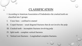
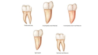
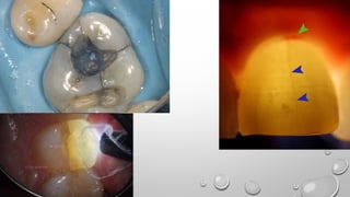
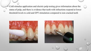
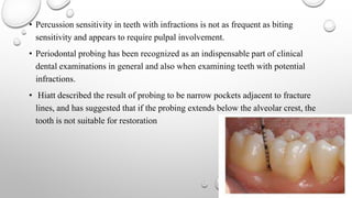
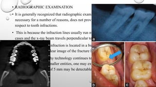
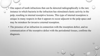
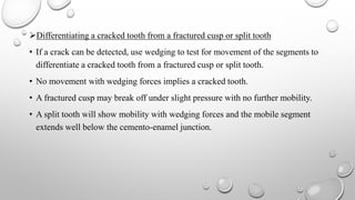
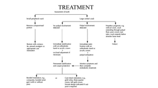
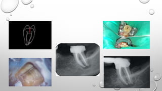
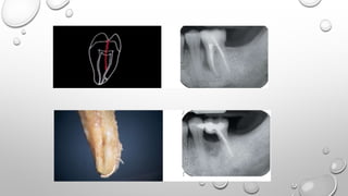
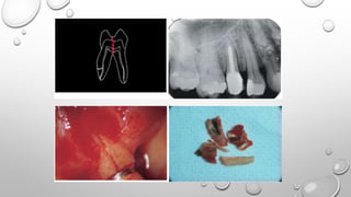
TOOTH INFARCTION
HEMA.M
1ST YR PG
DEPARTMENT OF CONSERVATIVE DENTISTRY AND ENDODOTICS
SRI SIDDHARTHA DENTAL COLLEGE
CONTENTS
• INTRODUCTION
• HISTORY
• DEFINITION
• CLASSIFICATION
• INCIDENCE
• ETIOLOGY
• SIGNS AND SYMPTOMS
• DIAGNOSTIC TESTS.
• TREATMENT
• CONCLUSION
INTRODUCTION
• Fractures are one of the major conditions in human teeth that cause pain.
• Tooth fractures include trauma related crown, crown-root and root fractures and a
broad group of cracked teeth.
• However, when a tooth fracture is incomplete, the presentation is more subtle
and frequently remains undiagnosed because signs and symptoms are often
confusing and are not sufficiently recogonized by clinicians.
HISTORY
• 1954 – Gibbs termed this condition as Cuspal fracture odontalgia
• 1956 – Melion called it as a Fractured cusp
• 1962 – Sutton termed it as Greenstick fractures of the tooth
• 1964 – Cameron called it as Cracked tooth syndrome
• 2001 – Ellis termed it as Incomplete tooth fractures.
DEFINITION
• Tooth infarction can be defined as an incomplete tooth fracture extending
partially through a tooth.
• It can be further described as originating in the tooth crown, originating from
pulp towards the dentinoenamel junction and propagating apically in the root.
• It can be identified as three fairly distinct types: 1) those confined to the enamel,
2) those related to cuspal fractures not involving the pulp and 3) those more
centrally located and involving pulp.
CLASSIFICATION
• According to American Association of Endodontics the cracked teeth are
classified into 5 groups:
I. Craze lines – confined to enamel
II. Cuspal fracture – usual diagonal fractures that do not involve the pulp
III. Cracked teeth – incomplete fractures involving pulp
IV. Split tooth – complete vertical fractures
V. Vertical root fractures – Longitudinal complete fractures.
• INCIDENCE:
• The incidence of tooth infarction is not extensively reported. A study conducted
by SnyderDE 1976 and Motsch A.1992 indicated that 2-3% of dental patients
have infarction in the molars and premolars; mostly asymptomatic, so incidence
of symptomatic teeth may be less than 1%.
• The location of fracture mostly occurs in the mesiodistal direction and only in
few cases buccolingual direction or combination of both.
• Brynjuslfsen et al noted that 10% of the infarction were centrally located,
meaning pulpal involvement.
• In their study maxillary teeth, majority(70%) of fracture lines were towards
buccal surface and in mandibular teeth (70%) were towards the lingual surface.
• Many reports appear to point to mandibular teeth, in particular the second molar
as being most frequently involved.
• In rare cases, of dental trauma the anterior teeth may show infarction.
ETIOLOGY:
• It is considered that the restored teeth are more often associated with tooth
infarction than the non restored teeth.
• It is said that excessively large and incorrect designed restoration lead to tooth
infarction
• Use of pin for supporting large restoration especially self-threading and friction-
lock pins,
• Abrasion, erosion, and caries may also contribute to it along with age changes in
dentin, oral habits and thermal cycling.
• The act of chewing food is also implicated as an predisposing factors like
repeated occlusal overload due to oral habits or accidental biting on hard things
which may lead to tooth infarction or vertical root fractures.
• Bruxism and often clenching of teeth combined with wedging effect of cusp on
opposing fossae can be stressful to the teeth.
• In molars upto 90kgs biting forces is noted hence are subject to extensive stress,
so is termed as ‘fatigue root fractures’ (Suggested by Yeh et al).
SIGNS AND SYMPTOMS
• Pain is the general complaint which draws the patients attention to the problem
teeth.
• Pain on chewing is the most common finding in symptomatic infarctions.
• It is unique in nature as the patient complains of pain after release of pressure.
Hence it is sometimes called as rebound/relief pain.
• Brannstrom has proposed that the masticatory pain is due to sudden movement of
dentinal fluid when fractured portion move independently, activating A-δ fibres
and creating rapid, acute pain response.
• Pain stimulate by change in temperature especially cold is also a common
feature.
• Ritchey et al noted the elevated response to cold condition attributed to the
pulpal inflammation caused by infarction communicating to the pulp and likely
contains bacteria .
• Branstrom suggested that this leads to inflammation of pulp and hence
stimulation of the C fibres that response to the temperature change.
• Localization of infarcted tooth is difficult until it reaches the periodontal region
or has periapical pathosis as the pulp lacks proprioception.
• CLINICAL EXAMINATION:
• The absence of a carious lesion, seemingly intact tooth.
• Visual aids like transillumination and dye examination using methylene blue can
be used.
• The use of transillumination should be done after the removal of the restoration
and if the fracture line is present the light passing through the dentin will bend at
that point and makes the other side of line to appear darker.
• Methylene blue dye in rubbed on to the teeth in suspected area with a cotton
pellet and the removed using a cotton pellet soaked in alcohol. The fracture lines
will retain the dye.
• Additionally, the use of microscope can help in identifying it.
• Another important test is biting test, various techniques has been recommended
such as biting on burlew wheels, rubber wheels, cotton tip applicators, moist
cotton rolls, and commercial biting applicators such as Tooth Slooth
(Professional Results Inc., Laguna Nigel, CA) and Fracfinder (Svenoka Dental
Instruments, Vasby, Sweden).
• To differentiate between biting pain from restorations with microleakage and
pain from tooth infraction, a Tooth Slooth can be positioned so that pressure is
first placed on the filling and then on tooth cusp. If the tooth shows the
rebound/relief pain it indicates the tooth infarction.
• Cold stimulus application and electric pulp testing gives information about the
status of pulp, and there is evidence that teeth with infractions respond at lower
threshold levels to cold and EPT stimulation compared to non cracked teeth
• Percussion sensitivity in teeth with infractions is not as frequent as biting
sensitivity and appears to require pulpal involvement.
• Periodontal probing has been recognized as an indispensable part of clinical
dental examinations in general and also when examining teeth with potential
infractions.
• Hiatt described the result of probing to be narrow pockets adjacent to fracture
lines, and has suggested that if the probing extends below the alveolar crest, the
tooth is not suitable for restoration
• RADIOGRAPHIC EXAMINATION
• It is generally recognized that radiographic examination, while obviously
necessary for a number of reasons, does not provide much information with
respect to tooth infractions.
• This is because the infraction lines usually run mesiodistally in the majority of
cases and the x-ray beam travels perpendicular to the fracture lines.
• If, however, the tooth infraction is located in a buccolingual direction, the x-ray
beam may produce a clear image of the fracture line on the film or the sensor.
• If computed tomography technology continues to improve with respect to
imaging smaller and smaller entities, one may expect that in the future,
infractions in the size of 5 mm may be detectable.
• One aspect of tooth infractions that can be detected radiographically is the rare
instance in which bacteria in the infraction has stimulated clastic activity in the
pulp, resulting in internal resorptive lesions. This type of internal resorption is
unique in many respects in that it appears to occur adjacent to the pulp space and
may be mistaken for invasive external resorption.
• The presence of an infraction in connection with the resorption defect, and no
communication of the resorptive defect with the periodontal tissues, confirms the
diagnosis.
Differentiating a cracked tooth from a fractured cusp or split tooth
• If a crack can be detected, use wedging to test for movement of the segments to
differentiate a cracked tooth from a fractured cusp or split tooth.
• No movement with wedging forces implies a cracked tooth.
• A fractured cusp may break off under slight pressure with no further mobility.
• A split tooth will show mobility with wedging forces and the mobile segment
extends well below the cemento-enamel junction.
Conditions that may be misdiagnosed as a cracked tooth involve the following:
• acute periodontal disease,
• reversible pulpitis,
• dentinal hypersensitivity,
• galvanic pain,
• postoperative sensitivity associated with microleakage from recently placed
composite resin restorations,
• fractured restorations,
• areas of hyperocclusion from dental restorations,
• pain from bruxism, orofacial pain, or atypical facial pain.
TREATMENT
CONCLUSION
• The possibility of tooth infarction must always be considered when a patient
complains of pain or discomfort on chewing or biting. In spite of it being a
diagnostic challenge, having knowledge and awareness of tooth infarction should
enable the dental practitioner to detect the same, thereby preventing further crack
propagation and complications associated with crack propagation
REFERENCE
• Ingle’s endodontics 6th edition
• The pathways of pulp-cohen, 12th edition
• Hasan et al, Int J App Basic Med Res 2015;5:164-8. Crack tooth syndrome : A
overview of literature.
• Batalha,-Silva et al, Operative dentistry, 2014, 39-5, 460-468. Cracked tooth
syndrome in an unrestored maxillary premolar: A case report.
• The crack tooth syndrome - 1May 2002, Dentistry Today
Tooth infarction

Tooth infarction

  • 2.
    TOOTH INFARCTION HEMA.M 1ST YRPG DEPARTMENT OF CONSERVATIVE DENTISTRY AND ENDODOTICS SRI SIDDHARTHA DENTAL COLLEGE
  • 3.
    CONTENTS • INTRODUCTION • HISTORY •DEFINITION • CLASSIFICATION • INCIDENCE • ETIOLOGY • SIGNS AND SYMPTOMS • DIAGNOSTIC TESTS. • TREATMENT • CONCLUSION
  • 4.
    INTRODUCTION • Fractures areone of the major conditions in human teeth that cause pain. • Tooth fractures include trauma related crown, crown-root and root fractures and a broad group of cracked teeth. • However, when a tooth fracture is incomplete, the presentation is more subtle and frequently remains undiagnosed because signs and symptoms are often confusing and are not sufficiently recogonized by clinicians.
  • 5.
    HISTORY • 1954 –Gibbs termed this condition as Cuspal fracture odontalgia • 1956 – Melion called it as a Fractured cusp • 1962 – Sutton termed it as Greenstick fractures of the tooth • 1964 – Cameron called it as Cracked tooth syndrome • 2001 – Ellis termed it as Incomplete tooth fractures.
  • 6.
    DEFINITION • Tooth infarctioncan be defined as an incomplete tooth fracture extending partially through a tooth. • It can be further described as originating in the tooth crown, originating from pulp towards the dentinoenamel junction and propagating apically in the root. • It can be identified as three fairly distinct types: 1) those confined to the enamel, 2) those related to cuspal fractures not involving the pulp and 3) those more centrally located and involving pulp.
  • 7.
    CLASSIFICATION • According toAmerican Association of Endodontics the cracked teeth are classified into 5 groups: I. Craze lines – confined to enamel II. Cuspal fracture – usual diagonal fractures that do not involve the pulp III. Cracked teeth – incomplete fractures involving pulp IV. Split tooth – complete vertical fractures V. Vertical root fractures – Longitudinal complete fractures.
  • 9.
    • INCIDENCE: • Theincidence of tooth infarction is not extensively reported. A study conducted by SnyderDE 1976 and Motsch A.1992 indicated that 2-3% of dental patients have infarction in the molars and premolars; mostly asymptomatic, so incidence of symptomatic teeth may be less than 1%. • The location of fracture mostly occurs in the mesiodistal direction and only in few cases buccolingual direction or combination of both. • Brynjuslfsen et al noted that 10% of the infarction were centrally located, meaning pulpal involvement.
  • 10.
    • In theirstudy maxillary teeth, majority(70%) of fracture lines were towards buccal surface and in mandibular teeth (70%) were towards the lingual surface. • Many reports appear to point to mandibular teeth, in particular the second molar as being most frequently involved. • In rare cases, of dental trauma the anterior teeth may show infarction.
  • 11.
    ETIOLOGY: • It isconsidered that the restored teeth are more often associated with tooth infarction than the non restored teeth. • It is said that excessively large and incorrect designed restoration lead to tooth infarction • Use of pin for supporting large restoration especially self-threading and friction- lock pins, • Abrasion, erosion, and caries may also contribute to it along with age changes in dentin, oral habits and thermal cycling.
  • 12.
    • The actof chewing food is also implicated as an predisposing factors like repeated occlusal overload due to oral habits or accidental biting on hard things which may lead to tooth infarction or vertical root fractures. • Bruxism and often clenching of teeth combined with wedging effect of cusp on opposing fossae can be stressful to the teeth. • In molars upto 90kgs biting forces is noted hence are subject to extensive stress, so is termed as ‘fatigue root fractures’ (Suggested by Yeh et al).
  • 14.
    SIGNS AND SYMPTOMS •Pain is the general complaint which draws the patients attention to the problem teeth. • Pain on chewing is the most common finding in symptomatic infarctions. • It is unique in nature as the patient complains of pain after release of pressure. Hence it is sometimes called as rebound/relief pain. • Brannstrom has proposed that the masticatory pain is due to sudden movement of dentinal fluid when fractured portion move independently, activating A-δ fibres and creating rapid, acute pain response. • Pain stimulate by change in temperature especially cold is also a common feature.
  • 15.
    • Ritchey etal noted the elevated response to cold condition attributed to the pulpal inflammation caused by infarction communicating to the pulp and likely contains bacteria . • Branstrom suggested that this leads to inflammation of pulp and hence stimulation of the C fibres that response to the temperature change. • Localization of infarcted tooth is difficult until it reaches the periodontal region or has periapical pathosis as the pulp lacks proprioception.
  • 16.
    • CLINICAL EXAMINATION: •The absence of a carious lesion, seemingly intact tooth. • Visual aids like transillumination and dye examination using methylene blue can be used. • The use of transillumination should be done after the removal of the restoration and if the fracture line is present the light passing through the dentin will bend at that point and makes the other side of line to appear darker. • Methylene blue dye in rubbed on to the teeth in suspected area with a cotton pellet and the removed using a cotton pellet soaked in alcohol. The fracture lines will retain the dye.
  • 18.
    • Additionally, theuse of microscope can help in identifying it. • Another important test is biting test, various techniques has been recommended such as biting on burlew wheels, rubber wheels, cotton tip applicators, moist cotton rolls, and commercial biting applicators such as Tooth Slooth (Professional Results Inc., Laguna Nigel, CA) and Fracfinder (Svenoka Dental Instruments, Vasby, Sweden). • To differentiate between biting pain from restorations with microleakage and pain from tooth infraction, a Tooth Slooth can be positioned so that pressure is first placed on the filling and then on tooth cusp. If the tooth shows the rebound/relief pain it indicates the tooth infarction.
  • 20.
    • Cold stimulusapplication and electric pulp testing gives information about the status of pulp, and there is evidence that teeth with infractions respond at lower threshold levels to cold and EPT stimulation compared to non cracked teeth
  • 21.
    • Percussion sensitivityin teeth with infractions is not as frequent as biting sensitivity and appears to require pulpal involvement. • Periodontal probing has been recognized as an indispensable part of clinical dental examinations in general and also when examining teeth with potential infractions. • Hiatt described the result of probing to be narrow pockets adjacent to fracture lines, and has suggested that if the probing extends below the alveolar crest, the tooth is not suitable for restoration
  • 22.
    • RADIOGRAPHIC EXAMINATION •It is generally recognized that radiographic examination, while obviously necessary for a number of reasons, does not provide much information with respect to tooth infractions. • This is because the infraction lines usually run mesiodistally in the majority of cases and the x-ray beam travels perpendicular to the fracture lines. • If, however, the tooth infraction is located in a buccolingual direction, the x-ray beam may produce a clear image of the fracture line on the film or the sensor. • If computed tomography technology continues to improve with respect to imaging smaller and smaller entities, one may expect that in the future, infractions in the size of 5 mm may be detectable.
  • 23.
    • One aspectof tooth infractions that can be detected radiographically is the rare instance in which bacteria in the infraction has stimulated clastic activity in the pulp, resulting in internal resorptive lesions. This type of internal resorption is unique in many respects in that it appears to occur adjacent to the pulp space and may be mistaken for invasive external resorption. • The presence of an infraction in connection with the resorption defect, and no communication of the resorptive defect with the periodontal tissues, confirms the diagnosis.
  • 24.
    Differentiating a crackedtooth from a fractured cusp or split tooth • If a crack can be detected, use wedging to test for movement of the segments to differentiate a cracked tooth from a fractured cusp or split tooth. • No movement with wedging forces implies a cracked tooth. • A fractured cusp may break off under slight pressure with no further mobility. • A split tooth will show mobility with wedging forces and the mobile segment extends well below the cemento-enamel junction.
  • 25.
    Conditions that maybe misdiagnosed as a cracked tooth involve the following: • acute periodontal disease, • reversible pulpitis, • dentinal hypersensitivity, • galvanic pain, • postoperative sensitivity associated with microleakage from recently placed composite resin restorations, • fractured restorations, • areas of hyperocclusion from dental restorations, • pain from bruxism, orofacial pain, or atypical facial pain.
  • 26.
  • 30.
    CONCLUSION • The possibilityof tooth infarction must always be considered when a patient complains of pain or discomfort on chewing or biting. In spite of it being a diagnostic challenge, having knowledge and awareness of tooth infarction should enable the dental practitioner to detect the same, thereby preventing further crack propagation and complications associated with crack propagation
  • 31.
    REFERENCE • Ingle’s endodontics6th edition • The pathways of pulp-cohen, 12th edition • Hasan et al, Int J App Basic Med Res 2015;5:164-8. Crack tooth syndrome : A overview of literature. • Batalha,-Silva et al, Operative dentistry, 2014, 39-5, 460-468. Cracked tooth syndrome in an unrestored maxillary premolar: A case report. • The crack tooth syndrome - 1May 2002, Dentistry Today