
Recommended
More Related Content
What's hot
What's hot (20)
Similar to CANINE HEPATITIS: CAUSES, SYMPTOMS AND PREVENTION
Similar to CANINE HEPATITIS: CAUSES, SYMPTOMS AND PREVENTION (20)
Recently uploaded
Recently uploaded (20)
CANINE HEPATITIS: CAUSES, SYMPTOMS AND PREVENTION
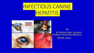
- 1. INFECTIOUS CANINE HEPATITIS By: Dr. Rashmi Singh, Assistant Professor Veterinary Medicine PGIVER, Jaipur
- 2. Brief Overview: • Introduction • Incidence and distribution • Etiology • Pathogenesis • Clinical Findings • Necropsy findings • Differential diagnosis • Treatment • Prevention and Control
- 3. Synonyms: • Rubarth’s disease • Hepatitis contagiosa canis • Contagious hepatitis • Fox encephalitis
- 4. Introduction : • An acute contagious viral disease of dogs. • Irrespective of their ages . • Characterized by severe depression, high rise of temperature, marked leukopenia, vomition, diarrhoea, convulsion and prolong bleeding time.
- 5. Distribution and Incidence : • Rubarth first time recorded the disease in 1947 in Sweden. • Disease is widely prevalent in many countries of the world. • The disease is prevalent in epizootic proportions in India. • Prior to the development of effective vaccines, seroprevalence of ICH among dogs was 30-60% resulting in 10-30% mortality in dogs and foxes.
- 6. Etiology : • Is caused by a DNA virus. • Which belongs to adenovirus group. • There are two adeno viruses: • Canine adeno virus -1-ICH • Canine adeno virus-2 – causes respiratory disease. • Virus can be preserved in 50% glycerine for several years. • Virus can resist either, chloroform and alcohol.
- 7. Viral properties… • Virus can be cultivated in the yolk sac of embryonated eggs. • Can be grown in kidney cell culture of dog, ferret, racoon.
- 8. Susceptible host : • All Canidae family is susceptible. • Dog and foxes are the important species to suffer from this infection. • In fox-the disease is manifested with neurological disturbances. • Under one year age dog –show acute form of the disease. • After which the dogs are inapparently infected with periodic relapses. • Experimentally disease can be produced in pups and foxes. • But not in ferret, coyote and other animals.
- 9. Mode of transmission : • Most significant method - excretion of the virus through urine. • Infectious canine hepatitis is not an airborne disease. • Virus is present in the faeces, blood, saliva and urine of affected dogs. • directly touching the snout or back end of infected/carrier, it can be transmitted • from contaminated feed or water bowl. • Recovered dog shed virus through urine for about 6 months.
- 10. Pathogenesis:
- 11. Clinical features: • Clinical features vary from a slight fever to death. • The disease may flare up as: • Acute form • Per-acute • Inapparent form
- 12. Acute form: • Disease starts with apathy, anorexia and high rise of temp. upto 105 degree F. • Vomition and diarrhoea. • Faeces blood tinged , abdominal pain. • First rise of body temp usually falls after 24 to 48 hours without levelling to normal temperature. • Rises again to form a saddle curve, which lasts for 6 days. • Leukopaenia-2nd day of temp., persist 3-4 days. • Buccal mucous membrane turn fiery red or haemorrhagic.
- 13. Cont…. • nose and mouth - reddened or covered with small bruises. • Animal shows pain on palpation of xiphoid region. • Dogs may show ‘tucked up’ condition of the abdomen. • After 1-3 weeks following disappearance of clinical signs-a transient corneal opacity may develop-this condition –hepatitis blue eye
- 14. Cont… • Haemorragic spots may be seen over the skin of the abomen. • Control of haemorrhage is difficult bcoz clotting mechanism of the blood may be impaired. • Prognosis is grave if the animal show profuse bleeding. • Following 4-7 days many dogs may recover and their appetite return to normal.
- 15. Cont… • Fraser(1986) : corneal oedema and inflammation of the anterior uveal tract -much specific manifestation. • Wright et al.(1971): recorded signs of pneumonia in pups, with rapid respiration, coughing, snapping and watery eye discharge. This may be due to another strain of adenovirus infection.
- 16. Per-acute form: • Animal die within 12-24 hours. • Signs like febrile stage, leucopaenia, conjunctivitis, haemorrhages, swollen tonsil, red buccal mucous membrane and tendered abdomen can not be appreciated. • The dog which is apparently normal in the night would die in the next morning.
- 17. Inapparent form: • Most common form. • Dog shows a very mild or subclinical attack with a passing temp, mild photophobia, enlarged tonsils and rapid recovery. • The wt loss is slow .
- 18. Lesions: • The liver is enlarged and friable on post-mortem examination. • Extensive centrilobular necrosis leads to a pale, mottled appearance, and widespread haemorrhage - on the serosal surface. • Hepatitis leads to ascites and fibrinous or Fibrino-haemorrhagic adhesions-b/w the lobes of the liver. • Large no. of intra nuclear inclusion bodies in Hepatic cells & Kupffer cells.
- 19. Cont… • Gall bladder- Enlarged, oedematous and thickened. • Lymph glands- Swollen and haemorrhagic. • Spleen- Enlarged, blood filled. • Kidneys- Interstitial nephritis and cortical necrosis. • Stomach & Intestine – Inflammed, paint brush haemorrhage of gastric mucosa. • Eyes-inflammation of iris and ciliary body. -Inclusion bodies in the corneal cells and iridal endothelial cells.
- 20. Diagnosis: • Clinical features. • Joshua (1962): In hepatitis there is a thin thread pulse with weak, rapid heart sounds. In tonsillitis or any general systemic infection, dog always has bounding pulse and heart rate. • Microscopic changes- intranuclear inclusion bodies in the liver, gall bladder, brain and cornea.
- 21. Cont… • Gel diffusion test- by this test diagnose the disease even when decomposition of the animal has occurred. • Compliment fixation test. • Neutralization test. • Animal inoculation test. • Liver function test.
- 22. Differential diagnosis: • Warfarin poisoning: Prolong clotting time, Presence of source of poison, absence of intranuclear inclusion bodies, absence of leukopaenia, absence of pain on palpation of liver. Tonsilitis: caused by strepto or staphylococcus, coughing and nasal discharge, difficulty in swallowing. It may be sec due to distemper or hepatitis virus infection.
- 23. Cont… • Leptospirosis : signs of jaundice and nephritis. -Presence of leptospira in urin under dark field illumination. -Fluorescent antibody test-rapid diagnosis by using specific antisera labelled with a fluorescent dye. • Distemper : Distinct clinical signs. -Biphasic temp reaction ,signs of neural disorders. No change in clotting time. absence of intranuclear inclusion bodies.
- 24. Treatment : • No specific treatment. • Symptomatic treatments are to be given. • Antiserum may be useful. • Severely affected case may require blood transfusion. Dose -5-8 ml/lb of b.w. by slow iv infusion. • Broad spectrum antibiotics –to control sec bacterial infection. • Fluid and electrolytes are to be given. • Protein hydrolysate –to restore vitality • Careful nursing is of great importance.
- 25. Prevention and Control: • Maternal antibodies dam to foetus in utero • In new born pup by nursing the colostrums. • Level of these antibodies declines to negligible concentration by 14- 16 wks. • Immunization successful when maternal antibody titer declines below 1:100, which occurs at age 5-7 weeks of age.
- 26. Cont… • Modified live CAV-1 vaccine are commercially available. • Also give protection to CAV-2 infection . • Modified live vaccines are widely used due to their high efficacy but they induce post vaccinal ocular lesions including corneal opacity, and virus excretion in urine in dogs post vaccination. • Therefore CAV-2 vaccine evaluated. • CAV-2 protects both against CAV-1 and CAV-2 infections. • And no other adverse clinical signs and no urinary shading.
- 27. Cont… • A bivalent vaccine containing modified canine distemper and CAV-2 or CAV-1 strain. • Trivalent vaccine comprising of Bordetella bronchiseptica, canine para influenza and CAV-1 . • Polyvalent vaccine having live modified CAV-2, canine distemper virus, canine parainfluenza virus and leptospira bacterin- is also safe and provide good immunity. • Another PV vaccine containing canine parvo virus, rabies, distemper, CAV-1/CAV-2, canine parainfluenza, leptospira canicola and L. icterohaemorrhagiae also provide 100% seroconversion with high antibody titer. dose 1ml,route-I/V
- 28. Cont… • Vaccination of dogs and the induction of active immunity have controlled ICH in the canine population very effectively. • Control measures in addition to vaccination are not necessary. • Though the infected dog should be isolated from other healthy dogs.
- 29. Conclusion : • Infectious canine hepatitis is a very contagious disease of dog • That can cause inflammation of liver. • It is not zoonotic but humans can be a source of its transmission if carry virus on skin and cloths. • It is also transmitted by contact with water and food bowls of infected dogs (as by some other methods) so not allow your dog to drink in communal water bowls. • Treatment is symptomatic and it is almost always be prevented by vaccination against the canine adenovirus.