
Recommended
More Related Content
What's hot
What's hot (20)
Similar to Marek's disease
Similar to Marek's disease (20)
More from Aashish Tanwar
More from Aashish Tanwar (18)
Recently uploaded
Recently uploaded (20)
Marek's disease
- 1. Dr Ashish Tanwer Teaching Associate V. C.C, C.V.A.S Bikaner
- 2. SYNONYMS AND INTRODUCTION Synonyms:- • Range paralysis • skin leucosis • Neural leucosis • Neural lymphomatosis • pearl eye
- 3. Introduction :- • Marek’s disease (MD) is a lymphoproliferative, highly contagious disease of poultry and caused by a highly cell associated gallid herpes virus, a DNA virus belonging to Herpes viridae family. • There are three serotypes of the virus recognized. Serotypes 1 and 2 are designated as virulent and avirulent chicken isolates respectively. • Serotype 3 designates the avirulent turkey herpes virus. • Serotypes 2 and 3 and attenuated serotype-I virus are used for vaccine production. The virus remains stable for about 24 hours at 30˚C. • The Marek’s disease virus (MDV) has been propagated and assayed in newly hatched chicks, tissue cultures (co-cultivation of lymphocytes with chicken kidney cells or duck embryo fibroblasts) and embryonated eggs. This disease is exists in poultry-producing countries throughout the world.
- 4. • The infection is transmitted through inhalation of infected material from the environment. • Virus particle can persist for a considerable period of time in the dandruff of feather follicles, which are released in environment. • The infective materials are oral, nasal and tracheal secretions and litter materials. The darkling beetle (Alphitrbius diaperinus) is acting as mechanical transmitter of the disease. • The chickens are the most important natural host and MD is very rare and probably of no real importance in other species with the possible exception of quail. • Chickens of 12-24 weeks of age are mostly susceptible to Marek’s disease and generally it does not occur in chickens below 6 weeks of age and older birds above 24 weeks of age.
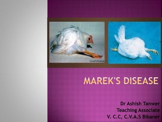
- 5. CLINICAL MANIFESTATIONS :- • Incubation period-ranges from 3 weeks to 9 weeks. • The disease appears in several forms. • Classical form or neural form Birds of 16-20 weeks age usually suffer. Signs are mostly concerned with the affection of nerves. Paralysis of legs, drooping wings. Nerves like sciatic nerve, brachial nerve, celiac and vagus nerve running through neck, thoracic and abdominal viscera are affected. Birds unable to stand remain in recumbent position, legs and wings may stretched in either direction. The “split leg” stance is the usual feature. Vents remain soiled with green diarrhea. Mortality rate is comparatively low and mostly noted at the onset of maturity.
- 6. • Acute or visceral form Generally birds at the age of 3-4 weeks are affected. Depression, droopiness, unthirfitness, dehydration, emaciation and anaemia. Internal organs of the birds affected. Mortality rate may go as high as 60%. Chicks may die suddenly without showing any clinical manifestation. Ovaries of the affected layers and pullets – looks like a cauliflower and mulberry respectively.
- 7. • Transitional paralytic form Occurs in chickens at the age of 5-18 weeks of age. Sudden development of paresis or paralysis of the legs, wings and neck. Signs usually disappear within 24-48 hours. • Ocular form Blindness in birds due to mononuclear cell infiltration in the iris causing “grey eye” or “pearl eye”. • Skin or cutaneous form Distinct white nodules on the skin and in extreme cases looks like brownish nodules.
- 8. • Muscular form Superficial and deep muscles like pectoral muscles affected. Muscles look lusterless, whitish grey and there are tiny white streaks to nodular tumours in the muscles.
- 10. LESIONS :- • Affected nerves thickened to more than 2-3 times than normal • Striation and glistening appearance of nerve is lost and looks oedematous • Celiac, cranial, mesenteric, brachial and sciatic plexes and greater splanchnic nerves are mostly affected • Tiny whitish streaks to nodular tumours in muscles • Atrophy of bursa • Ovary- cauliflower like appearance • Pale, single or multiple nodular tumours in myocardium • Skin- whitish nodule, scab with brownish colour.
- 13. DIAGNOSIS :- • Specimens to be collected Skin, dander, feather tips of infected chickens, blood • Based on the clinical signs and postmortem lesions • Identification of the agent Under field conditions, most chickens become infected with MDV during the first few weeks of life and then carry the infection throughout their lives, often without developing overt disease. The infection is usually detected by inoculating live buffy coat cells on to monolayer cultures of chicken kidney cells or duck embryo fibroblasts, in which characteristic viral plaques develop within a few days.
- 15. Two serotypes of MDV are recognized – 1 and 2 – and a third serotype is represented by the related herpes virus of turkeys (HVT). Serotype 1 includes the virulent strains and serotype 2 the naturally avirulent strains. MD viral antigen can be detected in the feather tips of infected birds using a radial precipitin test. •Serological tests Antibodies to MDV develop within 1–2 weeks of infection and are commonly recognized by the agar gel immunodiffusion (AGID) test, the indirect fluorescent antibody test, and by other serological tests such as enzyme-linked immunosorbent assay, virus neutralization tests.
- 16. PREVENTION AND CONTROL :- • Three classes of vaccines using Attenuated serotype 1 MDV, HVT and natural a virulent isolate of serotype 2. • Herpes virus turkey (HVT) – most extensively used because it is economical to produce and cell free virus extracted from infected cells. • The vaccine usually administered at 0 day age by intra nasal or intra ocular route.
- 17. Thanks