The SuperArgus state-of-the-art preclinical PET/CT system: An overview of the technical specifications, unique product features and novel applications

These systems are ideally suited for pre-clinical imaging of small animals such as mice and rats, all the way up to medium sized animals such as rabbits, non-human primates and other similarly sized animals. Some of the unique imaging capabilities include real-time imaging of awake animals, as well as multiplexed PET imaging of standard and non-standard isotopes. Key research applications and example images were reviewed. Positron Emission Tomography (PET) is the gold standard in metabolic imaging, providing high sensitivity to specific radiotracer used to detect specific metabolic activity or biomarkers in vivo. The most common uses for PET imaging in pre-clinical research include oncology, neurobiology, cardiology, as well as dynamic imaging. These systems are considered to be best in class imaging system with state of the art detectors and electronics. The systems have been designed to be self-shielded, requiring no additional shielding at the location selected for installation. The systems come in a three different bore sizes allowing for imaging of animals such as mice all the way up to rabbits and even non-human primates. The CT component of these systems has been optimized for reduced radiation exposure, rapid acquisition times, and high resolution images; all ideal for the longitudinal studies so commonly performed in pre-clinical research. The SuperArgus system is uniquely designed to provide consistent resolution across the entire field of view, while maintaining sensitivity and system performance. Reconstruction algorithms have also been implemented to rapidly process and display the acquired images. The system performs very well for standard imaging applications such as oncology, cardiology, etc. Additionally, the system has some unique features which allow for some unique imaging capabilities such as real-time awake animal imaging, self-gated cardiac imaging, as well as multiplex imaging of standard and non-standard isotopes.

Recommended
Recommended
More Related Content
What's hot
What's hot (20)
Similar to The SuperArgus state-of-the-art preclinical PET/CT system: An overview of the technical specifications, unique product features and novel applications
Similar to The SuperArgus state-of-the-art preclinical PET/CT system: An overview of the technical specifications, unique product features and novel applications (20)
More from Scintica Instrumentation
More from Scintica Instrumentation (20)
Recently uploaded
Recently uploaded (20)
The SuperArgus state-of-the-art preclinical PET/CT system: An overview of the technical specifications, unique product features and novel applications
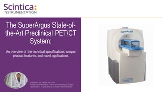
- 1. The SuperArgus State-of- the-Art Preclinical PET/CT System: An overview of the technical specifications, unique product features, and novel applications Presenter: Dr. Patrick McCunn Postdoctoral Research Fellow at University of Guelph Application’s Specialist at Scintica Instrumentation
- 2. The SuperArgus State-of- the-Art Preclinical PET/CT System: An overview of the technical specifications, unique product features, and novel applications Presenter: Dr. Patrick McCunn Postdoctoral Research Fellow at University of Guelph Application’s Specialist at Scintica Instrumentation
- 3. WWW.SCINTICA.COM Outline • Preclinical PET • Who is Sedecal? • Product Overview • What Makes the Sedecal System Unique? • Applications and Current Research 3
- 5. WWW.SCINTICA.COM Positron Emission Tomography (PET) Review 5 From: Minimization of parallax error in positron emission tomography using depth of interaction capable detectors: methods and apparatus I Mohammadi et al 2019 Biomed. Phys. Eng. Express 5 062001 doi:10.1088/2057-1976/ab4a1b
- 6. WWW.SCINTICA.COM Positron Emission Tomography (PET) Review 6 From: Small-animal positron emission tomography as a tool for neuropharmacology. Sophie Lancelot, Luc Zimmer. Trends in Pharmacological Sciences ; Volume 31, Issue 9, Pages 411-417 (September 2010); DOI: 10.1016/j.tips.2010.06.002 Isotope Half-Life 15O (oxygen) 2 min 13N (nitrogen) 10 min 11C (carbon) 20 min 18F (fluorine) 110 min
- 7. WWW.SCINTICA.COM Main Applications of Preclinical PET • Oncology • Neurology • Cardiology • Bone & disease imaging • Biodistribution • Dynamic imaging 7
- 9. WWW.SCINTICA.COM Who is Sedecal? • Founded in 2002; head office in Madrid, Spain, facilities in 6 other countries • OEM manufacturers for most of the largest clinical equipment suppliers • OEM manufacturers for several different preclinical PET suppliers in the past • GE’s eXplore Vista, BioScan’s BioPET/CT • Global service and support • Systems are compliant with all international quality and safety standards 9
- 10. WWW.SCINTICA.COM “First To Market” Within the PET/CT Market 10 • First to Market • Combined PET/CT for small animals • Small animal PET scanner with parallax correction using true depth of interaction (tDOI) technology – Phoswich detectors • Small animal PET scanner with full 3D OSEM reconstruction (ordered subset expectation maximization; iterative approach) • Ultra fast 3D OSEM reconstruction without a cluster of computers • Currently the only PET/CT scanner on the market with real-time PET imaging; minimum frame rate of 25msec.
- 11. Product Overview
- 12. WWW.SCINTICA.COM Sedecal SuperArgus PET/CT – Best in Class 12 • Unique Phoswich detector technology • Highest resolution on the market (≤1.0mm) • Resolution uniformity across the entire FOV, with FOV filling majority of the bore size due to the tDOI technology correcting the parallax error • Highest sensitivity (11%; at 100-700keV) • Real-time imaging (up to 2.5msec frame rate if desired) • Conscious/Awake Imaging • Multiplex PET Imaging
- 13. WWW.SCINTICA.COM Sedecal SuperArgus PET/CT - Models 13 • Super Argus Models (PET and/or CT) • r models – mouse, rat, marmoset • 90mm bore • FOV – 220mm axial (dynamic); 80mm transaxial • R models – up to 3kg rabbit • 160mm bore • FOV – 350mm axial (dynamic); 120mm transaxial • P models – up to non-human primate, canine, porcine, etc. • 260mm bore • FOV – 650mm axial (dynamic); 210mm transaxial • 2, 4, or 6 PET ring options for a 50mm, 100mm, or 150mm fixed axial FOV
- 14. WWW.SCINTICA.COM Self-Shielded • Self-shielded to meet FDA guidelines • No special room preparation, controls, or additional shielding required • Systems can easily be placed within existing laboratory environments, imaging cores, or within the animal facility • Can have the following integrated into animal handling: • Anesthesia – isofluorane with gas scavenging • Heating • Physiological monitoring • Image may be saved as DICOM, Interfile, or JPEG 14
- 15. WWW.SCINTICA.COM Rapid Reconstruction Algorithms • Choice of reconstruction algorithm to balance throughput and image quality • FBP (filtered back projection; arithmetic approach) ≤ 5sec • Analytical approach • 2D OSEM (ordered subset expectation maximization; iterative approach) ≤ 1min • Iterative approach in a single slice • 3D OSEM ≤ 2min • Iterative approach across a 3D volume 15 FORE+FBP 3D OSEM Courtesy of Prof. J.M. Udías (UCM, Madrid, Spain)
- 16. WWW.SCINTICA.COM Multi-Animal Handling System • Up to 4 mice can be scanned simultaneously in the R model (160mm bore) using the multi-animal handling system • Individually controlled anesthesia flow and heating controls • Separate animal preparation workstation is also included • Additional configurations are available for rats and/or mice depending on specific needs 16
- 17. WWW.SCINTICA.COM Multi-Animal Handling System Resolution and sensitivity are maintained throughout the entire FOV due to the unique tDOI Phoswich detectors – correcting for the parallax error 17
- 18. What Makes Sedecal’s System Unique?
- 19. Phoswich Detectors and Parallax Error Correction
- 20. WWW.SCINTICA.COM Parallax Error Correction and Phoswich Detectors 20 From: Minimization of parallax error in positron emission tomography using depth of interaction capable detectors: methods and apparatus I Mohammadi et al 2019 Biomed. Phys. Eng. Express 5 062001 doi:10.1088/2057-1976/ab4a1b DOI = Depth of Interaction LOR = Line of Response
- 21. WWW.SCINTICA.COM Parallax Error Correction and Phoswich Detectors Shadows represent all accessible LORs (Lines of Response) 21 From: Minimization of parallax error in positron emission tomography using depth of interaction capable detectors: methods and apparatus I Mohammadi et al 2019 Biomed. Phys. Eng. Express 5 062001 doi:10.1088/2057-1976/ab4a1b
- 22. WWW.SCINTICA.COM Parallax Error Correction and Phoswich Detectors Parallax Error increases as the bore diameter decreases 22 Radial Resolution Tangential Resolution ? One Detector Ring (10 mm) Scintillation crystals Positron-emitting source Lines-of-response between crystals in time coincidence Modified from: J. Seidel et al. Journal of Nuclear Medicine. 2006;47(11):1891. • Conventional solutions to maintain resolution • Reduce crystal height • Reduce angle of the crystals increase bore diameter • Both solutions dramatically reduce sensitivity
- 23. WWW.SCINTICA.COM Parallax Error Correction and Phoswich Detectors • Unique features of the SuperArgus virtually eliminates any parallax error: • Dual layer Phoswich crystals allowing for DOI • 15mm total crystal height • Four LOR • 3D OSEM reconstruction • Results in improved and consistent resolution across a larger FOV while maintaining high sensitivity 23 Modified from: J. Seidel et al. Journal of Nuclear Medicine. 2006;47(11):1891. GSO crystal LYSO crystal 8 mm 7 mm Phoswich scintillation crystals Positron-emitting sources Lines-of-response between crystals in time coincidence Argus Phoswich scintillation crystals Tangential Resolution Concentric Detector Rings 15 mm Radial Resolution
- 24. WWW.SCINTICA.COM Parallax Error Correction and Phoswich Detectors 24 DOI-CORRECTED NOT DOI-CORRECTED A B C A’ B’ C’ - 1.6 mm sector - streaking -1.2 mm sector LOR density - Perimeter activity Green MV, Ostrow HG, Seidel J, Pomper MG. Experimental evaluation of depth-of-interaction correction in a small-animal positron emission tomography scanner. Mol Imaging. 2010; 9(6):311-318 0 5 10 15 20 25 28 FBP- No DOI 1.5 1.6 2.1 2.7 3.0 3.7 3.9 FBP- DOI 1.4 1.6 1.7 1.8 2.1 2.3 2.4 2D-OSEM 0.9 1.0 1.1 1.3 1.7 1.9 2.1 3D-OSEM 0.7 0.7 0.7 0.8 0.8 0.9 0.9 0.0 0.5 1.0 1.5 2.0 2.5 3.0 3.5 4.0 Radial Spatial Resolution (mm) Radial Offset (mm) Radial Resolution of SuperArgus PET/CT FBP- No DOI FBP- DOI 2D-OSEM 3D-OSEM Courtesy of Prof. J.M. Udías (UCM, Madrid, Spain)
- 25. Multiplex PET (mPET) Imaging
- 26. WWW.SCINTICA.COM Multiplex PET (mPET) Imaging • Triple coincidences occur in PET – normally disregarded but could be used: • Increase sensitivity with standard isotopes • Allow for multiplex PET using non- standard PET isotopes 26 Non-standard PET Isotopes: 124I, 76Br, 94mTc, 60Cu, 44Sc … β+γ events Standard PET Isotopes: 18F, 11C ,15O, 13N … Inter-detector Scatter Random Triple
- 27. WWW.SCINTICA.COM Multiplex PET (mPET) Imaging • mPET provides a more comprehensive picture of a disease state in a single scan 27 Tracer A – Standard Isotope Tracer B – Non-Standard Isotope Anatomy from CT Courtesy of Dr. M. Desco & M. Soto et al, Hospital General Universitario Gregorio Marañón HGUGM (Madrid, Spain)
- 28. WWW.SCINTICA.COM Multiplex PET (mPET) Imaging - Neurology • Simultaneous images of glucose metabolism and dopamine transporters in the striate • Wistar Rat • Radiotracers 18F-FDG (200μCi) to study glucose metabolism • 124I-β-CIT (400μCi) to study dopamine transporters • 30min acquisition time 28 Standard PET/CT 18F-FDG+ 124I-β-CIT mPET 124I-β-CIT mPET 18F-FDG
- 30. WWW.SCINTICA.COM Real-Time Imaging 30 • 520g Wistar Rat • 410µCi dose, injected i.v. through the tail vein • 3D reconstruction with 0.33sec frames; slow motion replay x10
- 31. WWW.SCINTICA.COM Real-Time Imaging • Benefits of real-time imaging: • Confirmation of successful injection • Dynamic studies with time frame as low as 2.5msec • First-pass imaging • Sensorless cardiac gating • PET guided biopsy possibilities • Image guided radiotherapy possibilities 31
- 33. WWW.SCINTICA.COM Conscious/Awake Imaging • Motion tracked and corrected using small fiducial markers • Placed behind either ear and on either side of the nose • <1mm in diameter, saturated with radiotracer of choice, and glued in place • Red plastic tube used to reduce stress to animal; animal acclimatized to tube in home cage prior to imaging • Can acquire a static or dynamic image depending on study needs 33
- 35. WWW.SCINTICA.COM Conscious/Awake Imaging 255g Wistar Rat 20msec acquisition; allowing 50 fps real-time imaging of the conscious/awake rat 35 Sagittal Coronal Axial
- 36. Applications and Current Research
- 37. WWW.SCINTICA.COM Oncology • PET tracers have been developed to study: • Cell proliferation • Apoptosis • Angiogenesis • Metastasis • Gene expression • Receptor-ligand interactions • Substrate transportation • Metabolism of nutrients • PET may be used on the following tumor model types: • Orthotopic • Transgenic/spontaneous • Xenografts • Metastatic • Important system characteristics for oncology studies: • High sensitivity • High spatial resolution • Quantifiable 37
- 38. WWW.SCINTICA.COM Oncology • 168g Wistar Male Rat with large subcutaneous tumor on hind limb • Dose: 1.15 mCi (42.55 MBq) of 18F-FDG • Incubation period: 49 min • PET Acq. Time: 45 min; 3 FOV; 400-700 keV; 8 slices overlap • CT Acq: 150 μA; 45 kVp; 360 deg; 8 shots; 200 μm resolution; 10 min acq. 38 Courtesy of Dr. M. Desco & J.J. Vaquero, UMCE Hospital Gregorio Marañón HGUGM (Madrid, Spain)
- 39. WWW.SCINTICA.COM Oncology • Spatial resolution enables heterogeneity of the tumor • FDG biodistribution is visible, highlighting the necrotic core of the tumor 39 47g nude mouse; Pancreatic subcutaneous tumor Dose: 590 μCi (21.83 MBq) 18F-FDG; Incubation period: 47 min CT contrast: Iopamiro; 0.2 mL; ip CT Acq: 350 μA; 45 kVp; 360 projections; 8 shots; 200 μm resolution PET Acq: Static; 40 min; 400-700 keV
- 40. WWW.SCINTICA.COM Oncology – Recent Publications 40 Lesniak WG, Mease RC, Chatterjee S, Kumar D, Lisok A, Wharram B, Kalagadda VR, Emens LA, Pomper MG, Nimmagadda S. Development of [18F]FPy-WL12 as a PD-L1 Specific PET Imaging Peptide. Mol Imaging. 2019 Jan-Dec;18:1536012119852189. doi: 10.1177/1536012119852189. PMID: 31187691; PMCID: PMC6563393. • PD-L1 is expressed in many tumour types • Levels of PD-L1 may be used for patient stratification • This study showed the efficacy of [18F]FPy-WL12 as a PD-L1 specific PET radiotracer
- 41. WWW.SCINTICA.COM Oncology – Recent Publications 41 • CAR T cell therapy is a rapidly emerging immunotherapy approach • PET of CAR T cells through [18F]DCFPyL can address various shortcomings and difficulties in tracking CAR T cells Minn I, Huss DJ, Ahn HH, Chinn TM, Park A, Jones J, Brummet M, Rowe SP, Sysa-Shah P, Du Y, Levitsky HI, Pomper MG. Imaging CAR T cell therapy with PSMA-targeted positron emission tomography. Sci Adv. 2019 Jul 3;5(7):eaaw5096. doi: 10.1126/sciadv.aaw5096.
- 42. WWW.SCINTICA.COM Neurology • PET tracers have been developed to study: • Biodistribution of a specific target • Cerebral blood flow • Cerebral metabolic rate • Availability of specific receptors in the brain • Dopamine transmission • Plasma membrane transporters • Receptor binding sites • PET may be used on the following disease models: • Parkinson’s, Alzheimer’s, and Huntington’s Disease • Stroke • Epilepsy • Traumatic Brain Injury • Important system characteristics for neurology studies: • High sensitivity • High temporal resolution • High spatial resolution • Quantifiable 42
- 43. WWW.SCINTICA.COM Neurology • 190g Wistar Rat • 475 µCi 18F-FDG • 60 min uptake • 60 min acquisition 43 Axial cortex cortex Harderian glands Olfactory bulb spinal cord cortex Sagittal Coronal Courtesy of Dr. M. Desco & J.J. Vaquero, UMCE Hospital Gregorio Marañón HGUGM (Madrid, Spain)
- 44. WWW.SCINTICA.COM Neurology 44 • 11C-Methylphenidate (MP) tracer to study dopamine receptor activity in mice • MPTP known to markedly reduce dopaminergic activity in striatum • High spatial resolution allows both striata to be visualized • High sensitivity allows for high temporal resolution to generate time vs. activity curves • Binding Potential (BP) of the tracer is derived from the time-activity curves Control MPTP BP=0.89 BP=0.22 Courtesy of Dr. M. Pomper, JHMI (Baltimore, MD)
- 45. WWW.SCINTICA.COM Neurology 45 • mGlu5R is involved in regulation of synaptic plasticity and modulation of neural network activity – relevant in various psychiatric and neurological disorders • 18F tracer has a longer half life than 11C tracers, improving the logistics of doing such studies Courtesy of Dr. Stefanie Kramer ETH Zurich Quantitative positron emission tomography of mGluR5 in rat brain with [18F]PSS232 at minimal invasiveness and reduced model complexity
- 46. WWW.SCINTICA.COM Neurology 46 Designer Receptors Exclusively Activated by Designer Drugs (DREADDs) are a powerful new technique for manipulation of neuronal activity. This study published in Nature Communications presents the first dedicated 18F positron emission tomography (PET) DREADD radiotracer, [18F]JHU37107 Bonaventura, J., Eldridge, M.A.G., Hu, F. et al. High-potency ligands for DREADD imaging and activation in rodents and monkeys. Nat Commun 10, 4627 (2019). https://doi.org/10.1038/s41467-019-12236-z
- 47. WWW.SCINTICA.COM Cardiology • PET tracers have been developed to study: • Myocardial perfusion to examine extent of stenosis and severity of obstruction • Myocardial metabolism • Myocardial viability • Infarct assessment • Calcium scoring in coronary artery disease • Inflammation and plaque development for risk stratification • PET may be used on the following disease types: • Coronary artery disease • Myocardial infarction • Heart failure • Important system characteristics in cardiology studies: • High sensitivity • High temporal resolution • High spatial resolution • Gating capabilities • Quantifiable 47
- 48. WWW.SCINTICA.COM Cardiology 48 • Can visualize LV & RV walls in both rats and mice without gating • Resolution is so high it can visualize papillary muscles in left ventricle • Used to study a variety of cardiac models including ischemia/reperfusion, heart failure, etc. to look at perfusion, viability, hypoxia and reporter genes depending on tracer used 10mm 10mm 10mm LV RV LV LV RV Axial Papillary Muscle Coronal Sagittal • 250g rat • 2mCi (74MBq) 18FDG • 120min uptake • 30min acquisition
- 49. WWW.SCINTICA.COM Cardiology 49 ECG-BASED GATING Automatic Gating SYSTOLE DIASTOLE SYSTOLE DIASTOLE • 3D-OSEM reconstruction of a cardiac study of a 200g rat • Fully automated heartbeat detection • No wires or special positioning within the FOV • Possible with multi-animal handling system
- 50. WWW.SCINTICA.COM Other Studies • Applications of PET in drug development: • Target concentrations • Kinetics • Biodistribution • Dynamic Imaging - 3D images over time (4D) • Time activity curves • Radiotracer accumulation • Biodistribution kinetics • Metabolic Bone Diseases • Osteoporosis • Osteomalacia • Rickets • Rheumatoid arthritis • Metabolic Disorders • Brown fat identification and metabolism 50
- 51. WWW.SCINTICA.COM Bone Studies • Visualize • Triangular shape of the tibia • Fibulas • Medullary cavity of both the femur and tibia bones • Study bone cancers, metabolic bone diseases, rheumatoid arthritis 51 10mm 10mm 10mm Tibial bone Femoral bone PET MIP(Coronal) PET (Axial slice) X-Ray Courtesy of Dr. M. Wada, Nihon Medi-Physics Co. (Tokyo, Japan) • 308g rat • 1.5mCi (56MBq) 18F-NaF
- 52. WWW.SCINTICA.COM Inflammation Studies 52 • 108g rat • 1mCi (37MBq) 18F-FDG • 70 min uptake • 20 min acquisition PET (MIP) X-ray 10mm Control 10mm Arthritis Courtesy of Dr. M. Wada, Nihon Medi-Physics Co. (Tokyo, Japan)
- 53. WWW.SCINTICA.COM Infection Studies 53 • Clostridium difficile is a bacterium that causes an intestinal illness called Clostridium difficile infection (CDI) • Current diagnosis is not accurate enough to identify at risk patients • This study shows, for the first time, the utility of PET/CT as an in-vivo prognostic indicator Cussó L, Reigadas E, Muñoz P, Desco M, Bouza E. Evaluation of Clostridium difficile Infection with PET/CT Imaging in a Mouse Model. Mol Imaging Biol. 2020 Jun;22(3):587-592. doi: 10.1007/s11307-019-01408-4.
- 54. WWW.SCINTICA.COM Summary • Preclinical PET • Who is Sedecal? • Product Overview • What Makes the Sedecal System Unique? • Applications and Current Research 54
- 56. Questions? Please enter your questions in the Q&A section. Any questions not addressed during the webinar will be answered following the event. For additional information reach out to INFO@SCINTICA.COM or visit WWW.SCINTICA.COM