Molecular Imaging Techniques

This document summarizes various molecular imaging techniques. It discusses how nuclear medicine involves molecular imaging by combining detectable labels with biologically important molecules to assess cellular functions. Positron emission tomography (PET) and single photon emission computed tomography (SPECT) allow noninvasive assessment and quantification of small differences between patients. New molecular imaging agents currently in clinical trials will help transform imaging and therapy by quantifying targets like gene expression, receptors, and tumor metabolism. The document outlines different imaging modalities including PET, SPECT, magnetic resonance imaging, optical imaging, ultrasound, and identifies various biomarkers that can be used as imaging targets.

Recommended
More Related Content
What's hot
What's hot (20)
Similar to Molecular Imaging Techniques
Similar to Molecular Imaging Techniques (20)
More from azmal sarker
More from azmal sarker (20)
Recently uploaded
Recently uploaded (20)
Molecular Imaging Techniques
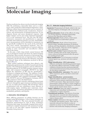
- 1. Chapter 2 Molecular Imaging Nuclear medicine has always involved molecular imaging (MI). By combining a detectable label, such as a radio-tracer, 16 with a molecule of physiological importance, many different cellular function parameters can be assessed. In fact, MI has been defined as the visualization, character-ization, and measurement of biological processes. It can diagnose disease and assess therapeutic response, long before changes can be seen with computed tomography (CT) at the anatomical level. The fact that MI allows noninvasive assessment and quantification is especially desirable when following patients over time. Small differ-ences among patients can be identified so treatments can be specifically modified for the needs of the individual. This move toward “personalized medicine” may also involve more accurate identification of research subjects, leading to more successful and cost-effective clinical trials in the future. In the past, a fairly sharp divide existed between MI uses in preclinical research and those for clinical imaging and therapy. More recently, the process of advancing trans-lational research has become an important focus of the MI community, and the line between the laboratory and clinic has blurred. Some of the definitions involved in MI are listed in Box 2-1. Many nuclear medicine techniques have played a role bridging the gap between the worlds of research and clinical practice. The widespread acceptance and use of fluorine-18 fluorodeoxyglucose (F-18 FDG) positron emission tomog-raphy with computed tomography (PET/CT) has high-lighted the importance of MI and has spearheaded a revolution in imaging and therapy. The addition of multi-modality equipment, such as single-photon emission com-puted tomography with computed tomography (SPECT/ CT), PET/CT, and now PET/magnetic resonance (PET/ MR), continues to drive the field forward. Multiple new agents currently undergoing clinical trials will move through the approval process and help transform the way we think about imaging. MI and therapy will be revolutionized with new quantitative assays directed at wide-ranging targets, including gene expression, mem-brane receptors, and protein upregulation, as well as tumor metabolism, perfusion, and hypoxia. IMAGING TECHNIQUES Many different parameters of cellular function can be imaged using MI techniques. A list of these techniques is provided in Table 2-1. The many potential benefits and limitations demonstrated by each of these techniques determine their usage. Increasingly, imaging forms may be combined as hybrid instruments with anatomical modali-ties such as CT that allow superior localization of the biomarker being detected. Box 2-1. Molecular Imaging Definitions Apoptosis: Programmed cell death, which is the way the body disposes of damaged, old, or unwanted cells. Pharmacodynamics: Study of the effects of a drug on a living organism, including relationship between the drug dose and its effect. Pharmacogenetics: Study of how a body reacts to a drug based on an individual’s genetic makeup. Pharmacokinetics: Study of how living tissues process drugs, including alterations in chemical makeup and drug absorption, distribution, metabo-lism, and excretion. This may involve tagging a drug with a probe or radiotracer. Reporter gene system: Engineered genes that encode a product that can be easily assayed to assess a process being monitored after the genes are transfected into cells. Signal amplification: Use of enzymes to activate contrast agent (e.g., protease activation optical agents). Target identification—DNA microarray: Efficient method for identifying potential targets by detecting mRNA expression. Further target validation needed because posttranscriptional and posttranslational processing means proteins are not always expressed. Target identification—genomics: The study of DNA sequences, genes, and their control and expression. Target identification—proteonomics: High-throughput methods to quantitatively determine tissue protein expression (alternative to DNA microarray). Mass spectrometry–based proteonomics using cell lines or tissue samples or immunohistochemistry of diseased or unaffected tissues can be used in tissue arrays. Target validation: Once the target is identified, expression and subcellular localization are evaluated in a variety of tissues. Translational medicine: The process of moving basic laboratory research into clinical practice, including necessary patient testing and clinical trials to ensure safety. Tumor marker: Substances that may be used to identify and monitor cancer. They may be materials released into blood or urine in response to cancer or may be labeled for identi-fication with molecular imaging techniques.
- 2. Molecular Imaging 17 Radionuclide Imaging SPECT and PET offer obvious advantages because of their sensitivity for radiolabeled probe detection. Although PET offers some potential advantages over SPECT, such as improved resolution and quantitative capabilities, SPECT is often more practical because it is less expensive and widely available. The combination of either technol-ogy with CT allows improved accuracy. Micro-PET and micro-SPECT systems are available for smaller animals. New dedicated clinical breast positron emission mammography (PEM) cameras recently have shown improved accuracy over whole-body PET-CT scanners in assessing primary breast tumors with F-18 FDG, and single-photon breast-specific gamma imaging (BSGI) or molecular breast imag-ing (MBI) cameras for technetium-99m sestamibi imaging have begun to find increased clinical demand. Magnetic Resonance Imaging Magnetic Resonance Spectroscopy Magnetic resonance spectroscopy (MRS) has been able to assess the molecular composition of tissues. Radiofre-quency pulses are applied that excite atoms such as hydro-gen- 1. It is possible to monitor concentrations of molecules, including choline, N-acetylaspartate, and creatinine, by the signals detected. Although other nuclei exhibit low signal-to- noise ratios with MRS, a process termed hyperpolarization allows the use of helium-3 and xenon-129 in lung perfusion examinations and carbon-13–labeled molecules, such as C-13 pyruvate, to map tumor metabolism. Applications of MRS are wide ranging, but its utility is limited in compari-son to the sensitivity of PET. Although PET can detect nanomolar concentrations of radiotracers, MR sensitivity is in the millimolar range. Diffusion Magnetic Resonance Imaging Not only are the high soft tissue contrast, anatomical reso-lution, and contrast enhancement capabilities of MRI use-ful but new imaging sequences are also able to assess some functional tissue characteristics. One of these, diffusion weighted MR (DW-MR), is being investigated as a means of characterizing, staging, and assessing therapy in some tumors. DW-MR makes use of the fact that water mole-cules are more mobile when tissues are less cellular. Based on the DW-MR, an apparent diffusion coefficient (ADC) value is calculated. Lower ADC values have been shown in tumors, such as glioblastomas, with poorer prognosis. Optical Imaging Because the low energy of light is attenuated and scattered by soft tissues, optical imaging of biological processes is limited to preclinical work with small animals (usually mice) or very superficial targets (such as in endoscopy). However, it offers several advantages by being inexpen-sive, flexible, and sensitive. Technical advancements in detectors have resulted in improved sensitivity and resolu-tion, as have multimodality cameras combining SPECT, PET, and CT. The two major categories of optical imaging are bioluminescence and fluorescent imaging. An example of bioluminescence involves the enzyme luciferase, which is responsible for the glow in insects such as the firefly, jellyfish, and some bacteria. This enzyme is placed into the DNA of cells, animals, or models of disease as a reporter gene. The substrate, d-luciferin, is adminis-tered, and a chemical reaction results in low levels of a spectrum of emissions. However, background noise is almost nonexistent because little light is emitted from the imaged tissues. Despite its disadvantages, this has become an integral technique for preclinical imaging. Fluorescence imaging uses a fluorescent protein—a fluorophore—that is excited by an external light source. The fluorescent protein can be genetically engineered into an animal, or a molecule of interest can be labeled with fluorophore fluorescent particles. Unlike biolumines-cence, in which the signal detected is proportional to the number of luminescent cells, fluorescence signal maybe more difficult to quantify because it is affected by the number of fluorescent cells and the intensity of the exter-nal light used for excitation. The quantum yield of the sig-nal in fluorescence is orders of magnitude higher than for bioluminescence and does not require administration of a substrate. Photoproteins such as green fluorescent protein (GFP) have been available for years. New proteins with emission spectra peaks in the near-infrared (NIR) wave-lengths, or NIR fluorochromes, have expanded the range of this technique, showing less absorption. It is possible to image multiple targets of interest at one time. Use of this technique has been extended beyond small animal pre-clinical research. For example, diffuse optical imaging and Table 2-1 Functional and Molecular Imaging Modalities Modality Advantages Disadvantages PET High sensitivity Highly quantitative Temporal monitoring possible Many translational agents under development Radiation Cyclotron on site for short-lived agents Spatial resolution relatively low SPECT Widely available Many probes Lower spatial resolution and less quantitative than PET Some radiation Optical imaging High spatial resolution possible Good sensitivity Quick and inexpensive Limited detection depth Limited clinical use MRS Sensitive Native molecules, no contrast needed Limited region examined MRI High resolution Lower temporal resolution Sensitivity lower Ultrasound with contrast Portable No radiation Low cost High frequency with microbubbles provides good spatial resolution Real-time temporal monitoring Microbubbles research only Sensitivity lower Quantitative ability low MRI, Magnetic resonance imaging; MRS, magnetic resonance spectroscopy; PET, positron emission tomography; SPECT, single-photon emission computed tomography.
- 3. 18 Nuclear Medicine: The Requisites diffuse optical spectroscopy using NIR light has been used to detect tumors and monitor effects of neoadjuvant che-motherapy in breast cancer. Ultrasound Recent advances in ultrasound contrast enhancement include the use of microbubble technology and high-frequency ultrasound. Small gas bubbles can be stabilized with a lipid, albumin, or polymer shell. For imaging, micro-bubbles have been conjugated onto many molecules, such as peptides and antibodies. Also, the microbubbles can be used to deliver a therapeutic payload. This could include gene therapy and cancer treatments. Perfluorocarbon-filled microbubbles allow optimal conditions for targeting and drug delivery. BIOMARKERS Background To assess cellular function noninvasively, it is important to identify biomarkers, specific characteristics of a disease or cellular process that can be measured. Biomarkers can help predict which patients are likely to respond to a spe-cific therapy, such as patients who possess a necessary receptor. Response biomarkers can be used to follow changes in a disease over time. The development of stan-dardized imaging biomarkers is growing. Many multi-center and corporate trials now incorporate measurements of various biomarkers by CT, ultrasound, or MR. Criteria such as the revised Response Evaluation Criteria in Solid Tumors (RECIST) have been put into general use to standardize the way size and tumor volume measure-ments are obtained and reported. The inclusion of F-18 FDG PET-CT criteria into clinical trials is rapidly grow-ing, and recommendations about standardized PET reporting as a biomarker (i.e., PET RECIST [PRECIST]) have been made. In addition to F-18 FDG, many other cellular function parameters are being investigated as potential biomarker targets. These include cell proliferation, peptide and membrane biosynthesis, receptor expression, hypoxia, angiogenesis, apoptosis, and gene transfection (Table 2-2). When developing a nuclear probe, target selection is criti-cal. For imaging, the target must be specific for the dis-ease, reflect disease extent, and be accessible to the probe. Probe accumulation, on the other hand, must reflect the extent of disease. Both intracellular and extracellular tar-gets have been successfully used, and many probes employ existing receptor ligands, antibodies, or enzymes as their foundation. Unlike targets such as receptors, in which one imaging molecule per target will be localized, the use of an enzyme approach results in signal amplification with many imaging molecules per target molecule. Cell Metabolism: Florine-18 Fluorodeoxyglucose The glucose analog F-18 FDG is a sensitive marker for many tumors, reflecting increased tumor glycolysis. F-18 FDG has been proven useful for tumor diagnosis, staging, and therapy monitoring. However, in some situations, F-18 FDG is not the optimal imaging agent. Many cancers, such as well-differentiated and slowly growing tumors, do not accumulate F-18 FDG to a significant extent. F-18 FDG also lacks specificity, showing accumulation in inflamma-tory and infectious processes. Cell Proliferation: Florine-18 Fluorothymidine A biomarker mirroring DNA synthesis could show increased specificity for tumors compared to F-18 FDG. The pyrimidine nucleoside thymidine is the logical choice because it is taken up proportionally to DNA synthesis but is not a precursor of mRNA. Thymidine kinase 1 (TK1) phosphorylates thymidine taken up into the cell, and the activity of TK1 roughly serves as a marker of DNA synthe-sis. However, the picture is complicated by the fact that Table 2-2 Functional Imaging Assays with Positron Emission Tomography and Single-Photon Emission Computed Tomography Cellular Parameter Agent Clinical (C) Translational (T) Preclinical (P) Metabolism F-18 fluorodeoxyglucose C Proliferation F-18 fluorothymidine (FLT) T Biosynthesis C-11 choline C-11 acetate TT Amino acid transport, metabolism, and peptide synthesis C-11 methionine F-18 fluoroethyltyrosine (FET) F-18 DOPA TT T Receptor expression In-111 pentetreotide Ga-68 DOTA-TOC Ga-68-DOTA-F(ab′)2- herceptin In-111-DTPA-trastuzumab F-18 16α-17β- fluoroestradiol (FES) F-18 fluorodihydrotes-tosterone CTT T T T Antigen expression In-111 ibritumomab tiuxetan (CD 20+) In-111 capromab pendetide (prostate-specific membrane antigen) C C Blood flow O-15 water P, T Angiogenesis F-18 galacto-RGD T Epidermal growth In-111-DTPA-EGF factor receptor Ga-68-DOTA-EGF (EGFR) TT Apoptosis Tc-99m annexin-V T Hypoxia F-18 fluoromisonidazole (FMISO) Cu-64 ATSM T T Transgene expression F-18 FHBG monitor gene Rx P FHBG, Fluoropencicyclovir; RGD, arginine-glycine-aspartic acid. *Some sites outside of the United States clinically applied.
- 4. Molecular Imaging 19 thymidine is also incorporated into DNA via a second pathway involving de novo synthesis from the nucleotide deoxyuridine (Fig. 2-1). Cells and tumors can vary in the use of the targeted extrinsic salvage pathway versus de novo synthesis. In addition, mitochondrial uptake, which occurs independently of cellular division but at a relatively low background level, adds to the complexity. Despite difficul-ties caused by complicated thymidine metabolism, TK1 activity significantly increases in cancer, which is critical for the success of an imaging probe. Several different thymidine analog compounds have been developed. The first radiotracer evaluated in vivo for the imaging of cellular proliferation was carbon-11 thymi-dine. The short half-life of the C-11 label and rapid catab-olism, resulting in labeled metabolites, limited utility. Different F-18–labeled thymidine analogs have been investigated. Currently, the most widely used and promis-ing proliferation agent is F-18 fluorothymidine (FLT). Once phosphorylated by TK1, F-18 FLT is essentially trapped within the cell. Unlike thymidine, hydrogen-3 thymidine, or C-11 thymidine, F-18 FLT is not further metabolized and is not incorporated into DNA. The normal distribution of FLT differs from that of F-18 FDG. Activity is lower in the mediastinum and bowel and very low in the brain. Marked marrow uptake and signifi-cant background liver activity are seen (Fig. 2-2, A). Obvi-ously, the areas with high background activity create challenges for the visualization of metastases, particularly in the bones (Fig. 2-2, B). In addition, F-18 FLT does not accumulate to any significant extent when the blood–brain barrier is intact, so sensitivity is poor for low-grade tumors Thymidine TK Thymidine TK TS Thymidine M UMP Thymidine D Thymidine T P P P FLT-M FLT-D FLT-T P P P DNA FLT FLT DNA Figure 2-1. Thymidine as an imaging biomarker. Upper, Thymidine is taken up into the cell and phosphorylated by thymidine kinase 1 (TK) in the external salvage pathway. Cells also perform endogenous de novo synthesis with deoxyuridine (UMP) and the enzyme thymidylate syn-thase (TS). Lower, F-18–labeled thymidine (FLT) is taken up into the cell, phosphorylated, and trapped similar to thymidine. However, as the dashed arrows suggest, FLT is not further metabolized and is not incor-porated into DNA. FLT-D, difluorothymidine; FLT-M, monofluorothy-midine; FLT-T, trifluorothymidine; P (circled), phosphate. A B C Figure 2-2. F-18 fluorothymidine (F-18 FLT) in newly diagnosed breast carcinoma. A, Maximum intensity projection image shows expected intense uptake in the bones, moderate activity in the liver, and very low uptake in the brain. B, Axial PET and CT images in the same patient show a lack of radiotracer activity in a sclerotic osseous metastasis. C, Radiotracer activity was present in the primary tumor in the left medial breast.
- 5. 20 Nuclear Medicine: The Requisites and those that do not show significant contrast enhance-ment on MRI or CT. It has been determined by examining parameters such as Ki-67 in many tumors, including breast cancer, non–small cell lung cancer, and high-grade brain tumors (Fig. 2-3), that F-18 FLT uptake does in fact corre-late with DNA synthesis (Fig 2-2, C ). Promising early stud-ies suggest FLT will be useful in monitoring chemotherapy response in breast cancer and possibly other tumors. Considered an investigational drug by the U.S. Food and Drug Administration (FDA), examinations are not reimbursable under private or government insurance plans and use of FLT requires an investigational new drug (IND) application be in place. To promote development of FLT as a potential clinical tool, the National Cancer Institute (NCI) of the National Institutes of Health (NIH) developed an IND application for F-18 FLT. Multicenter trials using FLT are under way through the American Col-lege of Radiology Imaging Network (ACRIN). FLT can now be purchased through commercial vendors. Biosynthesis Amino Acid Transport and Metabolism and Peptide Synthesis In addition to cellular proliferation, other parameters may better reflect tumor growth in some cases than F-18 FDG. Using amino acids as radiolabeled probes allows assessment of amino acid transport and metabolism and peptide biosynthesis. Many studies have been published examining the utility of radiolabeled amino acids in eval-uation of brain tumors, an area of limitation for F-18 FDG. Some agents, such as C-11 methionine and F-18 fluoroethyltyrosine (F-18 FET), have shown increased sensitivity for gliomas over FLT. This is particularly true in the case of low-grade gliomas and tumors failing to exhibit MR enhancement. However, C-11 methionine has been seen to accumulate in cases of infection, par-ticularly when severe, limiting specificity. C-11 methio-nine has also been used to examine prostate cancer with some success. F-18 DOPA not only has significantly higher sensitivities and specificities for various brain tumors than F-18 FDG but is also superior for detection of neuroendocrine cancers. Lipid Metabolism and Phospholipid Synthesis In addition to increased glycolytic activity, tumors can show increased fatty acid metabolism and lipid biosynthesis during the production of membranes. These dividing cells show an increased expression of fatty acid synthase (FAS) and choline kinase in two related paths for phospholipid production. Several trials have been done with radiolabeled C-11 acetate, C-11 choline, and more recently F-18 choline in prostate cancer. This is an area of particular interest given the limitations of F-18 FDG in this disease. These agents show uptake in primary prostate cancer with good discrimi-nation from the bladder because there is no urinary excre-tion, and they have been used with some success to assess metastasis. Choline uptake does not appear to correlate with tumor grade, and false positive findings could result from accumulation in benign conditions of the prostate. Uses for these agents in other cancers continue to be investigated. Hypoxia Tumor hypoxia is an important prognostic factor in a wide range of tumors; its presence predicts recurrence, metasta-sis, and decreased survival. Tumor hypoxia is an estab-lished resistance factor for radiotherapy and is increasingly recognized as promoting resistance to systemic cancer therapies. Hypoxia promotes a more aggressive and resis-tant cancer phenotype, mediated by the transcription fac-tor hypoxia-inducible factor 1 (HIF-1), which leads to cell cycle arrest, angiogenesis, and accelerated glycolysis. Non-invasive imaging studies have been examining tumor hypoxia for years; currently, two promising PET agents are undergoing multicenter clinical trials in the United States—F-18 fluoromisonidazole (FMISO) and copper-64 diacetyl-bis(N4-methylthiosemicarbazone) or Cu-64 ATSM. Florine-18 Fluoromisonidazole The nitroimidazoles are a class of hypoxia compounds that have been studied for years. In the viable cell, they are reduced to the RNO2 radical without regard to oxygen concentrations. However, when oxygen is present, the radical is reoxidized and uncharged misonidazole diffuses out of the cell. In situations in which oxygen levels are low, the radical is further reduced and is trapped after binding to intracellular molecules. F-18 fluoromisonidazole (FMISO) is the most exten-sively studied nitroimidazole for in vivo imaging. It read-ily diffuses into cells because of its lipophilic nature. Tissue nitroreductases lead to the generation of radical anions, which are quickly eliminated in the presence of Figure 2-3. F-18 fluorothymidine in glioma. A, T1-weighted, gadolinium-enhanced MRI of the brain shows a large, enhancing tumor in the right frontoparietal cortex. B, F-18 FDG PET image at the same level is deceptive, showing little activity. C, However, significant accumu-lation of F-18 FLT more accurately represents tumor activity. (Courtesy Mark Muzi.) A B C
- 6. Molecular Imaging 21 A B C D Figure 2-4. F-18 fluoromisonidazole (FMISO) tumor hypoxia. T1-weighted, gadolinium (A) and flare (B) MRI images of the brain reveal an aggressive-appearing enhancing tumor with mass effect and edema in the right cortex. C, F-18 FDG PET does show a peripheral ring of increased metabolic activity peripherally (arrow). D, F-18 FMISO images of the same area show significant hypoxic areas in the tumor (arrow), some more prominent than on F-18 FDG. Hypoxic areas are likely to be more resistant to chemotherapy and radiation. oxygen; however, in the absence of oxygen, these radicals bind to tissue macromolecules and are retained. Thus, after equilibration, typically around 2 hours after injec-tion, the accumulation of F-18 FMISO indicates tissue sites lacking oxygen. F-18 FMISO has been evaluated in several tumors, including head and neck cancer and glio-blastoma (Fig. 2-4), and, like F-18 FLT, is the subject of an NCI IND to promote research investigations. Copper-64 ATSM The other major class of hypoxia imaging agent is based on metal chelates of dithiocarbazones. Copper(II)-diacetyl-bis( N4-methylthiosemicarbazone) (Cu-ATSM) can be radiolabeled with copper-60 (T½ = 23.7 minutes), copper-62 (T½ = 9.74 minutes), or Cu-64 (T½ = 12.7 hours). The half-life of Cu-64 ATSM is better for clinical use and commercial distribution. Like F-18 FMISO, Cu-64 ATSM is reduced after entering the cell. The resulting unstable compound is reoxidized in the presence of oxygen and freely diffuses from the cell. In hypoxic tissues, the copper dissociates from the chelate and becomes irreversibly trapped. Hypoxia Imaging Applications Studies using the hypoxia agents include Cu-64 ATSM in cervical cancer and F-18 FMISO in head and neck cancer, non–small cell lung cancer, and gliomas. In glioblastoma, hypoxia is of particular interest given the necrotic nature of the tumor and a typical hypercellular rim that has been shown to be hypoxic. Aggressiveness of gliomas has been related to the presence of hypoxia. Although it is unclear whether F-18 FMISO will be able to predict outcomes, limited studies suggest it may guide therapy in different ways. For example, the antineoplastic therapeutic agent tirapazamine is activated by reductases to form free radi-cals in hypoxic cells, inducing DNA damage and sensitiz-ing tumors to other therapy. Given the side effects of this agent, identifying appropriate candidates is essential. Hypoxia imaging has also been used in some trials examin-ing ways to improve external beam radiation planning. Angiogenesis Vascular endothelial growth factor (VEGF) is overexpressed in many cancer cells, reflecting the ability of tumors to induce neovascularity. Treatment with bevacizumab (rhuMAb VEGF, Avastin), the first FDA-approved antian-giogenesis drug, has shown success at treating non–small cell lung cancer, metastatic colorectal cancer, and breast cancer when combined with conventional chemotherapy drugs. Noninvasive monitoring of angiogenesis is still in the early stages of development. Several radiolabeled forms of the VEGF molecule, including the SPECT agent iodine-123 VEGF and PET agents with such as zirco-nium- 89 VEGF or the Cu-64 labeled epidermal growth factor agent, Cu-64 DOTA-EGF, have been used to moni-tor therapy in preclinical and limited clinical trials. Imaging new vessel formation extends beyond VEGF. Although hypoxia induces expression of VEGF, regulation is a multistep process and occurs through a variety of fac-tors. One of these is ανβ3 integrin, which mediates acti-vated endothelial cell migration of during vessel formation and is a specific marker for neovessels. Therefore monitor-ing ανβ33 expression through the use of nuclear techniques has gained interest. Several proteins, such a fibrinogen, interact with ανβ3 integrin through the amino sequence arginine-glycine-aspartic acid (RGD). Strategies to radiolabel RDG and to develop improved imaging agents have been under way for several years. Glycosylation of the RGD has been one approach to show benefit. A product of this work, F-18 galacto-RGD shows improved retention in tumors and overall kinetics. Other PET, SPECT, and therapy agents also have been formed using the RGD molecule. Receptor Expression Somatostatin Receptors Somatostatin receptors (SSTRs) are found in a variety of tissues, including the gastrointestinal tract and brain. SSTRs are also expressed in numerous tumors, such as neuroendocrine tumors (carcinoid, insulinoma, pheochro-mocytoma, etc.), lung cancer, meningioma, and lymphoma. Most of the work to date involves targeting one of the most commonly expressed of the six known receptor subtypes— type 2. In addition to clinical imaging with indium-111 pentetreotide (Octreoscan), an 8-amino-acid somatostatin analog, improved accuracy has been shown with several PET-labeled peptides including: Ga-68 DOTA-Tyr3 octreotide (Ga-68 DOTA-TOC), Ga-68 DOTA-Tyr3
- 7. 22 Nuclear Medicine: The Requisites octreotate, and Ga-68 DOTA-1-NaI3 octreotide (Ga-68 DOTA-NOC). These agents all bind to SSTR receptor 2, but Ga-68 DOTA-NOC also has affinity for subtypes 3 and 5 and Ga-68 DOTA-TOC also binds to subtype 5. Great interest has been shown in therapeutic aspects of somatostatin receptor binding. In addition to trials using In-111 pentetreotide, good response has been shown with beta-emitters, including yttrium-90 and lutetieum-177. Trials with these agents are more prevalent in Europe, where the regulatory environment can be more conducive to early clinical trials. Hormone Receptors Given the fact that hormonal therapy plays an important role in treatment of breast and prostate cancer, it is no sur-prise that targeting these receptors with radiolabeled probes has become of interest. Although receptor status is easily determined by tissue biopsy, it is more difficult to assess the status of metastases. This is of particular impor-tance because receptor-positive primary tumors may show variable expression after metastasizing or recurring. F-18 16β-fluoro-5α-dihydrotestosterone (F-18 FDHT) has been used to detect androgen receptors in primary prostate cancer tumors and metastatic disease in early clin-ical trials. More work has been done in breast cancer recep-tor targeting. F-18 16α-17β-fluoroestradiol (F-18 FES) has shown the greatest promise for estrogen receptor labeling. F-18 FES uptake has been correlated with patient progno-sis and response to aromatase inhibitors. For HER2 (ErB2) receptor assessment, antibody-based probes using both SPECT and PET radiolabels have shown the greatest util-ity thus far. Several different agents, including In-111 trastuzumab, Zr-89 trastuzumab, and a Ga-68 labeled F(ab′)2 fragment, are under investigation. Apoptosis Apoptosis, or programmed cell death, is the primary method by which old or unneeded cells are removed from the body and is believed to be a major mechanism by which anticancer treatments work. It is a very different process from necrotic cell death associated with tumors, trauma, or infection. Apoptosis depends on signals, such as extrinsic tumor necrosis factor, to initiate a cascade response related to a series of caspases. Apoptotic cells will express surface phosphatidylserine, which can serve as a target for imaging probes. The first imaging probe for apop-tosis involved Tc-99m labeling of the peptide annexin V. Additional probes have been investigated, and nonnuclear techniques such as MRS examining factors such as changes in choline have been explored. Because chemotherapy agents often induce tumor cell death through apoptosis, imaging could identify areas at risk for therapy resistance. Additionally, apoptosis plays an important role in cardio-vascular disease. However, imaging remains a preclinical investigational tool at this time. Future Applications: Nanotechnology Nanoparticles are a rapidly developing and exciting area of investigation. These tiny organic and inorganic particles, ranging in size from 1 to 100 nm, are another area blurring the boundaries between imaging and therapy. Numerous different nanoparticles have been used in imaging and therapeutic applications. They can be used as imaging contrast agents and also can deliver therapy, with many being responsive to conditions associated with tumor expression or even factors such as pH. Rare earth–labeled nanoparticles can be used for optical imaging and MRI, and PET imaging is possible using radiolabels such as F-18 and Cu-64. REPORTER GENE IMAGING AND GENE THERAPY Recombinant DNA technology has resulted in the ability to insert genes into DNA to study basic biology and as an approach for gene-based therapy. Often, it is not possible to directly image a target of interest such as levels of expression of a therapeutic gene. The idea of using reporter genes—that is, diagnostic genes linked to another gene of interest designed to indicate when a particular transgene is expressed—was developed for basic cellular and molecular research using approaches such as GFP. The idea has been translated to imaging using optical reporters and reporters designed for use with radionuclide labels, such as the viral thymidine kinase gene from the herpes simplex virus type 1 (HSVtk1). This technique can be used to monitor gene therapy in humans with a reporter gene linked to genes intended for gene therapy that could noninvasively report on the success of transfection of the therapeutic gene in target tissues. Strategy Many different viral vectors have been used to transfer genetic material into a host cell, although the most common is the herpes simplex virus type I (HSV1). Several HSV1 characteristics make it a useful vector. It is highly infectious, with a broad range of targets on the host cell. It also pos-sesses many nonessential genes, which can be deleted with-out compromising its ability to infect and replicate, making room for genes of interest. Researchers can construct a plas-mid and use viral vector transport to insert a reporter gene into the system being observed. Imaging can be done with a targeted reporter probe that is trapped within a cell carry-ing one of these reporter genes. For example, in preclinical work, the gene for luciferase can be inserted into cells and then optical imaging can monitor expression in transfected cells. Two main categories of reporter gene strategies exist— those using receptors and those using enzymes. It is possible to insert reporter genes to produce recep-tors, and the degree of receptor expression can be imaged, reflecting the cellular activity. Although challenges exist, such as developing probes with sufficient binding affinity, these receptors make excellent imaging targets, easily accessible on the cell surface. Several well-characterized reporter systems are being used in clinical trials including D2 dopaminergic and somatostatin receptors. Enzyme-based reporter systems are more commonly used than receptor systems, providing the advantage of sig-nal amplification. Rather than the one-to-one relationship seen in receptor imaging, one enzyme molecule can act on numerous substrate molecules. The enzyme most widely used in reporter gene imaging is based on HSV1-tk. Once a cell is transfected, expression of HSV1-tk results in an
- 8. Molecular Imaging 23 enzyme with several potential substrates, including ganci-clovir, 5-iododeoxyuridine, and 1-(2′-deoxy-2′fluoro-1-β- d-arabinofuranosyl-5-iodouracil (FIAU). These can be radiolabeled with agents ranging from iodine (iodine-124 FIAU, I-123 FIAU) to F-18 (F-18 fluoroganciclovir). Monitoring Gene Therapy One exciting area of research concerns the possibility to use recombinant gene technology as a mechanism for ther-apy. These therapies could provide novel solutions for treating disease such as cancer. However, to be able to develop such protocols, accurate monitoring methods are needed. By linking a therapeutic gene with an imaging reporter gene, this would be possible in vivo using nonin-vasive means with PET or SPECT. In treatment, for example, a cell transfected with HSV1- tk could be killed by administering a prodrug substrate, such as ganciclovir, which would form a toxic compound inside the cell when acted on by HSV1-tk. Alternatively, cells could be transfected with the gene for a receptor, such as the somatostatin receptor, along with a therapeutic gene. The distribution of the gene could be assessed with In-111 octreotide or Ga-68 DOTA-TOC and activity followed over time to assess therapy effect. IMAGING BIOMARKERS AND NEW DRUG DEVELOPMENT Until recently, methods used to identify a target for a can-cer therapy agent or to monitor its effects have relied on assays using tissue or blood samples. A noninvasive, imaging- based assay offers several potential benefits. This includes the quantitation possible with PET without perturbing the system being analyzed as with biopsy so that accurate serial measurements are possible. In addi-tion, imaging biomarkers are an important component in the move toward “personalized” cancer therapy. To determine if a therapy will be successful, MI targets can be identified that will predict whether a patient will respond. One example of this would be identifying HER2 overexpression in a patient with breast cancer to decide if therapies such as trastuzumab directed against HER2 will be effective. In addition to the uses of MI in determining potential therapy targets, it also can help assess drug pharmacodynamics and the response, if any, to a certain drug. This knowledge can help prevent unnecessary treatments and undesirable delays in starting appropriate therapies. As potential new drugs move through the development process into clinical trials, many factors need to be consid-ered (Fig. 2-5). In phase I and II (early phase) trials, small numbers of patients are evaluated at a limited number of sites to confirm the effects of the drug and that appropriate patients are selected for the therapy. These trials often involve evaluating complex kinetics of the drug. MI tech-niques using short-lived labels such as C-11 are useful for rapid, serial studies needed to assess kinetics and drug transport. These early-phase trials also look at the effects of a drug on the tumor and on normal tissue to assess safety. Finally, it is critical to determine if the drug will affect the biodistribution or clearance of the imaging probe being used, because this could alter measurements. Preclinical research Target identification Candidate screen Clinical trials: Phase I + II Phase III FDA approval Discovery Preclinical Translational Clinical use Figure 2-5. Stages of new drug development. In phase II and III (late phase) trials, imaging biomark-ers can be helpful indicators of early response or might even act as surrogate end points. In many cases, tumors will show a response rapidly with an MI agent, even when the tumor mass appears unchanged on conventional imag-ing, such as CT. These larger trials require tightly con-trolled protocols at multiple centers, so the imaging markers used must be more widely available. PET agents labeled with F-18, Cu-64, or I-124 have sufficiently long half-lives to can be easily shipped from regional cyclotron and production centers. The increasing expense of taking a new agent through the FDA approval process into clinical use demands care-ful selection and monitoring protocols. From preliminary target identification, discovering sensitive populations, and monitoring therapeutic effects in clinical trials, noninvasive imaging techniques are playing an increasingly important role in this process. Suggested Reading Blankenberg FG, Norfray JF. Multimodality molecular imaging of apoptosis in oncology. AJR Am J Roentgenol. 2011;197(2):308-317. Contag CH. In vivo pathology: seeing with molecular specificity and cellular resolution in the living body. Ann Rev Pathol Mech Dis. 2007;2:277-305. Dunphy MP, Lewis JS. Radiopharmaceuticals in preclinical and clinical development for monitoring therapy with PET. J Nucl Med. 2009;50(suppl 1): 106S-121S. Ferrara K, Pollard R, Borden M. Ultrasound microbubble contrast agents: fundamentals and application to gene and drug delivery. Annu Rev Biomed Eng. 2007;9:415-447. Hylton N. Dynamic contrast-enhanced magnetic resonance imaging as an imaging biomarker. J Clin Oncol. 2006;24(10):3293-3298. Kang JH, Chung JK. Molecular-genetic imaging based on reporter gene expression. J Nucl Med. 2008;49(suppl 2):164S-179S. Ledezma CJ, Chen W, Sai V, et al. 18F-FDOPA PET/MRI fusion in patients with primary/recurrent brain gliomas: initial experience. Eur J Radiol. 2009;71(2):242- 248. Mankoff DA, Link JM, Linden HM, Sundararajn L, Krohn KA. Tumor receptor imaging. J Nucl Med. 2008;49(suppl 2):149S-163S. Nishino M, Jagannathan JP, Ramaiya NH, Van den Abbeele AD. Revised RECIST guideline version 1.1: what oncologists want to know and what radiologists need to know. AJR Am J Roentgenol. 2010;195(2):281-289. Plathow C, Weber WA. Tumor cell metabolism imaging. J Nucl Med. 2008;49(suppl 2):43S-63S. Rohren EM, Macapinlac HA. PET imaging of prostate cancer: other tracers. PET Clin. 2009;4:185-192. Salsov A, Tammisetti VS, Grierson J, Vesselle H, FLT. Measuring tumor cell proliferation in vivo with positron emission tomography and 3′-deoxy-3′[18F] fluorothymidine. Semin Nucl Med. 2007;37(6):429-439. Tromberg BJ, Cerussi A, Shah N, et al. Imaging in breast cancer: diffuse optics in breast cancer: detecting tumors in premenopausal women and monitoring neoadjuvant chemotherapy. Breast Cancer Res. 2005;7(6):279-285. Vallabhajosula S, Solnes L, Vallabhajosula B. A broad overview of positron emission tomography radiopharmaceutical and clinical applications: what is new? Semin Nucl Med. 2011;41(4):246-264. Virgolini I, Ambrosini V, Bomanji JB, et al. Procedure guidelines for PET/CT imaging with 68Ga-DOTA-conjugated peptides: 68Ga-DOTA-TOC, 68Ga-DOTA-NOC, 68Ga-DOTA-TATE. Eur J Nucl Med Mol Imaging. 2010;37(10):2004-2010. Wahl RL, Jacene H, Kasamon Y, Lodge MA. From RECIST to PERCIST: evolving considerations for PET response criteria in solid tumors. J Nucl Med. 2009;50(suppl 1):122S-150S.