Download to read offline
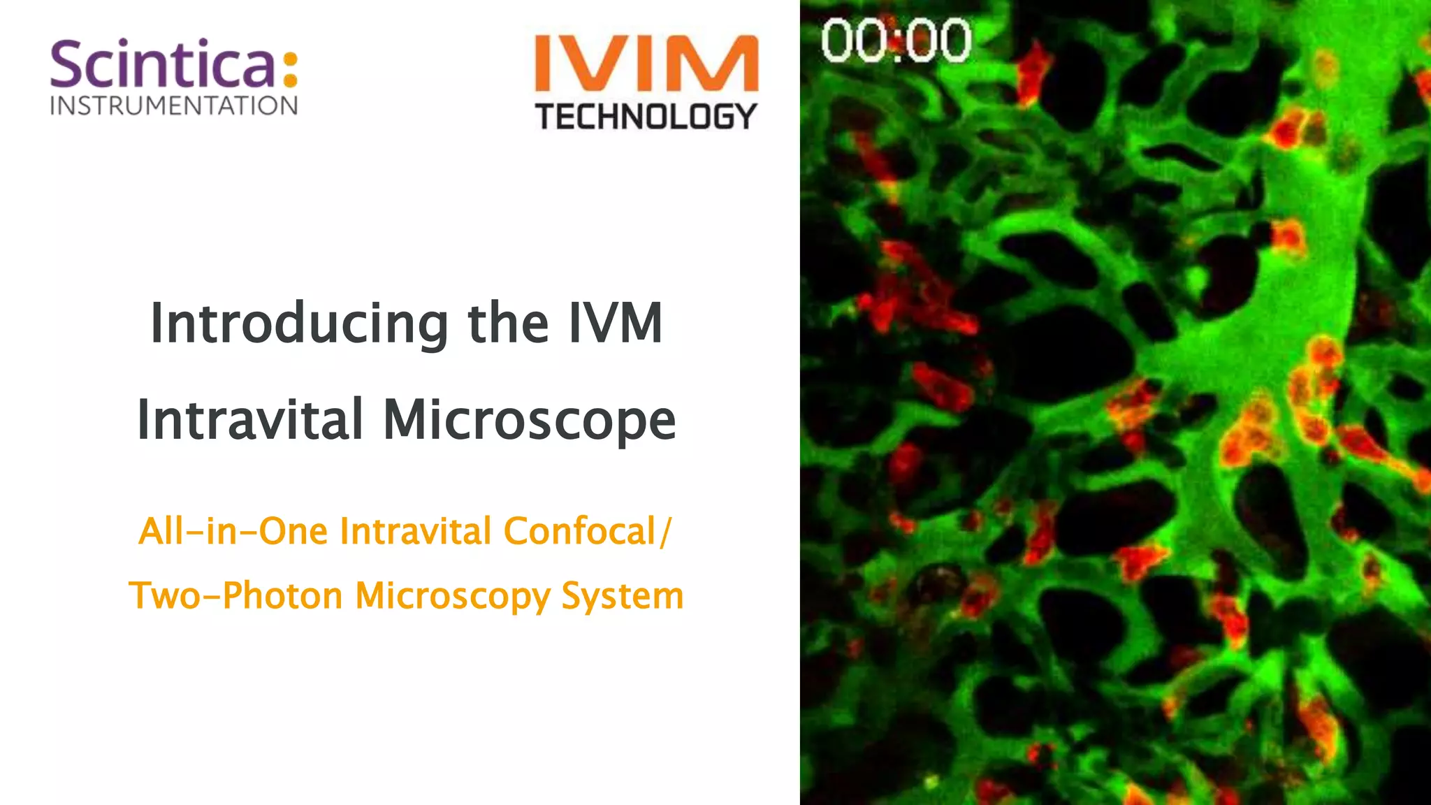
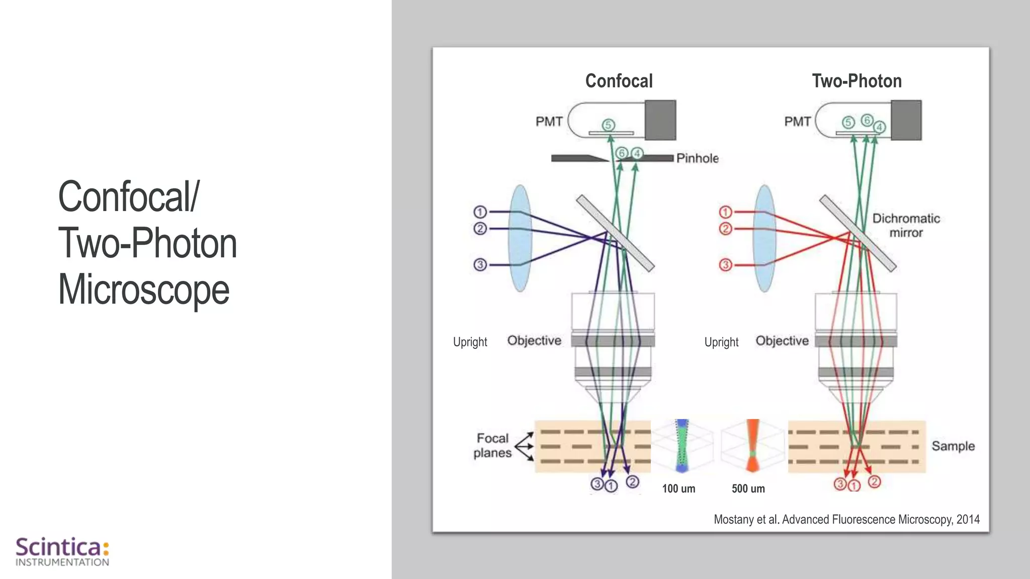
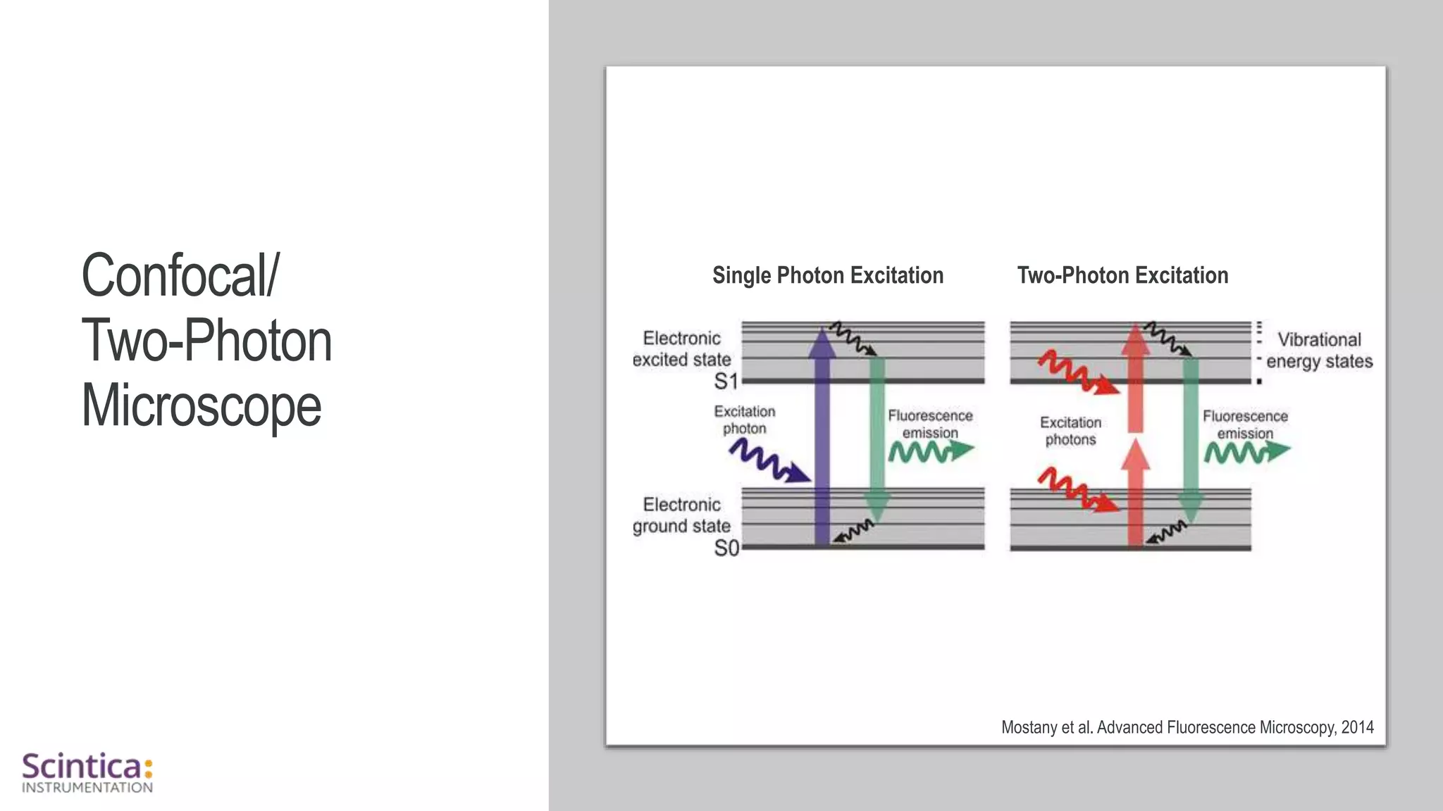
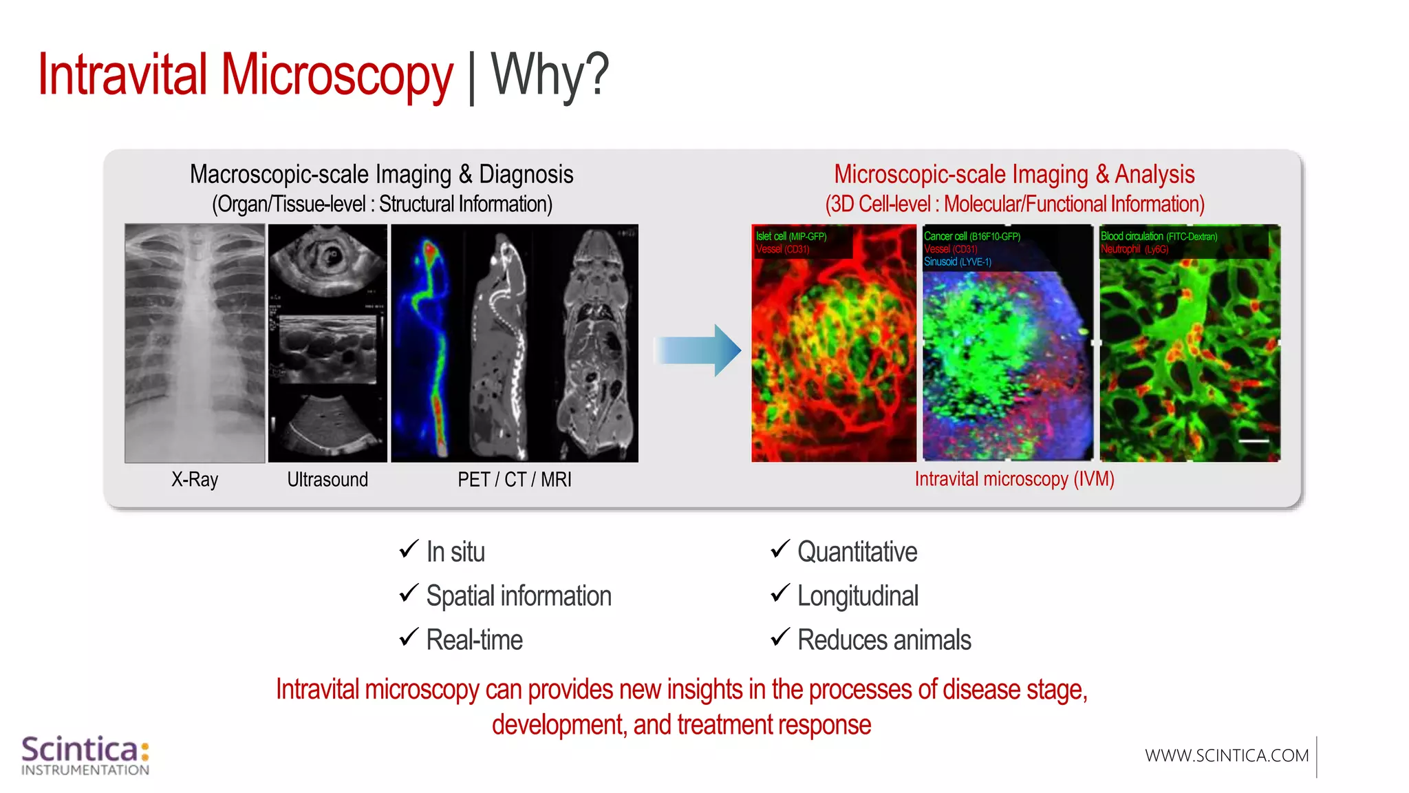
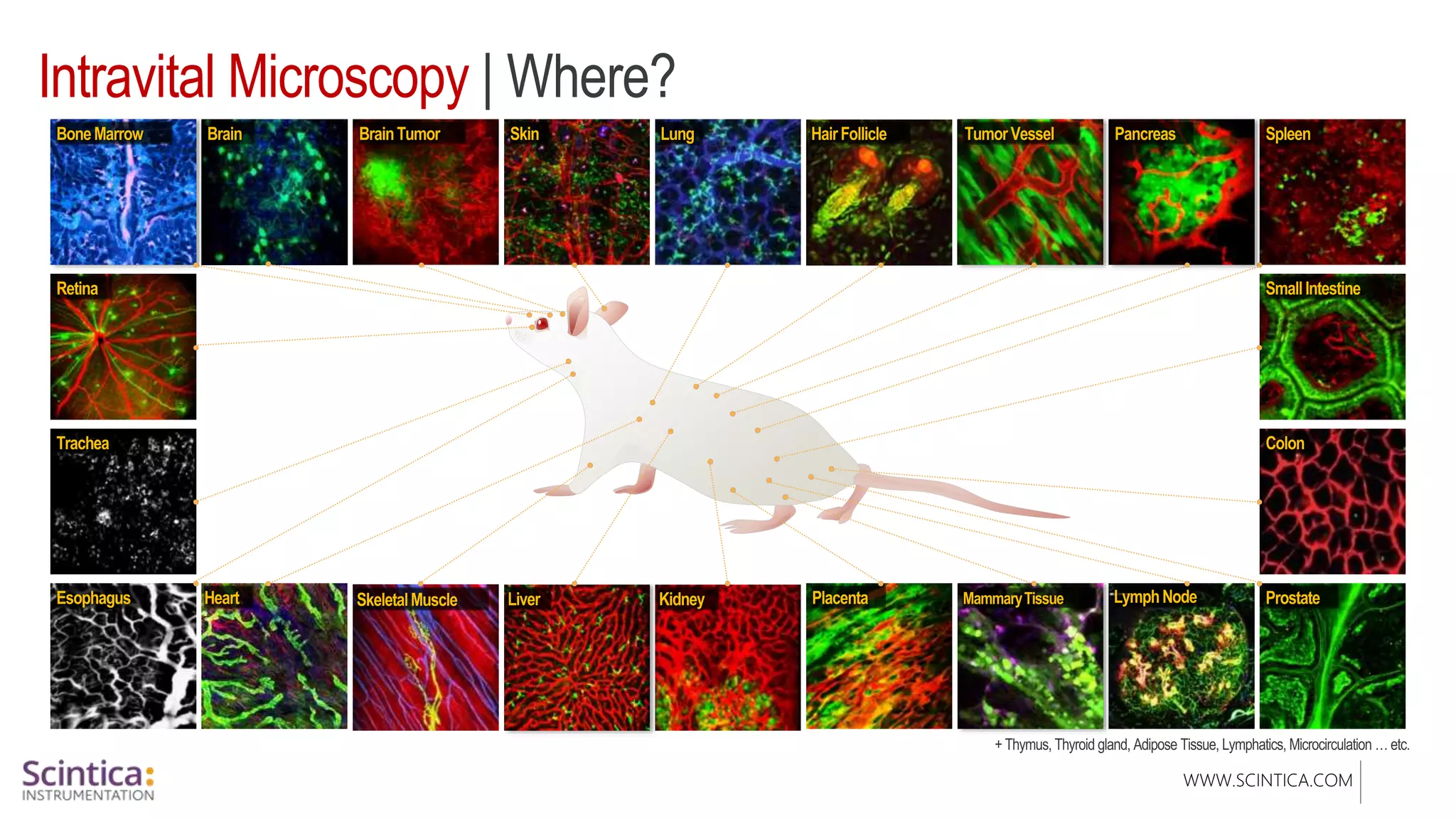
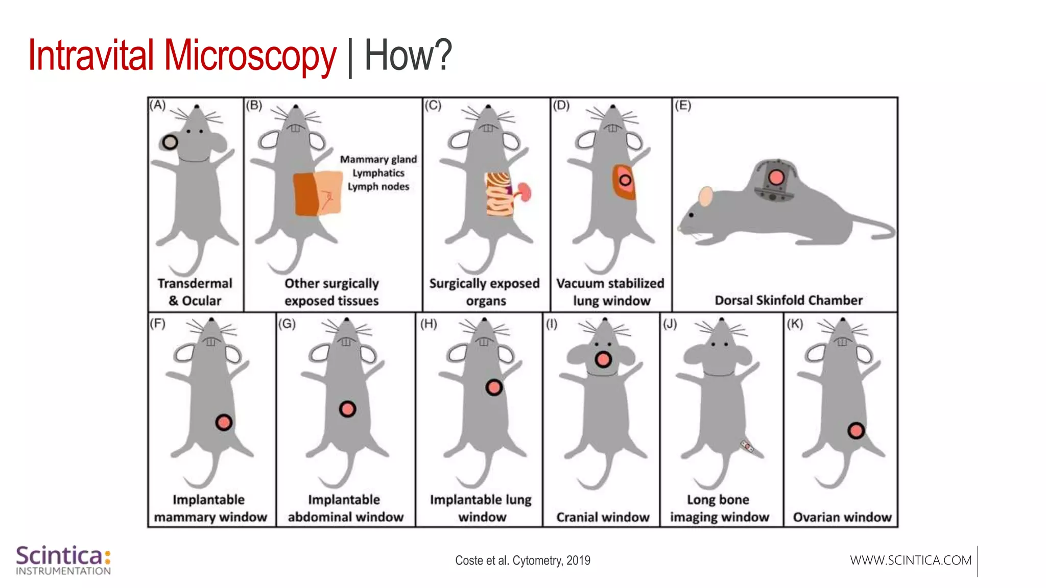
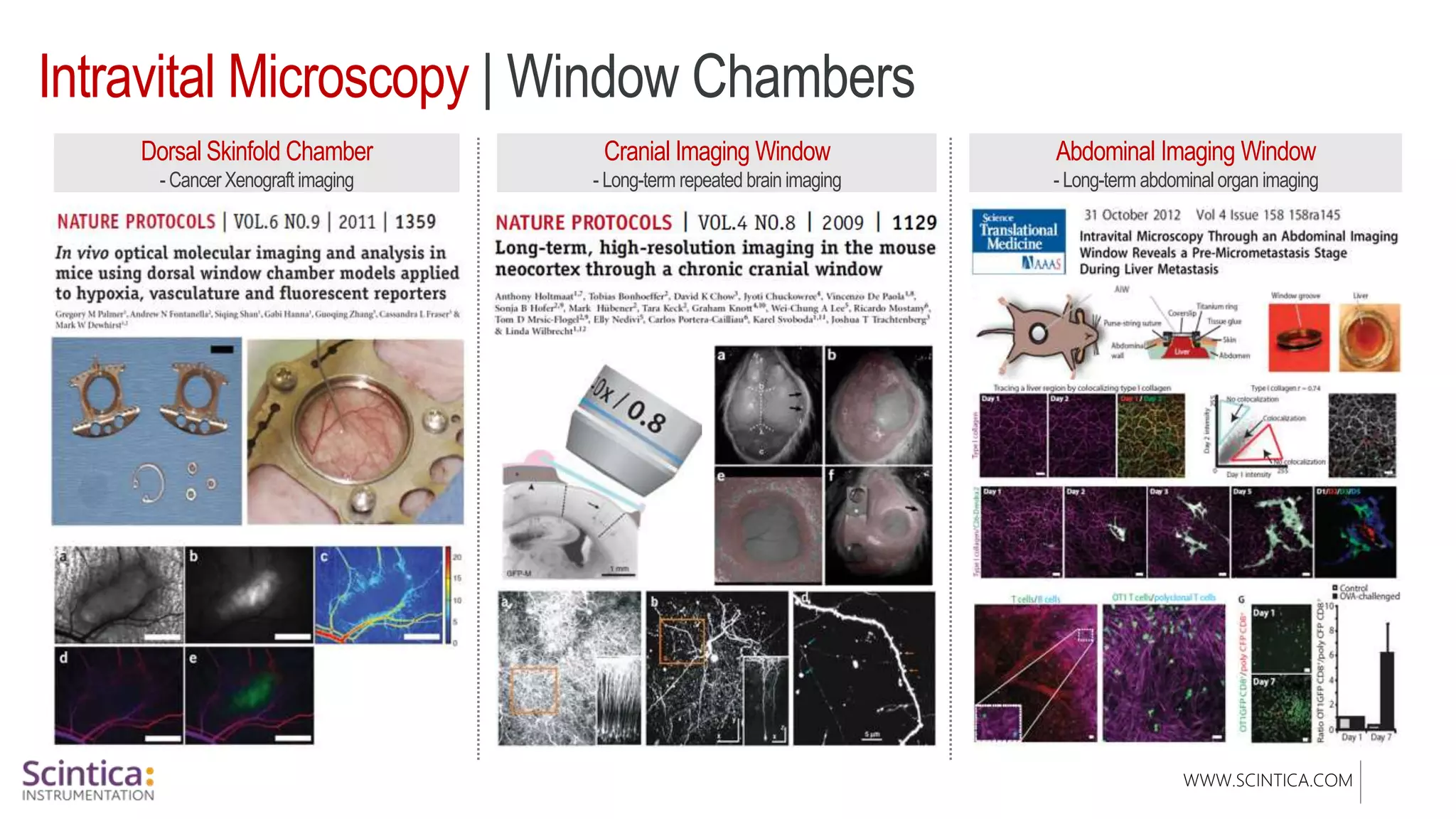
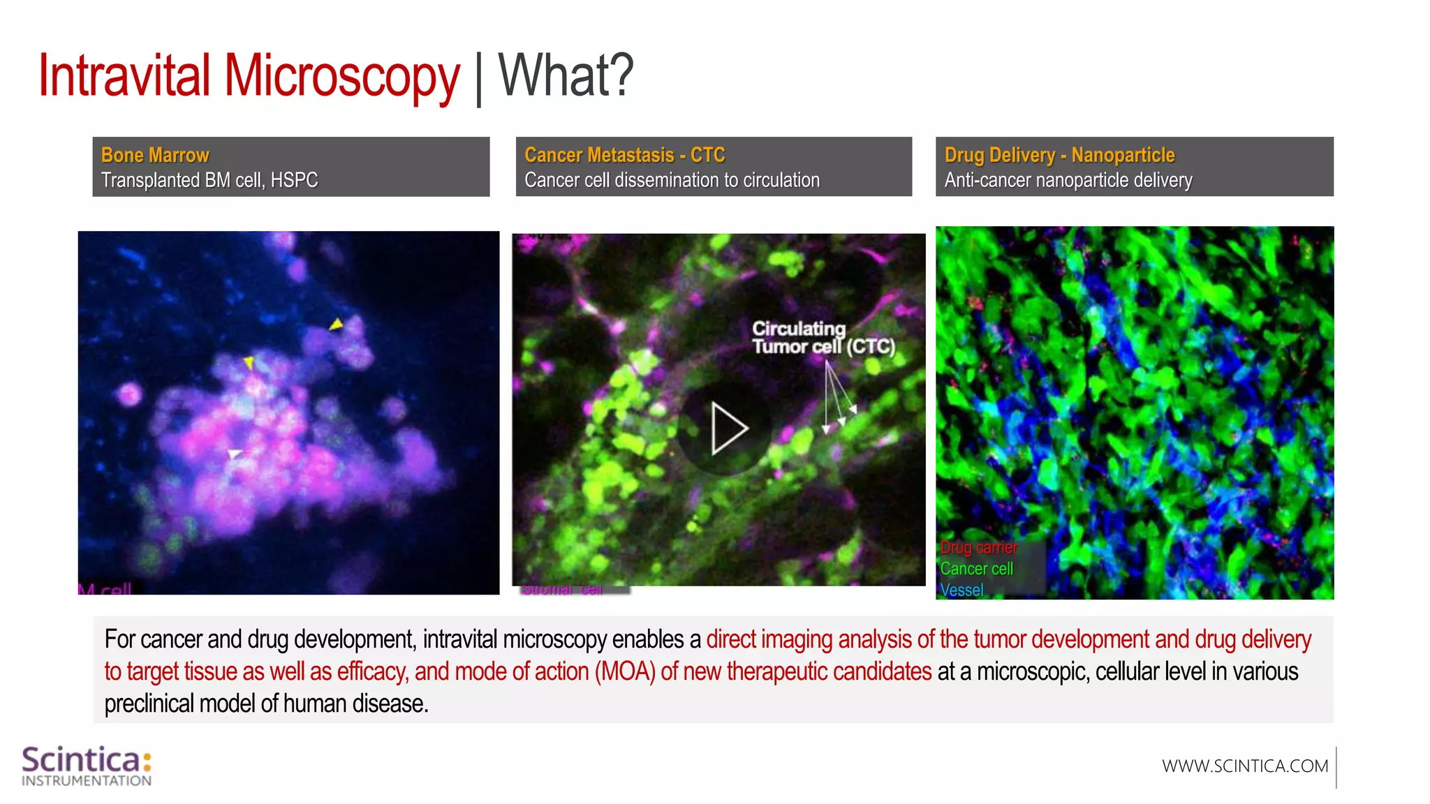
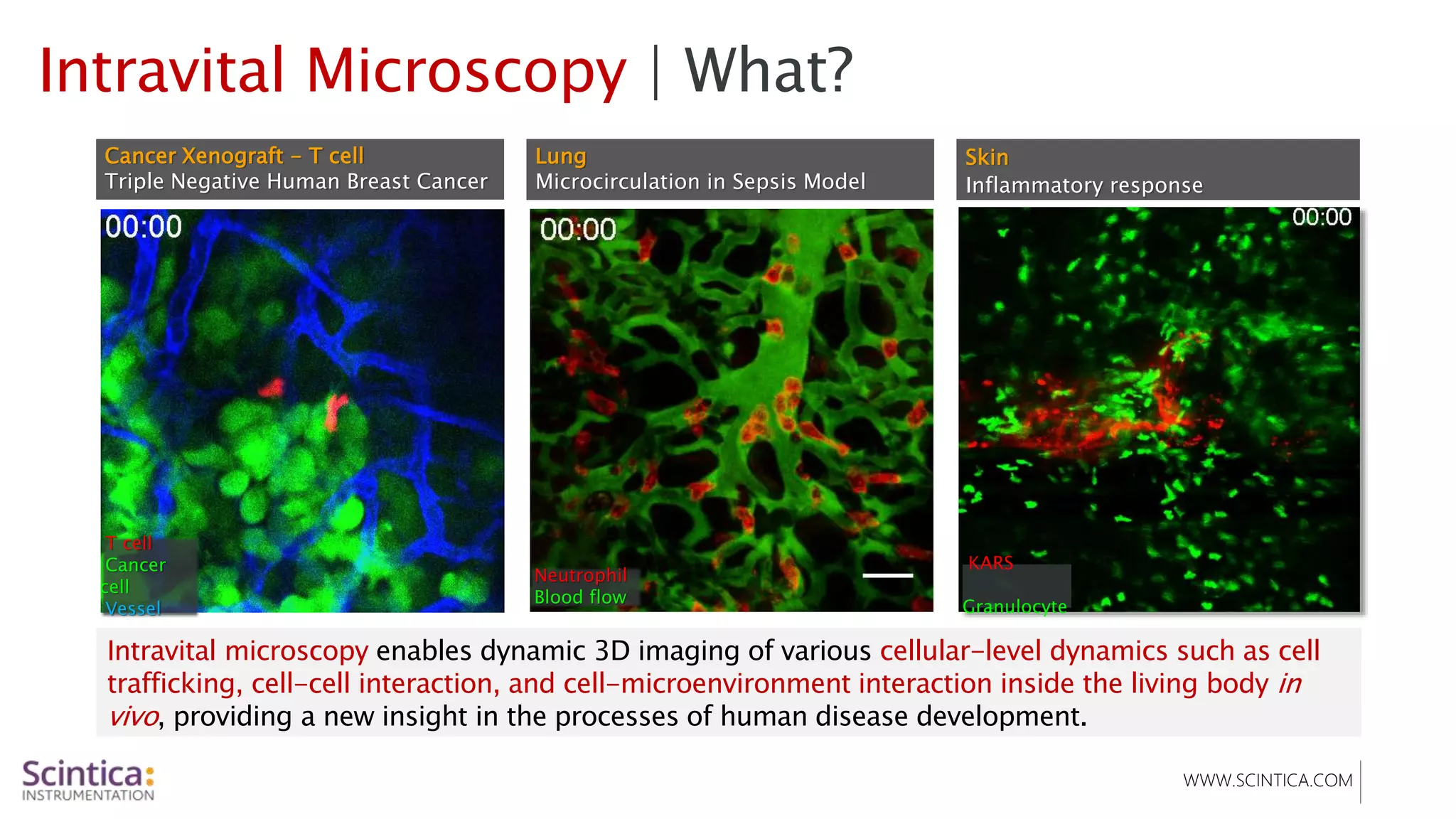
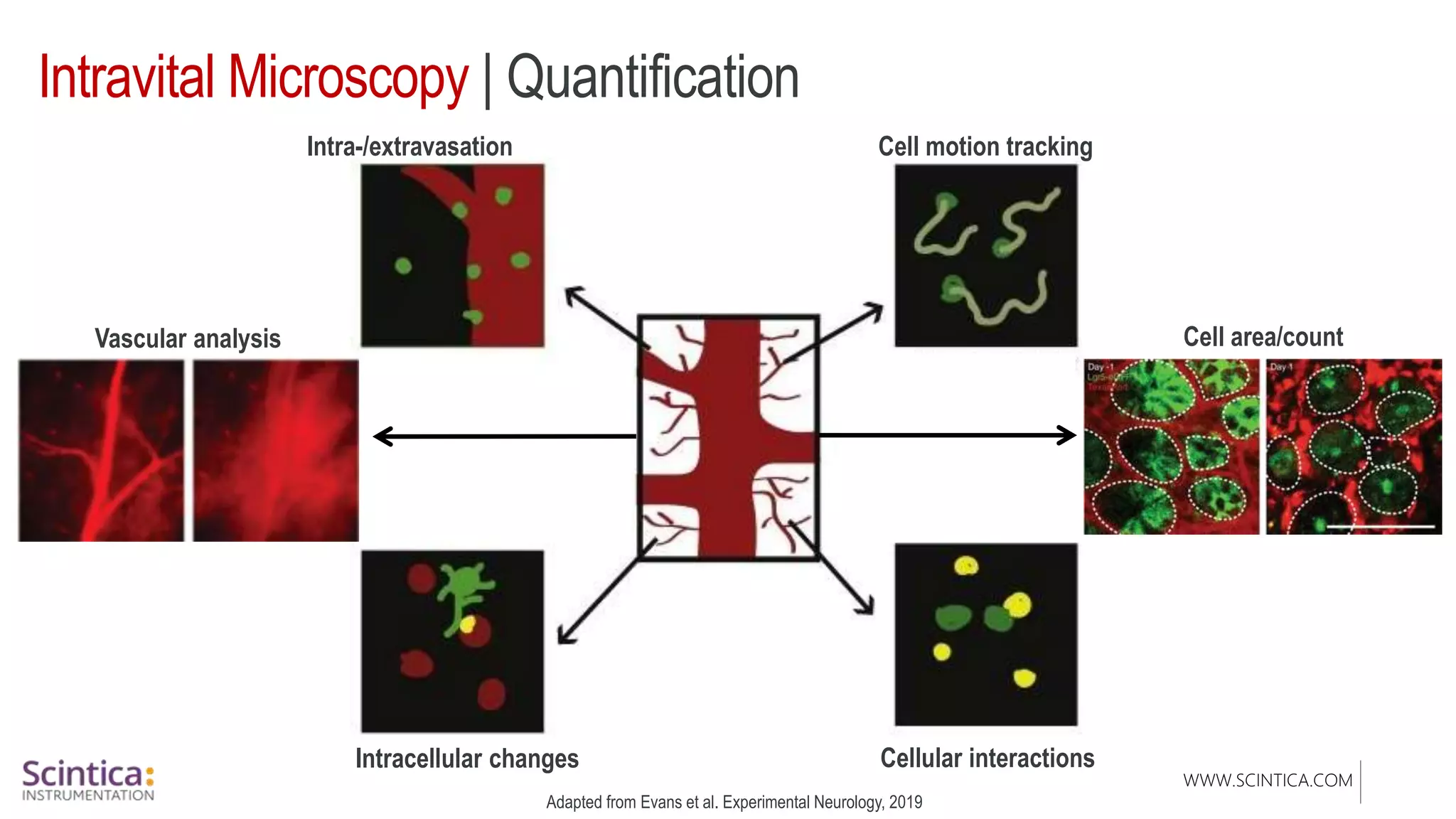
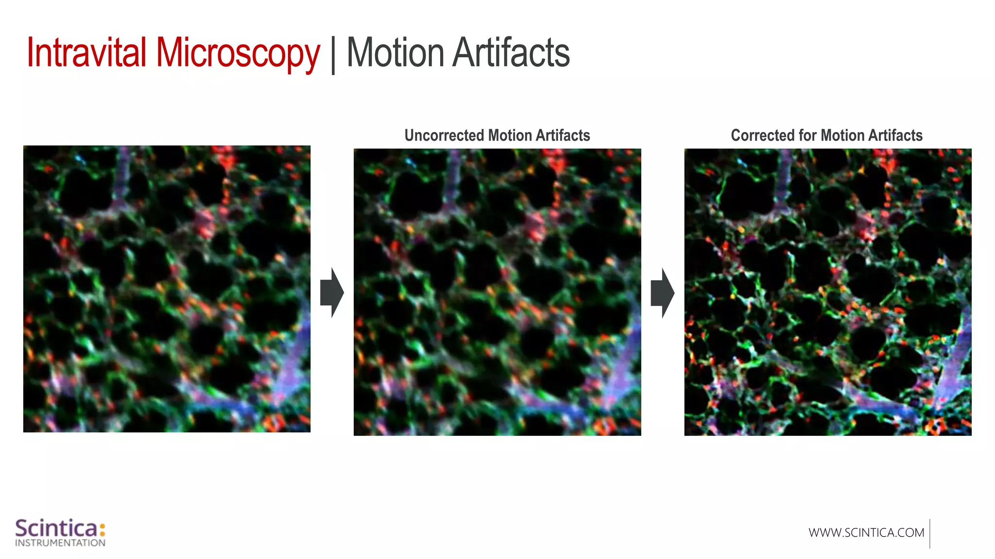
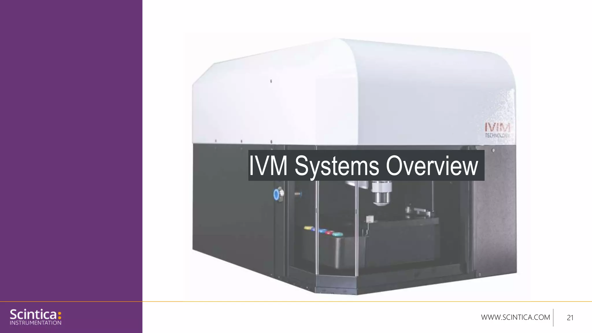
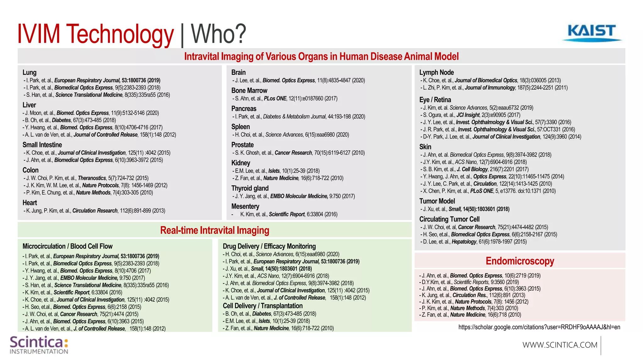
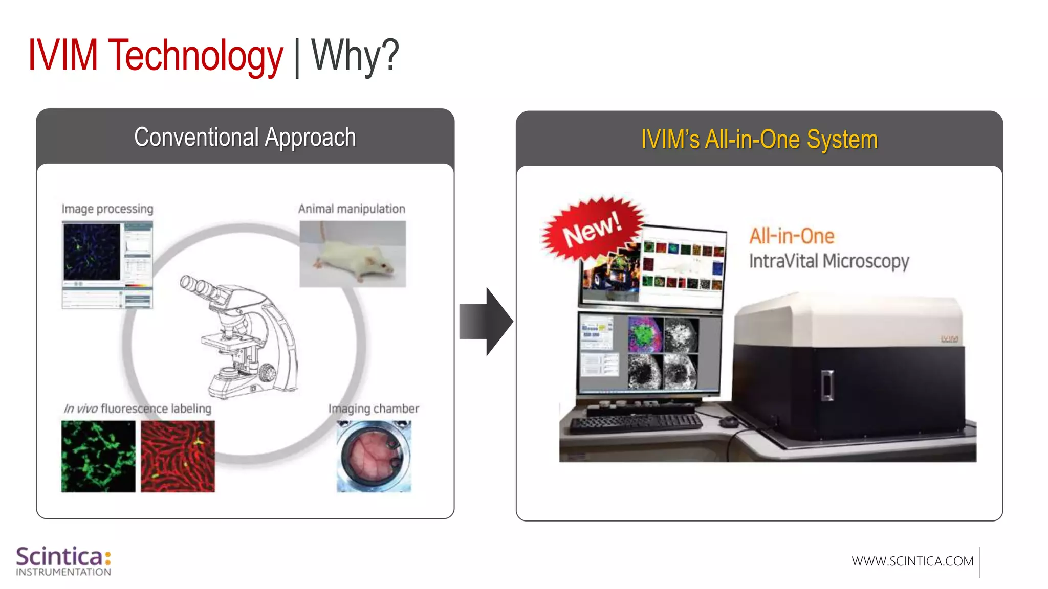
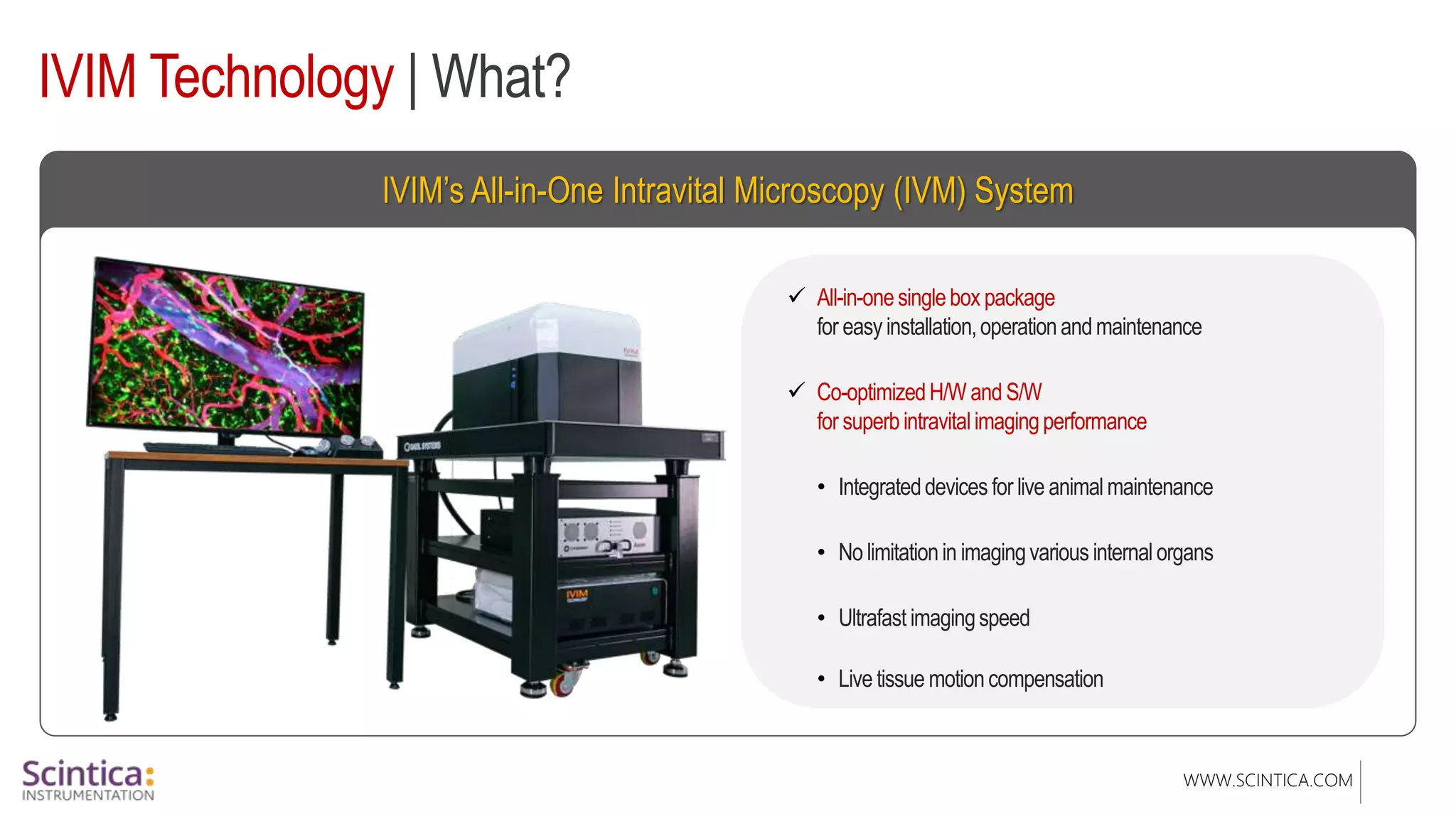
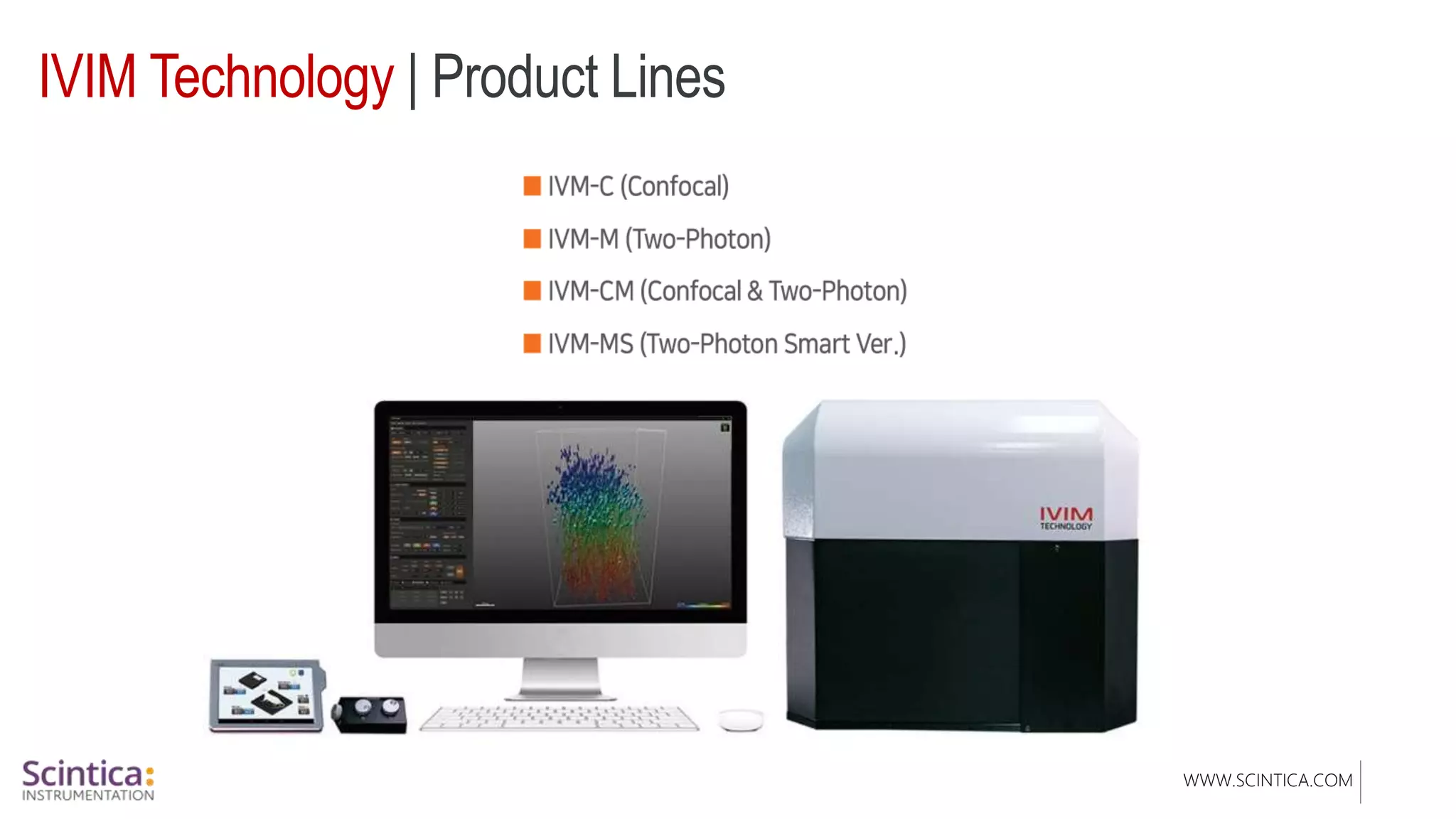
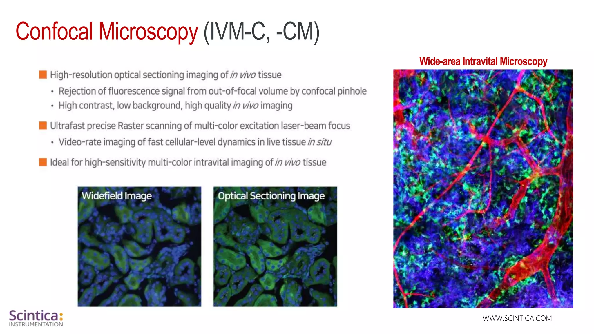
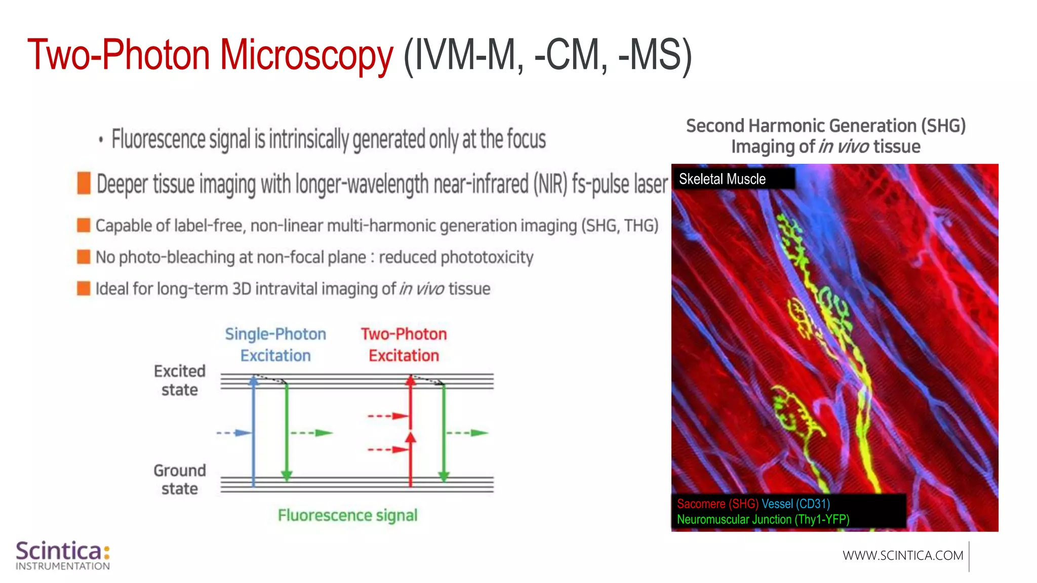

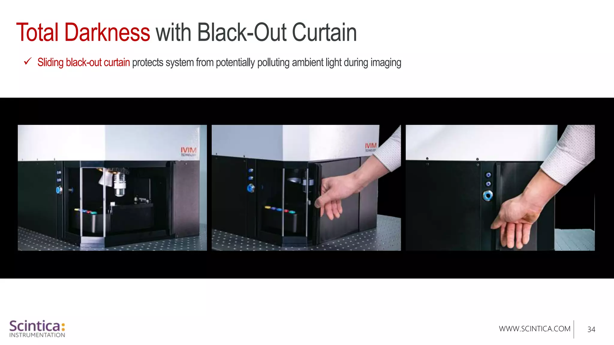

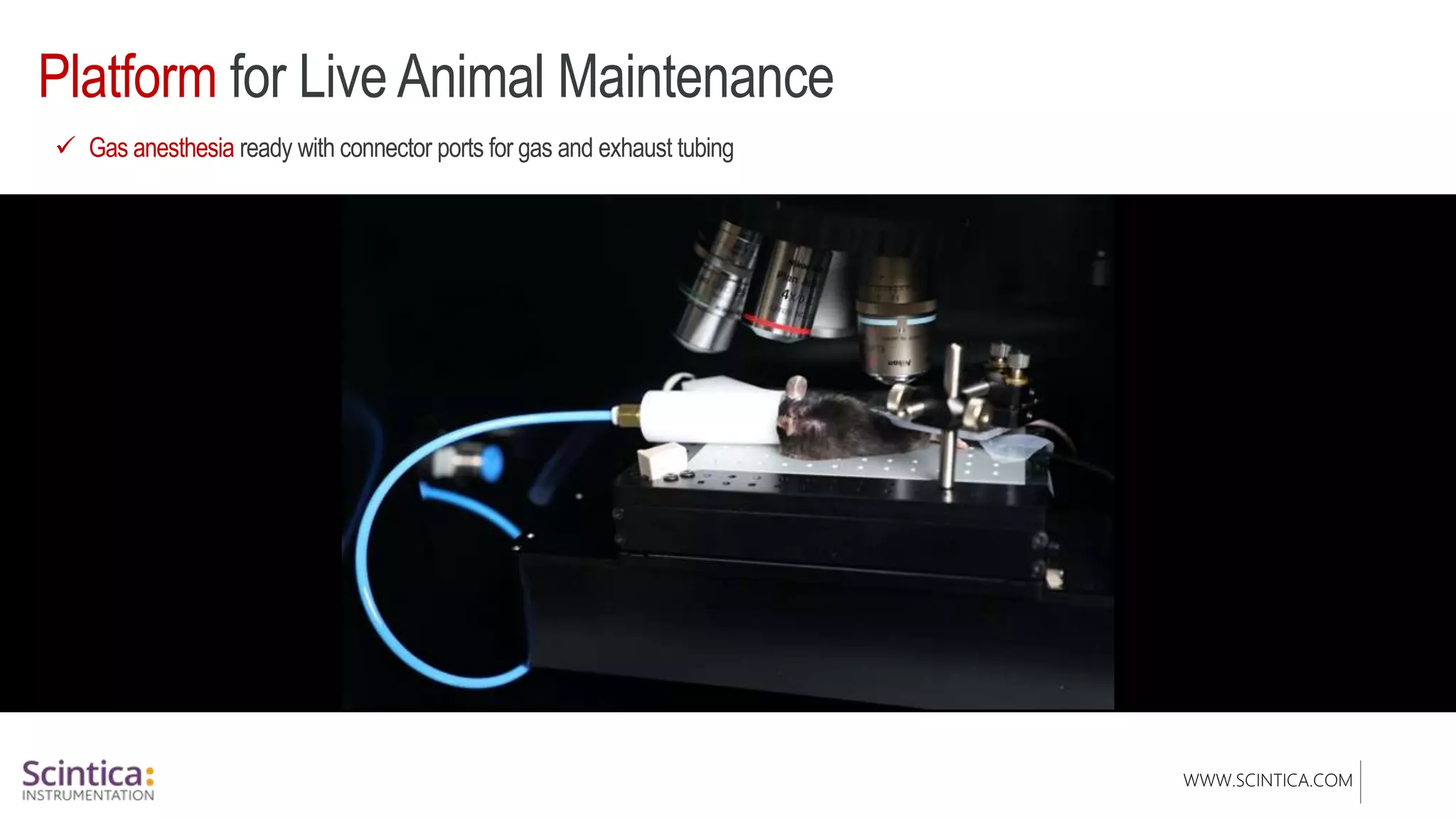
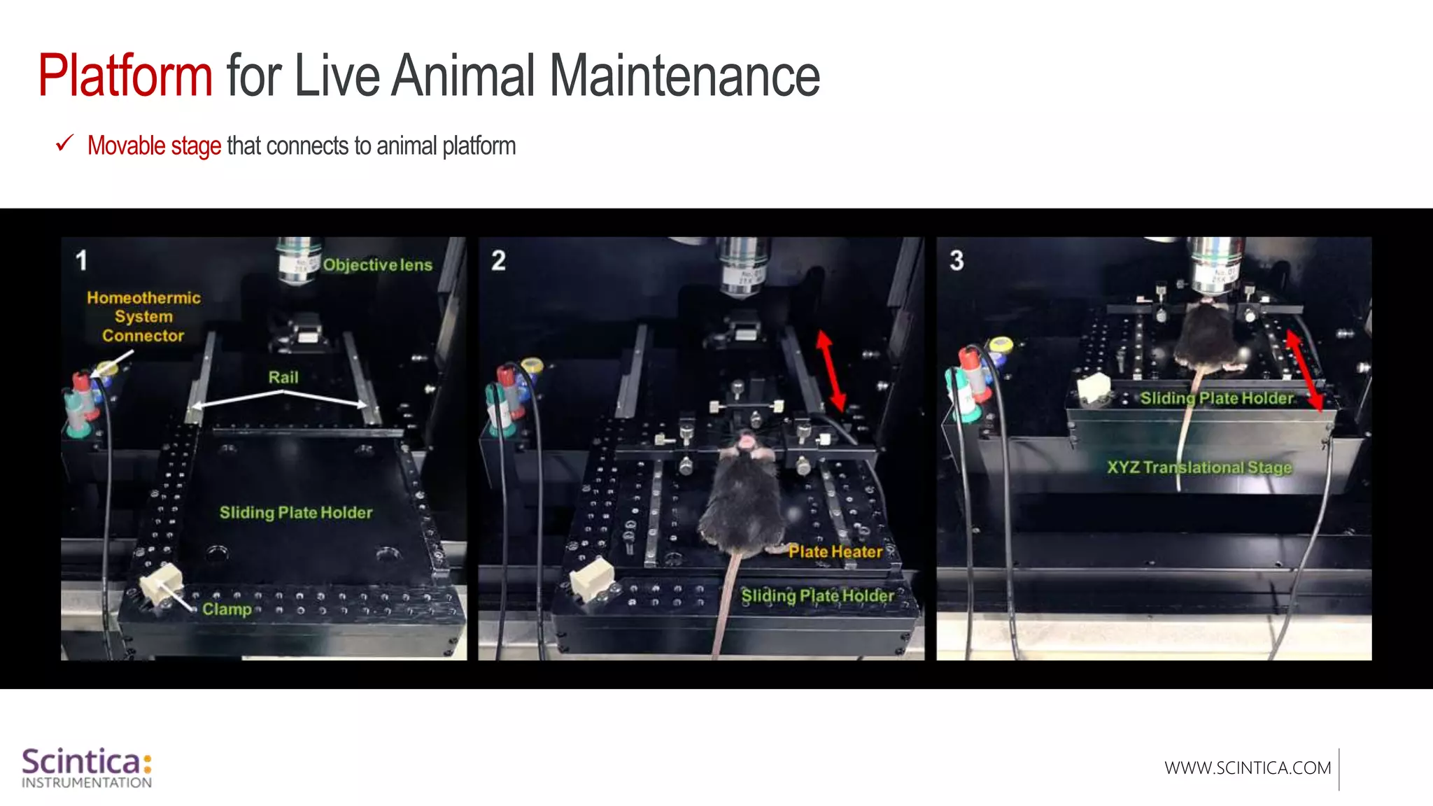
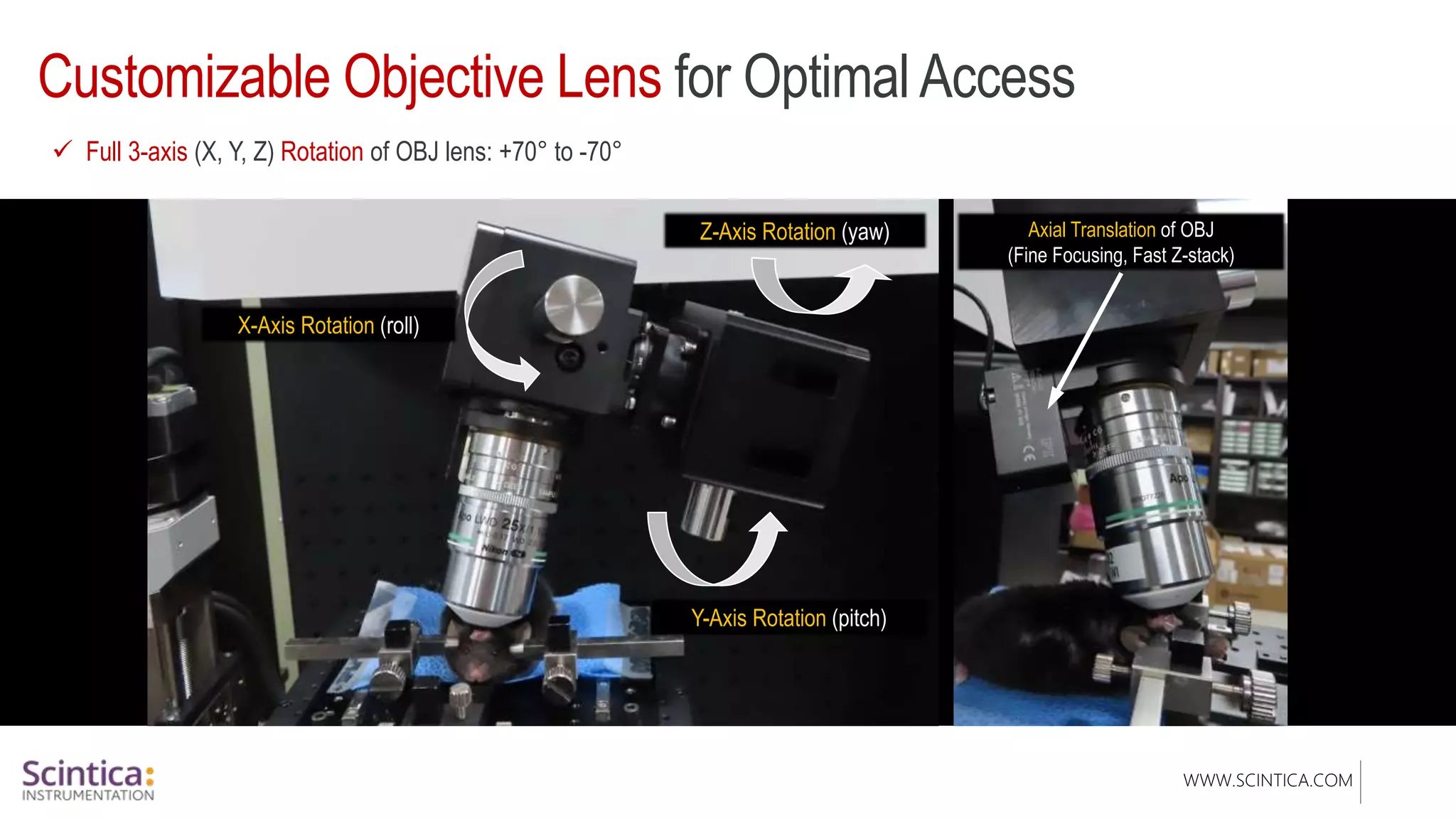
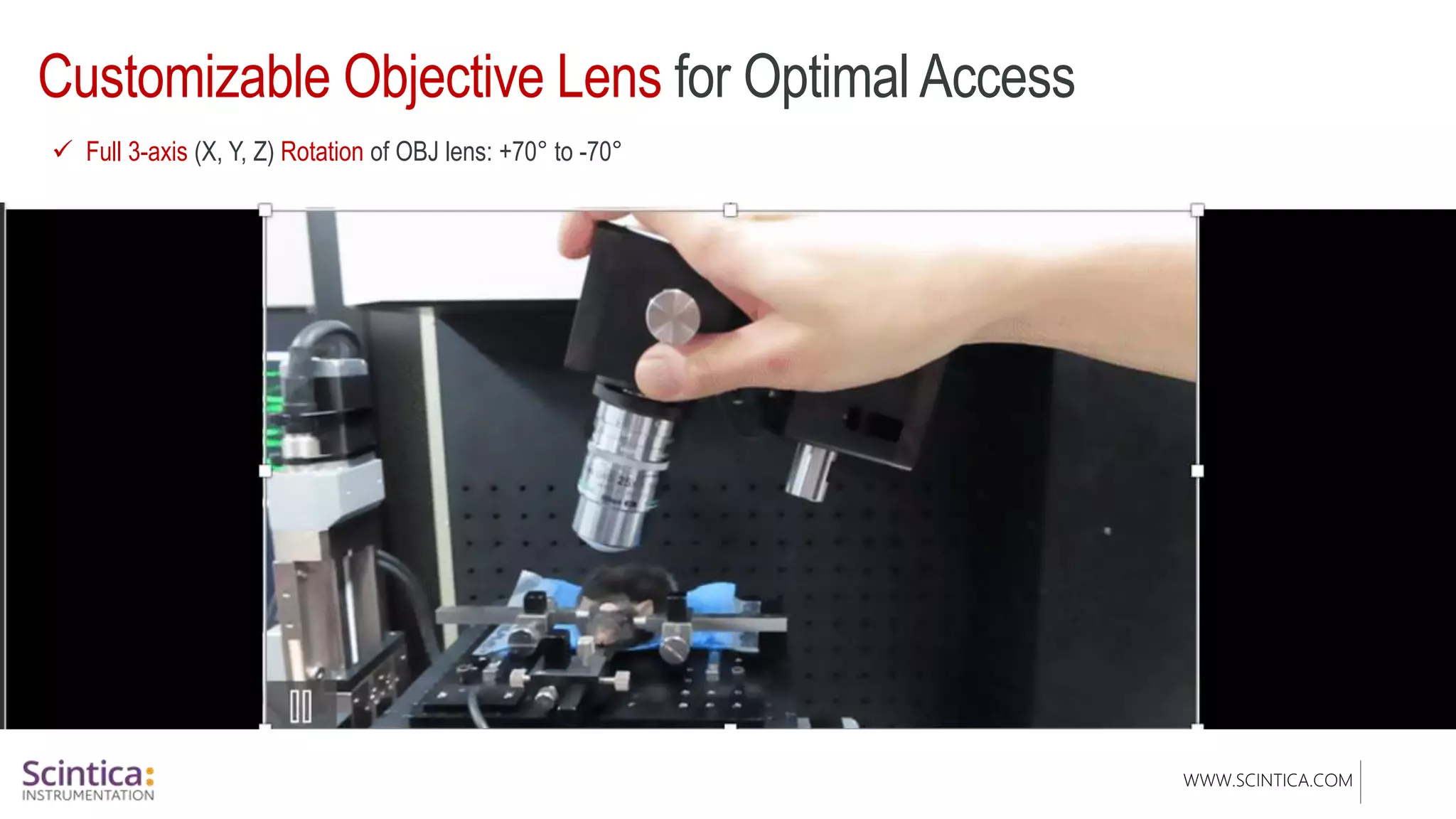
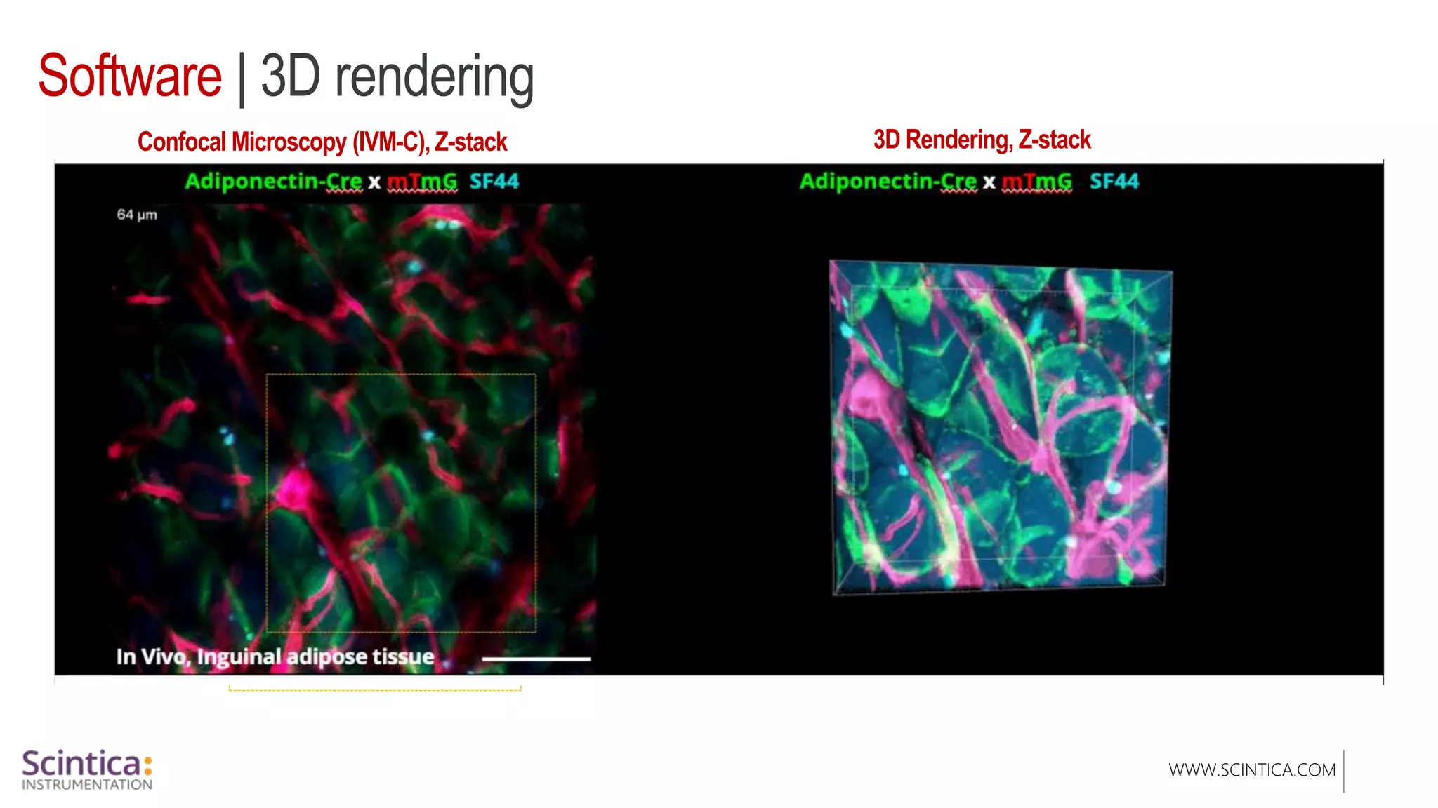
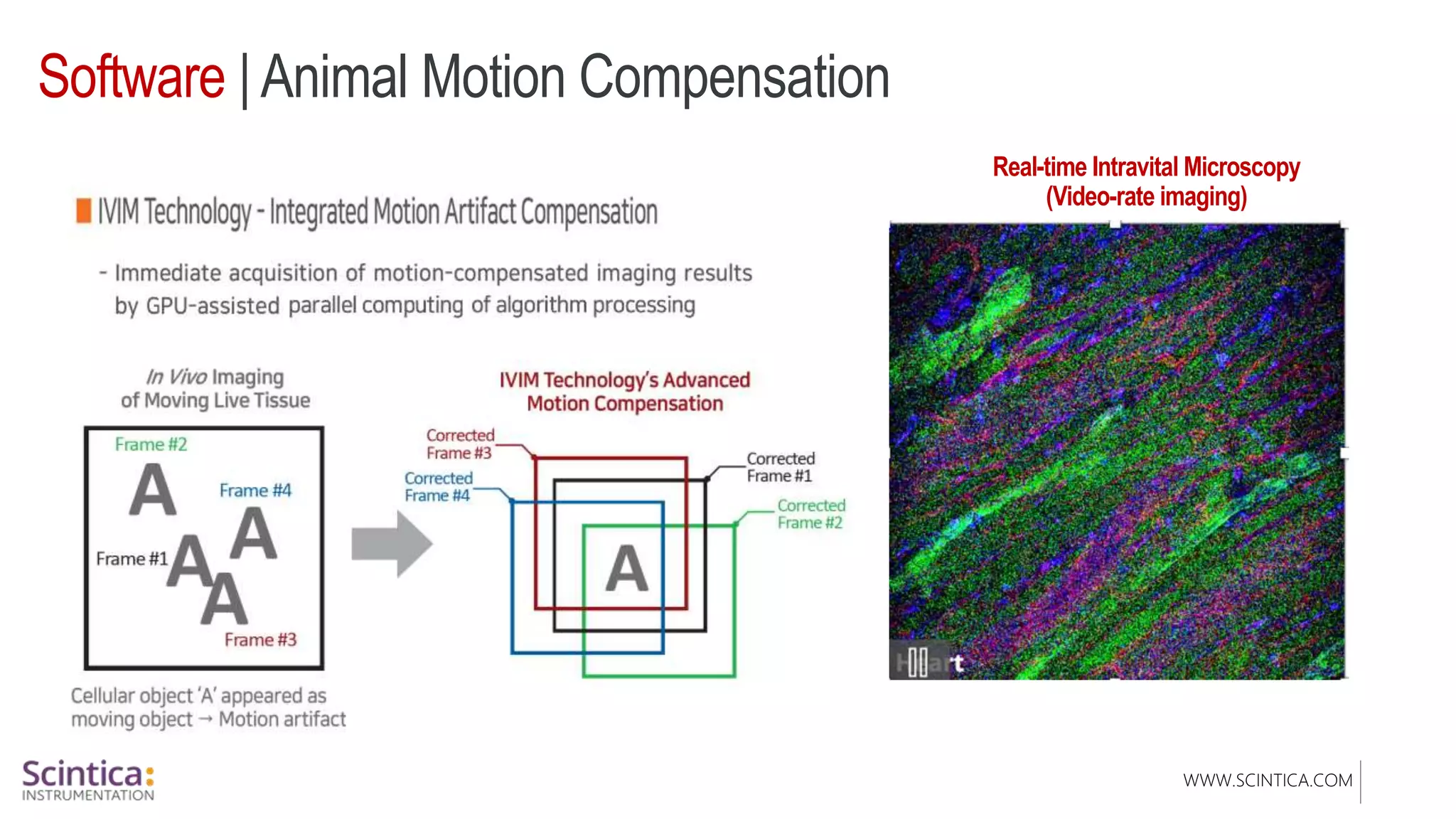
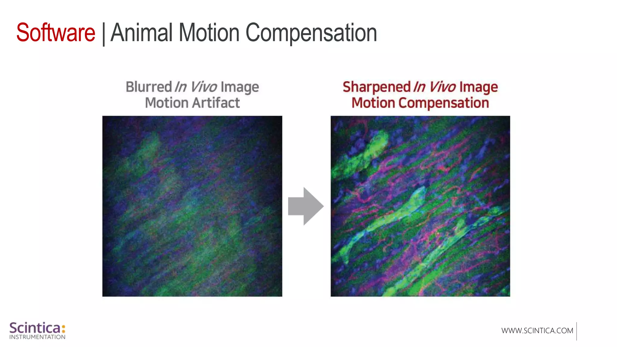
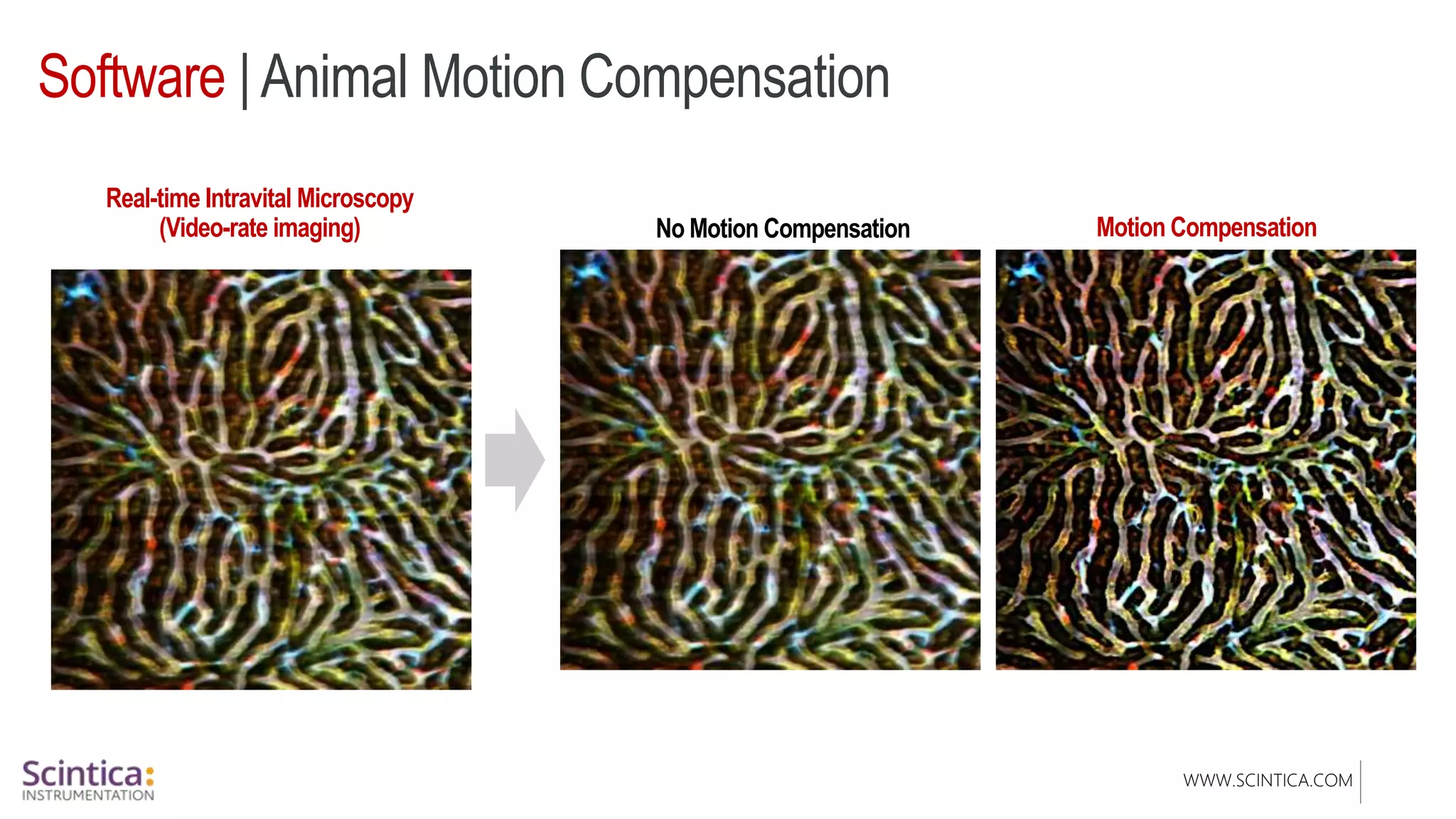
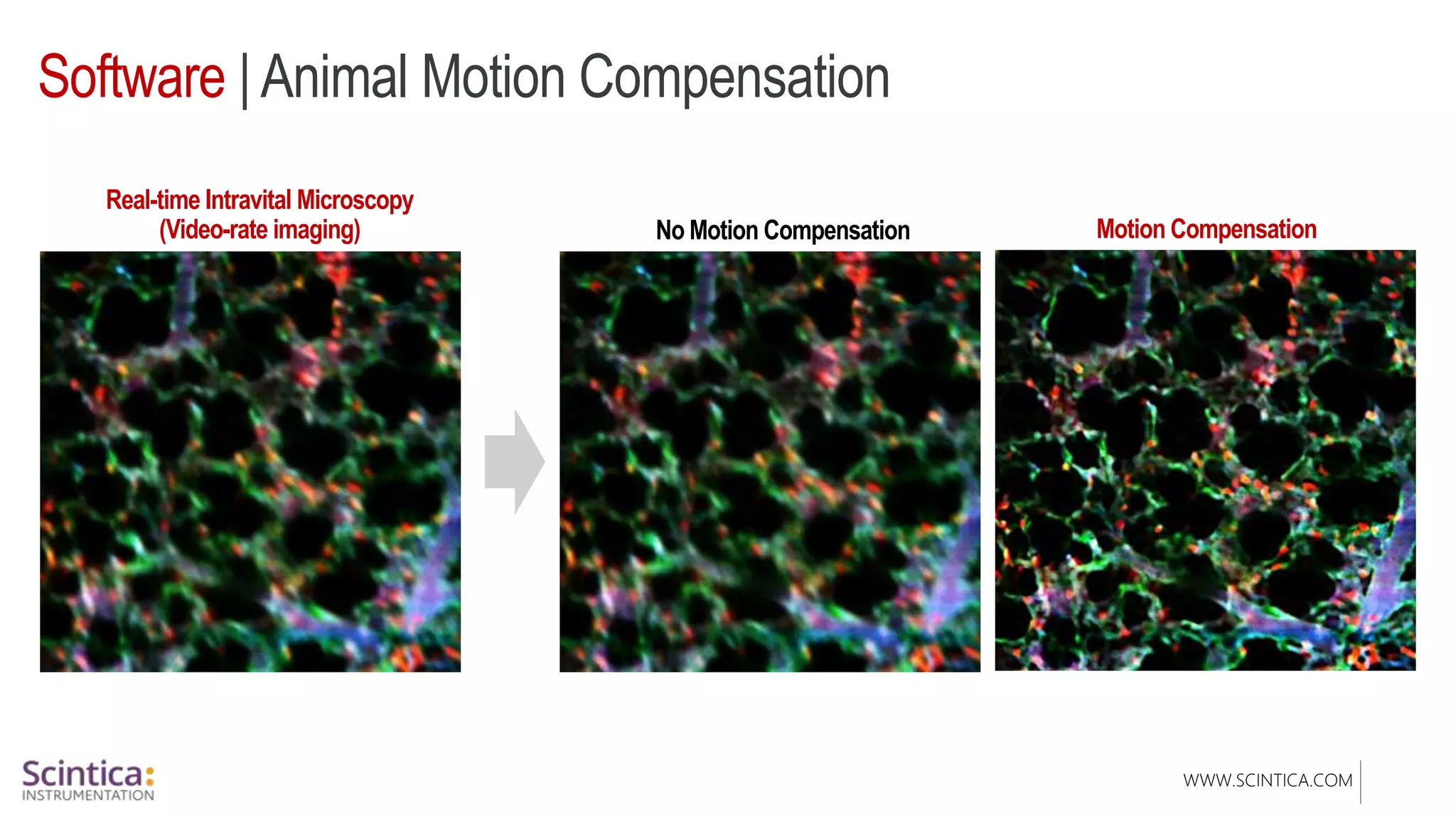
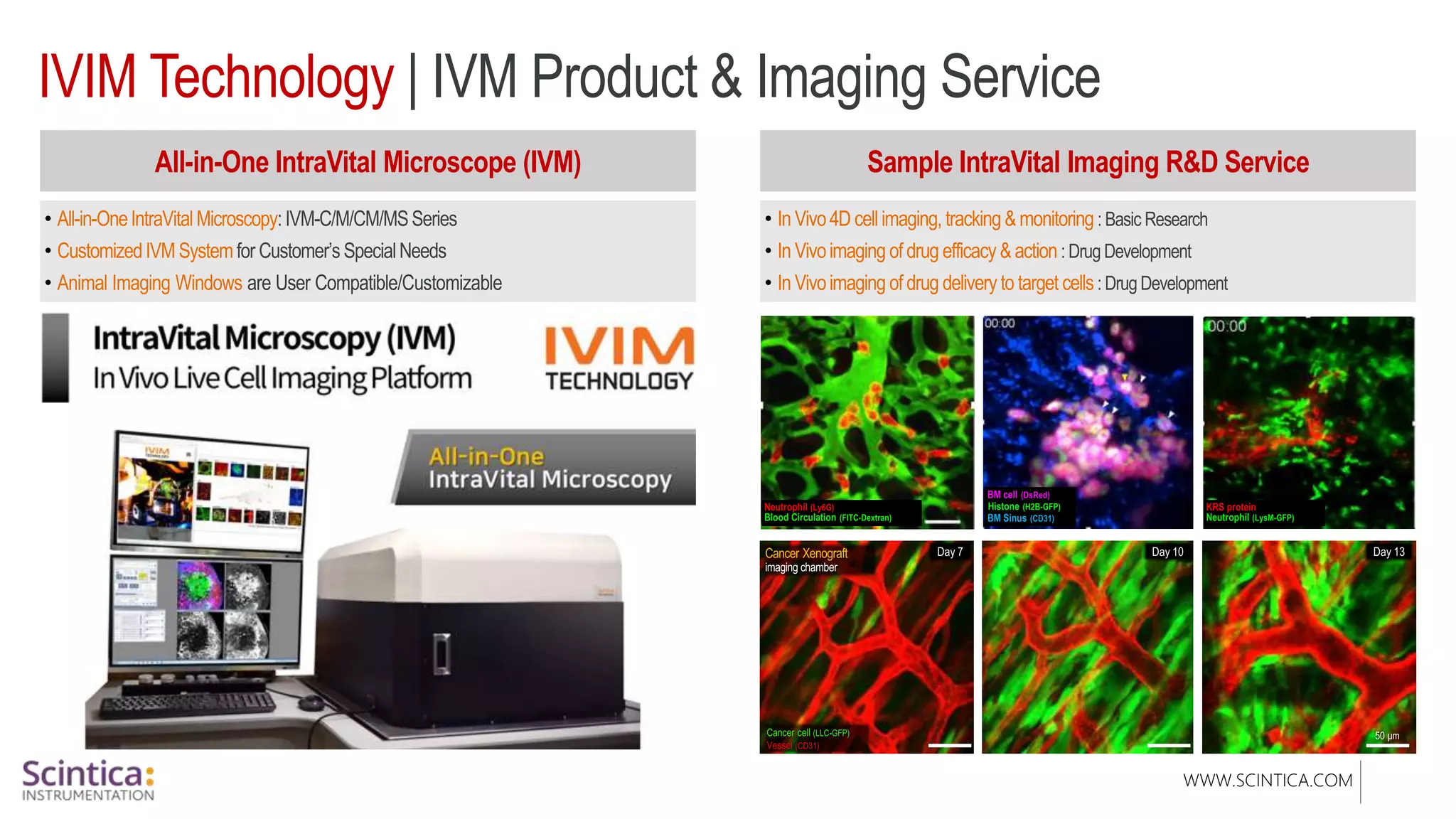
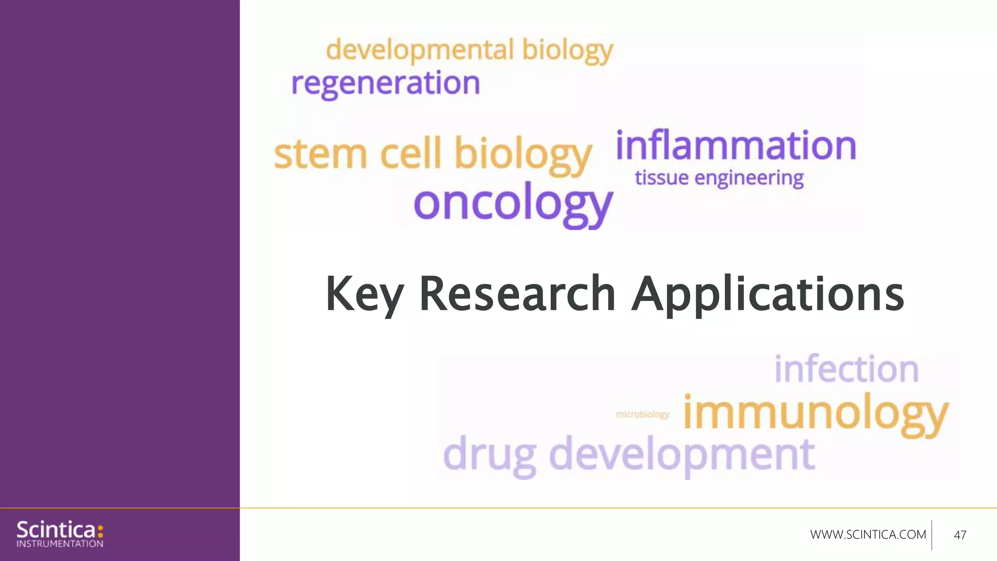

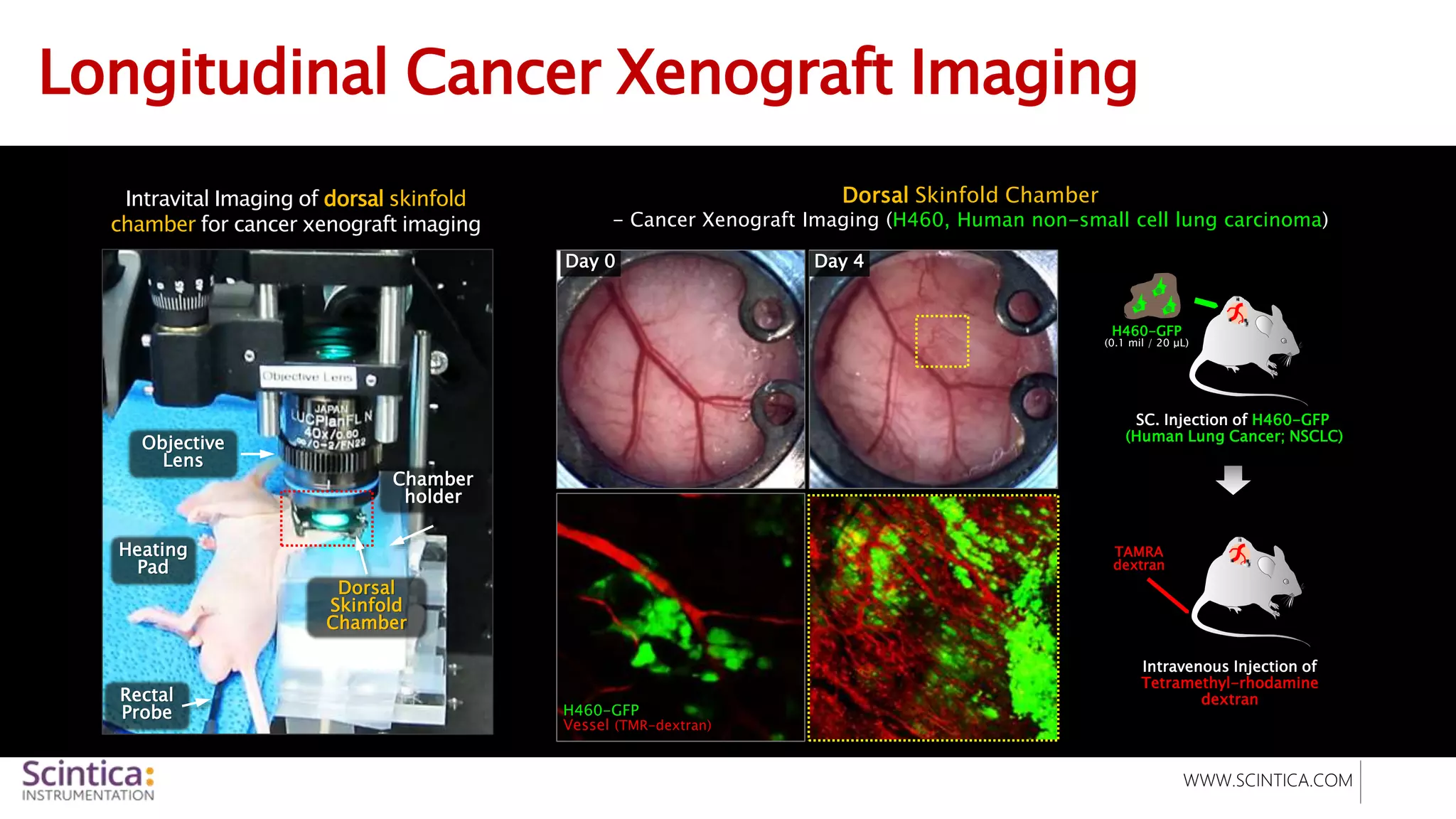
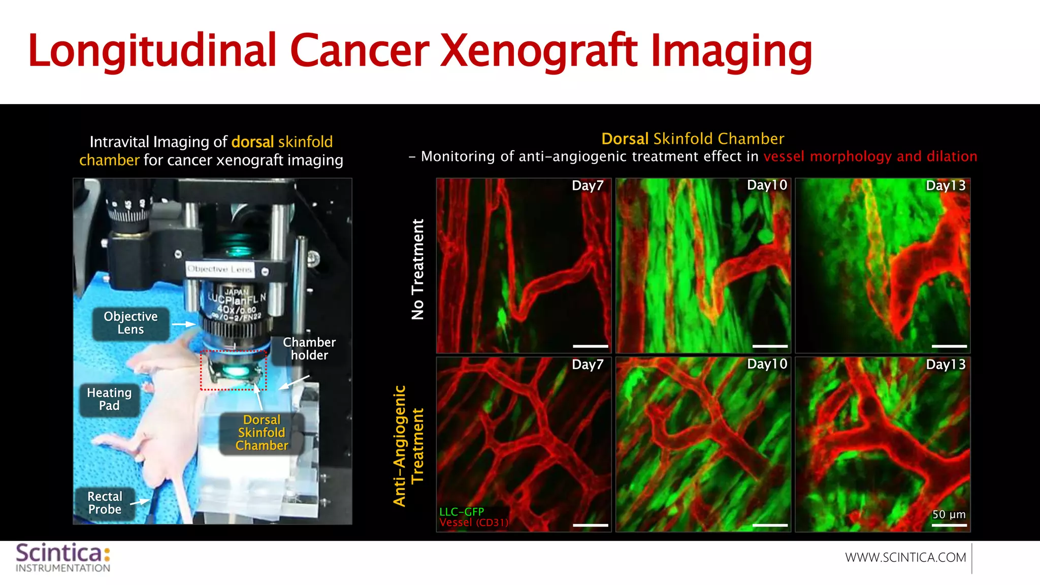
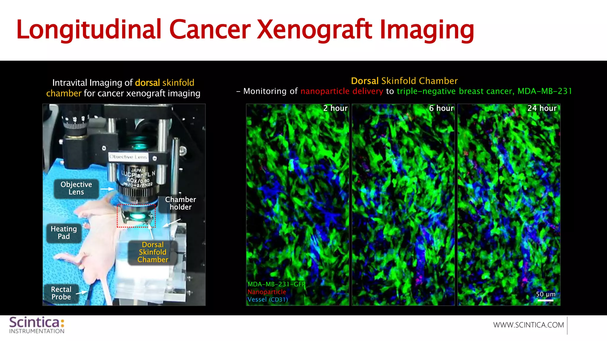
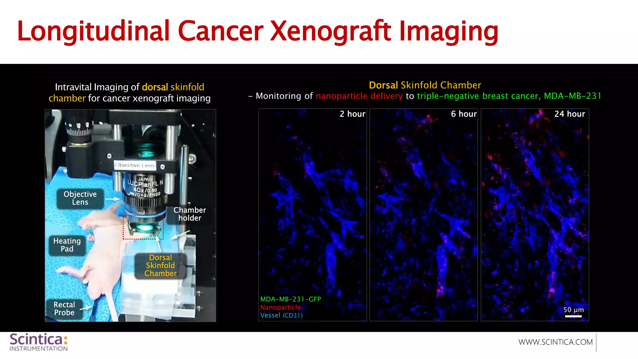

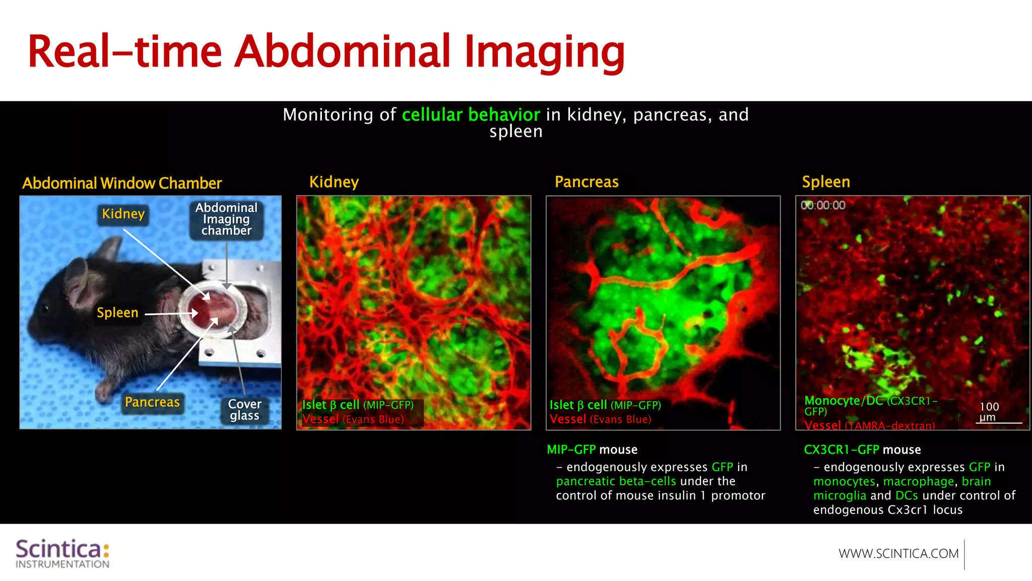
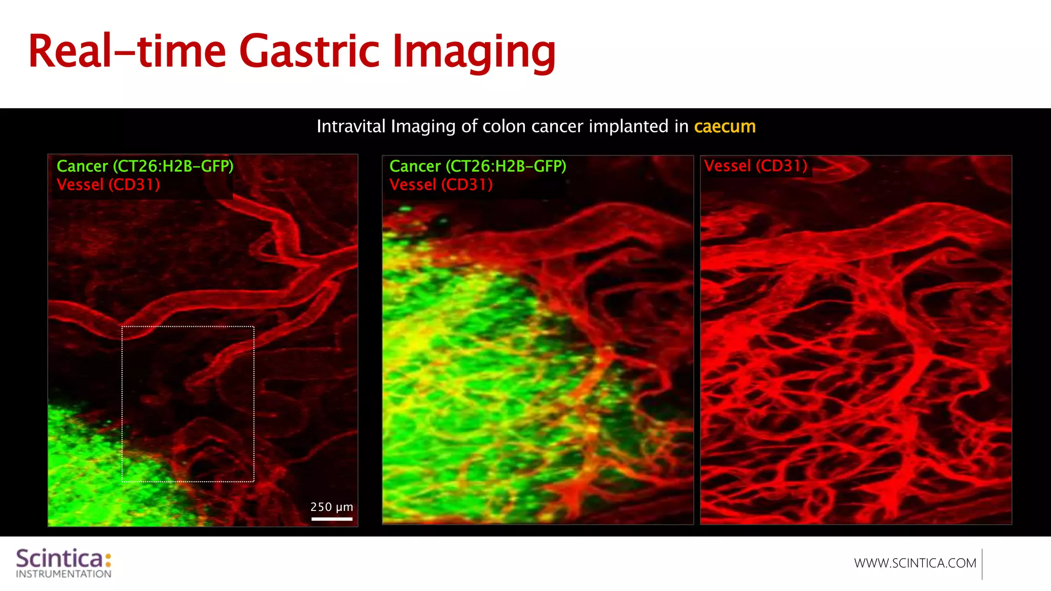
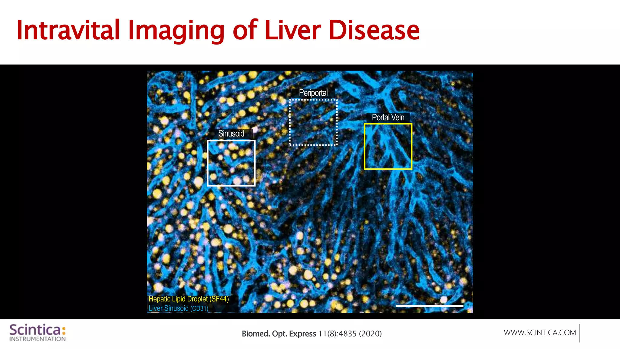
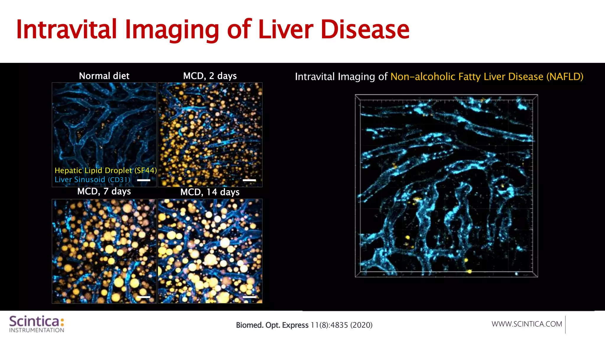

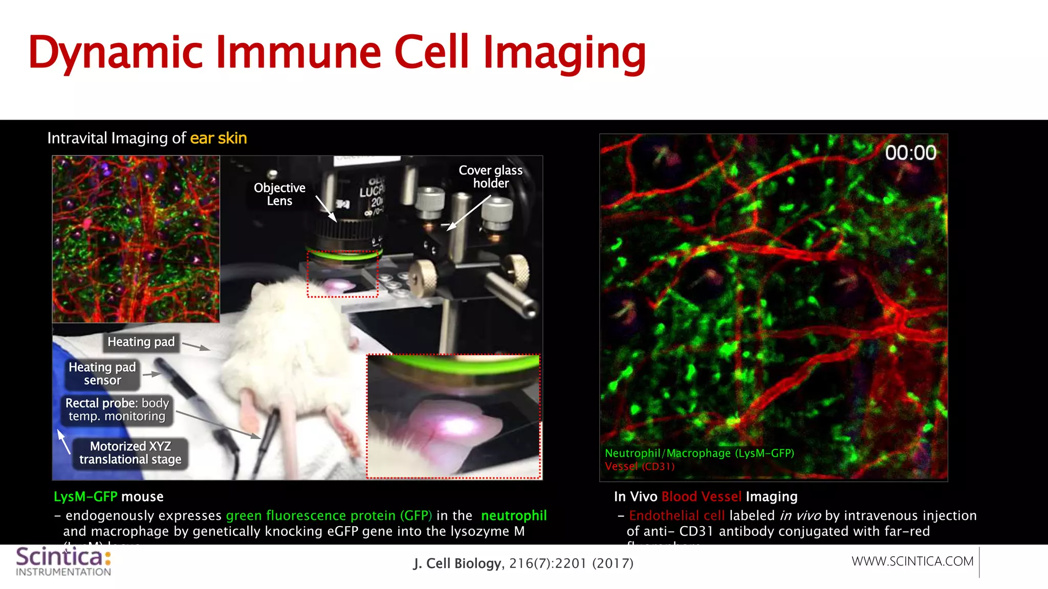
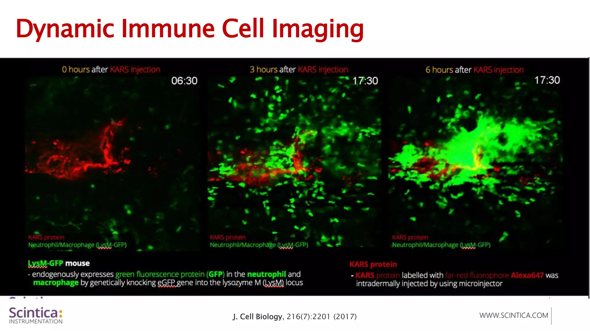
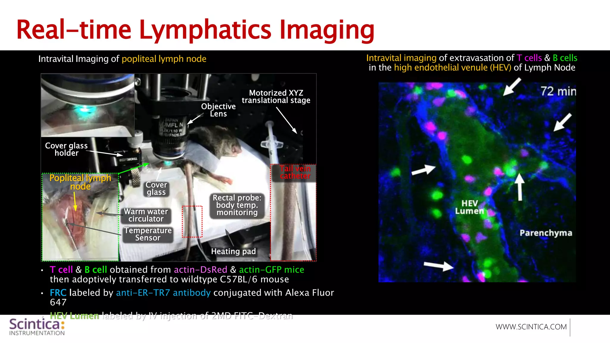
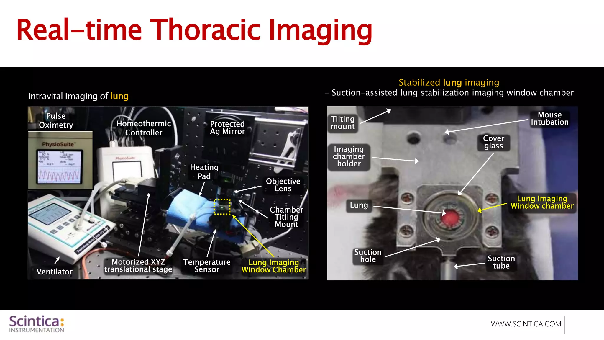


The document discusses the IVM (intravital microscopy) system, an advanced imaging technology that combines confocal and two-photon microscopy for in vivo imaging of biological processes. It highlights key applications in cancer research, drug development, and immunology, emphasizing its capability for real-time, high-resolution imaging of cellular dynamics within living organisms. The IVM system features a compact design with integrated live animal maintenance platforms, allowing for versatile imaging of various internal organs.


