SlideShare a Scribd company logo
1 of 41
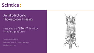
An Introduction to
Photoacoustic Imaging
Featuring the TriTom™ in-vivo
imaging platform
September 20, 2023
Lawrence Yip PhD, Product Manager
lyip@scintica.com
WWW.SCINTICA.COM
Topics of Discussion
• Motivation for photoacoustic imaging (PAI)
• Overview of the modality
• PA and fluorescence tomography as complementary modalities
• Preclinical applications
2
WWW.SCINTICA.COM
Selecting a Modality
Sensitivity
PAI
WWW.SCINTICA.COM
Motivation for PAI
• In vivo functional imaging analogous to functional MRI
• In vivo metabolic imaging analogous to PET
• In vivo label-free histologic imaging of cancer without excision
• In vivo molecular imaging of disease markers
Audience Poll
WWW.SCINTICA.COM
Topics of Discussion
• Motivation for photoacoustic imaging
• Overview of the modality
• PA and fluorescence tomography as complementary modalities
• Preclinical applications
6
WWW.SCINTICA.COM
Photoacoustic Tomography (PAT)
N-shaped pulse
Amplitude: absorption of light
Width: size of object
Includes elements from J. Lee et al., (2012) J. Breast Cancer
WWW.SCINTICA.COM
Photoacoustic Tomography (PAT)
Biswas, D., Roy, S. & Vasudevan, S. Biomedical Application of Photoacoustics: A Plethora
of Opportunities. Micromachines 13, 1900 (2022).
A typical PA signal from circular numerical
target where a is amplitude, T is time of flight,
τ is width and χ is the relaxation time of the
PA time domain signal.
WWW.SCINTICA.COM
Optical Absorption Spectra
Scholkmann, Kleiser, Metz, et al., (2014) NeuroImage; Zackrisson and Gambhir, (2014) Cancer Res
WWW.SCINTICA.COM
Array Geometries
LCM Yip et al., "Approaching closed spherical, full-view detection for photoacoustic tomography," J. Biomed. Opt. 27(8)
086004 (2022) https://doi.org/10.1117/1.JBO.27.8.086004
WWW.SCINTICA.COM
Topics of Discussion
• Motivation for photoacoustic imaging
• Overview of the modality
• PA and fluorescence tomography as complementary modalities
• Preclinical applications
11
WWW.SCINTICA.COM
Fluorescence Imaging (FLI)
• Fluorescent molecules absorb light and are excited to a higher
electronic state
• The additional energy can be released as photons at a different
(longer) wavelength.
• Widely used, both for whole-animal imaging and in microscopy
Vilber Newton 7.0 FT500 Brochure
WWW.SCINTICA.COM
FLI vs PAI
• FLI typically has higher molecular sensitivity than PAI due to the lack of
background signal from endogenous chromophores
• FLI has excellent spatial resolution for superficial tissue but degrades rapidly
with depth
Fluorophores generate both fluorescence emission and PA signals when
excited by the same nanosecond laser pulse
Together, FLI provides high sensitivity while PAT provides deep,
high-resolution imaging of fluorophores.
Audience Poll
WWW.SCINTICA.COM
TriTom Technology
• Compact tabletop design
• Integrated gas anesthesia line
• Temperature-controlled imaging
chamber
• High-sensitivity sCMOS camera for
optical imaging
• Standard emission filters covering
popular fluorophores
• PA excitation from visible to NIR II
range
15
TriTom™ small animal imaging platform
WWW.SCINTICA.COM
TriTom Technology
16
TriTom composite imaging of blood vasculature and
regional lymphatics in a live mouse model of
metastatic breast cancer.
Red – 895 nm excitation (deep vasculature)
Yellow – 532 nm excitation (superficial vasculature)
Scanned
region
True 3D with sub-millimeter spatial resolution:
160 µm x 160 µm in transverse planes
160 µm x 470 µm in sagittal and coronal planes
36 sec per multi-modality molecular imaging scan
WWW.SCINTICA.COM
Quick & Easy Sample Handling
• Reliable Gas Anesthesia:
• Delivered through the hollow shaft
• Bite bar within nose cone secures position
• Convenient and Repeatable Procedure:
• Mouse’s legs and head are fixed in a
consistent position with minimal stress
• Preparation time < 5 min
17
Mouse Holder –In Vivo Imaging
(1) Hollow shaft to fix the mouse holder inside
TriTom while reliably delivering anesthesia gas
(2) Bite bar for secure and repeatable mouse
positioning
(3) Plastic support rods
(4) Cushioned paw mounts
(5) Adjustable support block for mouse’s hind legs
1
2
3
4
5
WWW.SCINTICA.COM
High Efficiency Optical Excitation
• Wavelength range
• All popular visible (460-659 nm)
• Standard NIR I/II excitation (650-1320 nm)
• Extended NIR II excitation (1065-2300 nm)
• High-energy (>350 mJ) 1064 nm mode
• High-resolution deep anatomical images
• 20 Hz pulse repetition frequency
• Fast wavelength switching (each pulse, arbitrary sequence)
• Excitation linewidth < 0.5 nm (equivalent to 1,280 excitation filters
• Integrated energy meter (quantitative imaging)
18
Excitation Source: Tunable Pulsed Nd:YAG Laser
WWW.SCINTICA.COM
Topics of Discussion
• Motivation for photoacoustic imaging (PAI)
• Overview of the modality
• PA and fluorescence tomography as complementary modalities
• Preclinical applications
19
WWW.SCINTICA.COM
Applications
• Anatomical Imaging
• Cancer
• Contrast Agent Development
• Tissue Engineering &
Regeneration
• Neuroimaging
• Developmental Biology
• Therapeutic Monitoring
• Molecular Imaging
20
Audience Poll
WWW.SCINTICA.COM
Anatomical Imaging
22
High energy 1064 nm excitation of mouse
thorax and upper abdomen. (Click video to
play)
(1) Heart; (2)Liver; (3) Aorta; (4) Spleen;
(5) Left Kidney; (6) Right Kidney; (7)Intestines.
Whole-body imaging Deep-tissue structures
800 nm PAT reconstruction of a nu/nu nude
mouse
Kidney; Scale bar – 1
mm
1 – abdominal aorta
2 – interior vena cava
3 – renal vein
4 – renal cortex
5 – renal artery
6 – interlobular artery
(vein)
7 – medulla (renal
pyramids)
Superior Anatomical Reference
WWW.SCINTICA.COM
Cancer
23
Monitor Tumor Growth, Metastasis, and Microenvironment
Human breast ductal carcinoma
xenograft (BT474 cells) in nu/nu
mice
(Left) Maximum intensity projection
of 532 nm (yellow) and 890 nm
(red/grey) showing superficial and
deep tumor vasculature
(Right) Composite skin (532 nm;
grey) and deep tissue (890 nm; red)
3D images.
Tumor size = 10.6 x 4.7 x 11.6 mm3
Tumor
Dumani et al, Proceedings SPIE 10878, Photons Plus Ultrasound: Imaging and Sensing, 108784Y (2019)
WWW.SCINTICA.COM
Regional Lymphatic Drainage
24
Tracking Metastasis Development
Normal mouse, 24-hrs post-injection
Subcutaneous injection of glycol-chitosan-coated gold nanospheres (50 µg/ml) mixed with ICG (50 µg/ml)
Total injection volume 50 µl
Fluorescence image
Injection site
Lymph node
Lower abdomen of a healthy
mouse
Red – deep blood vessels,
890 nm excitation scan
Yellow – superficial
vasculature (skin), 532 nm
excitation scan
1 – right subiliac lymph node
2 – injection site (right
mammary fat pad)
1
2
Photoacoustic image
WWW.SCINTICA.COM
Quick & Easy Sample Handling
• High Throughput:
• Interrogate up to 10 samples per scan
• Preparation time < 5 min
• Small Sample Volume:
• 50 μL sample volumes
• Convenient and Repeatable Procedure:
• Radially oriented slots consistently hold samples in
place
• Allows for quick setup and removal of cuvettes
• Cuvette holder is instantly placed inside the TriTom
via magnetic connections
25
Cuvette Holder – Development of Contrast Agents
(1) Port for administering liquid scattering background
(2) Plastic support rods
(3) Radial slots for quick setup and removal of cuvettes
1
2
3
WWW.SCINTICA.COM
Contrast Agent Development
26
Phantom T1 T2 T3 T4
3-IR800 Control IR800-A (200 µM) IR800-B (100 µM) IR800-C (50 µM)
4-IR800 Control IR800-D (25 µM) IR800-E (12.5 µM) IR800-F (6.25 µM)
760 nm 780 nm 800 nm
Phantom
3
Phantom
4
Cross-sectional (axial) views of 0.8 mm tubes with IRDye800CW samples
3D PAT reconstruction of
Phantom 3; 780 nm excitation.
WWW.SCINTICA.COM
Contrast Agent Biodistribution
27
Molecular unmixing of an ICG-based contrast agent (green) overlaid on the 800 nm excitation scan of anatomy (white)
at 0, and 90-min post-injection. Circulation half-life of ICG contrast agent compared to ICG.
t = 0 min t = 90 min
Circulation
Half-life
Singh, S., et al., (2023). Size-tunable ICG-based contrast agent platform for targeted near-infrared photoacoustic imaging.
Photoacoustics, 29. https://doi.org/10.1016/j.pacs.2022.100437
WWW.SCINTICA.COM
Tissue Engineering & Regeneration
28
STEM Cell Tracking and Therapy Monitoring
3D photoacoustic image of gold nanosphere-labeled mesenchymal stem
cells locally injected in a rat spinal cord. The linear relationship between PA
signal and injection volume demonstrates the ability to track and monitor
cell migration in vivo. Scale bar = 10 mm.
Axial View Coronal View
The TriTom’s submillimeter spatial resolution in all three
anatomical planes makes image analysis and quantification easier
for applications with hard to identify anatomy.
Donnelly, E. M., et al., (2018). Photoacoustic Image-Guided Delivery of Plasmonic-Nanoparticle-Labeled Mesenchymal Stem
Cells to the Spinal Cord. Nano Letters, 18(10), 6625–6632. https://doi.org/10.1021/acs.nanolett.8b03305
WWW.SCINTICA.COM
Neuroimaging
29
10 mm-thick (A) coronal and (B) transverse maximum intensity projection
slabs of a PAT volume reconstructed from the 750 nm scan. 1) Superior
sagittal sinus, 2) transverse sinus, 3) confluence of sinus, 4) cerebral artery,
5)auricular artery, 6) jugular vein, 7) brachial artery, and 8) ophthalmic
artery. The scale bar is 5 mm in length.
PAT composite image of the upper torso
and brain of a female BALB/c mouse
acquired post-mortem with 532 nm
(orange) and 750 nm excitation (gray). (Click
video to play)
A B
WWW.SCINTICA.COM
Developmental Biology
30
Monitor Placental Function and Detect Pathologies in Pregnancy and
Development
Reconstructed PAT images of a pregnant mouse on gestational day 12 in (A) superficial, (B) deep tissue, and (C) composite of
superficial (orange) and deep tissue (gray) images. D) Reconstructed PAT images of a pregnant mouse on gestational day 17 in
deep tissue. The 1) common iliac arteries, 2) placentas, and 3) abdominal aorta are labelled. The scale bar is 10 mm in length.
A B C D
Huda, K., Wu, C., Sider, J. G., & Bayer, C. L. (2020). Spherical-view photoacoustic tomography for monitoring in vivo placental
function. Photoacoustics, 20, 100209. https://doi.org/10.1016/j.pacs.2020.100209
WWW.SCINTICA.COM
Developmental Biology
31
Reconstructed PAT images of a pregnant mouse on gestational day 17 (A) pre-injection, (B) 5 minutes post-
injection, and (C) 90 minutes post-injection of FA-PEG-ICG. The scale bar is 10 mm in length and the 1)
placenta and 2) spiral artery are labelled.
A B C
Huda, K., Wu, C., Sider, J. G., & Bayer, C. L. (2020). Spherical-view photoacoustic tomography for monitoring in vivo placental
function. Photoacoustics, 20, 100209. https://doi.org/10.1016/j.pacs.2020.100209
WWW.SCINTICA.COM
Therapeutic Monitoring
32
Deep tissue (808 nm) PAT reconstruction of the abdomen of a pregnant mouse
(A) pre-injection and (B) 13 minutes post-injection of G protein-coupled
estrogen receptor (GPER) agonist, G1. 1) Denotes the iliac artery and 2) denotes
the placenta. The scale bar is 10 mm in length.
A B
Huda, K., Lawrence, D. J., Lindsey, S. H., & Bayer, C. (2022). Photoacoustic tomography to assess acute vasoactivity of
systemic vasculature. Proceedings of SPIE, 11960, 1196007. https://doi.org/10.1117/12.2612093
WWW.SCINTICA.COM
Molecular Unmixing
33
NiSO4
CuSO4
720 nm 810 nm
The samples were scanned at A) 720 nm and B) 810 nm before the
unmixing algorithm was applied. The samples containing C) NiSO4 and
D) CuSO4 are shown after the unmixing algorithm was applied.
A B
C D
NiSO4
[3:1] NiSO4:CuSO4
CuSO
4
[1:1] NiSO4:CuSO4
[1:3] NiSO4:CuSO4
Sample NiSO4 (%) CuSO4 (%) CNR
[0:1] NiSO4 : CuSO4 0 100 3.08
[1:3] NiSO4 : CuSO4 25 75 2.74
[1:1] NiSO4 : CuSO4 50 50 2.23
[3:1] NiSO4 : CuSO4 75 25 2.96
[0:1] NiSO4 : CuSO4 100 0 2.73
WWW.SCINTICA.COM
Q&A Session
WWW.SCINTICA.COM
INFO@SCINTICA.COM
Please enter your questions
in the Q&A section.
Thank You!
Globally linking scientists with
precision tools for research
through expertise in science,
engineering and support
WWW.SCINTICA.COM
TriTom Patented Technology
Patents:
• USA –US 10,709,333
16/897,506
• China –CN 201780057506.8
• EU –EP 3,488,224
• Germany -602017039807.1
• Canada –CA 3,031,905
36
WWW.SCINTICA.COM
TriTom vs Alternative Platforms
37
TriTom Whole-body PAT Reconstructions
Subcutaneous imaging
532 nm
Deep-tissue imaging
800 nm
WWW.SCINTICA.COM
Outstanding imaging specifications
38
TRITOM compared to state-of-the-art whole-body photoacoustic and optical small animal imaging systems
Parameter TriTom Photoacoustic Optical FMT
Company(s) PhotoSound iThera Medical Perkin Elmer
Vilber
Perkin Elmer
TriFoil
Model(s) TriTom Premium MSOT inVision 512-echo IVIS SpectrumCT
Newton 7.0 FT500
FMT 4000
InSyTe FLECT/CT
High-throughput
(<5 min/animal total)
yes no yes no
FL / PA excitation range (nm) 460 - 1300 660 - 1300 415 – 850
420 - 800
635, 670, 745, 790
642, 705, 730, 780
Excitation line width (nm) < 0.5 < 0.5 30
30
< 3
< 3
Skin-level res (mm) (b)
Axial / Coronal / Sagittal
PA: 0.16 / 0.47 / 0.47
FL: 5 / 0.1 / 0.1
PA: 0.15 / 5 / 5
FL: n/a
PA: n/a
FL: 5 / 0.1 / 0.1
PA: n/a
FL: 2 / 0.3 / 0.3
Deep-tissue res, >2 mm depth (mm)
(b)
Axial / Coronal / Sagittal
PA: 0.16 / 0.47 / 0.47
FL: 5 / 5 / 5
PA: 0.15 / 5 / 5
FL: n/a
PA: n/a
FL: 5 / 5 / 5
PA: n/a
FL: 2 / 2 / 2
Registration over hard-tissue
anatomy (bone/cartilage)
n/a yes (IVIS)
no (Newton)
no (FMT 4000)
yes (InSyTe)
Registration over soft-tissue
anatomy
All views: skin, blood vessels, blood-
rich organs
Axial view only: skin, blood vessels,
blood-rich organs, B-mode US
Skin
Functional (physiological) imaging
without contrast agents
Available with spectral unmixing for blood content, perfusion and
hypoxia (sO2), water and lipid content
n/a
Molecular imaging sensitivity High Medium High Medium
Molecular imaging unmixing Accurate, quantitative, various types
of molecular sensors
Accurate, quantitative Qualitative Less accurate, quantitative
Bioluminescence Hardware enabled n/a yes n/a
WWW.SCINTICA.COM
TriTom:
Photoacoustic & Fluorescence Volumetric Imaging
39
TriTom™ small animal imaging platform
• The TriTom integrates photoacoustic and fluorescence
tomography to enable multimodal 3D imaging of small
animal models in vivo
• High sensitivity co-registered imaging due to
simultaneous PA and FL excitation using a high-efficiency
tunable laser
• Anatomical, functional, and molecular imaging with
submillimeter resolution in deep tissue in a single
benchtop instrument
• Molecular imaging platform optimized for longitudinal
studies
WWW.SCINTICA.COM
Neuroimaging
40
2D Transverse slices of the 750 nm
PAT scans acquired at several
vertical displacements. 1) Arms, 2)
cerebellum, 3) auricular artery, 4)
cerebral artery, 5) medulla, 6)
outline of brain, 7) transverse
sinus, 8) confluence of sinus, 9)
sublingual vein, 10) facial vein, 11)
superficial temporal vein, 12)
subarachnoid space, 13) right eye,
14) left eye, and 15) optic track.
The scale bar is 5 mm in length.
A B
D E
C
F
Audience Poll

More Related Content

Similar to (September 20, 2023) Webinar: An Introduction to Photoacoustic Imaging

Hybrid imaging ashik sctimst
Hybrid imaging ashik sctimstHybrid imaging ashik sctimst
Hybrid imaging ashik sctimstASHIK E H
 
SuperArgus PET/CT: Advanced Pre-Clinical Imaging for Small to Medium Animals
SuperArgus PET/CT: Advanced Pre-Clinical Imaging for Small to Medium AnimalsSuperArgus PET/CT: Advanced Pre-Clinical Imaging for Small to Medium Animals
SuperArgus PET/CT: Advanced Pre-Clinical Imaging for Small to Medium AnimalsInsideScientific
 
Radiology_Equipment_Lec-1_Dr. Emad Taleb.pdf
Radiology_Equipment_Lec-1_Dr. Emad Taleb.pdfRadiology_Equipment_Lec-1_Dr. Emad Taleb.pdf
Radiology_Equipment_Lec-1_Dr. Emad Taleb.pdfEmadTaleb1
 
cse Optical coherence tomography ppt.pptx
cse Optical coherence tomography ppt.pptxcse Optical coherence tomography ppt.pptx
cse Optical coherence tomography ppt.pptxssuser3b0320
 
Positron emission tomography and its application in veterinary field
Positron emission tomography and its application in veterinary fieldPositron emission tomography and its application in veterinary field
Positron emission tomography and its application in veterinary fieldIrawati Sarode
 
Introducing the SuperArgus PET/CT Systems; a Best In Class Imaging System wi...
 Introducing the SuperArgus PET/CT Systems; a Best In Class Imaging System wi... Introducing the SuperArgus PET/CT Systems; a Best In Class Imaging System wi...
Introducing the SuperArgus PET/CT Systems; a Best In Class Imaging System wi...Scintica Instrumentation
 
A New Frontier of Precision Medicine: Using PET for Image-Guided Neurointerve...
A New Frontier of Precision Medicine: Using PET for Image-Guided Neurointerve...A New Frontier of Precision Medicine: Using PET for Image-Guided Neurointerve...
A New Frontier of Precision Medicine: Using PET for Image-Guided Neurointerve...InsideScientific
 
Molecular Imaging
Molecular ImagingMolecular Imaging
Molecular Imaginggumccomm
 
OPTICAL COHERENCE TOMOGRAPHY
OPTICAL COHERENCE TOMOGRAPHY  OPTICAL COHERENCE TOMOGRAPHY
OPTICAL COHERENCE TOMOGRAPHY Nilesh Tawade
 
Method and apparatus for pet in hadrontherapy
Method and apparatus for pet in hadrontherapyMethod and apparatus for pet in hadrontherapy
Method and apparatus for pet in hadrontherapyToscana Open Research
 
Employing Electrophysiology and Optogenetics to Measure and Manipulate Neuron...
Employing Electrophysiology and Optogenetics to Measure and Manipulate Neuron...Employing Electrophysiology and Optogenetics to Measure and Manipulate Neuron...
Employing Electrophysiology and Optogenetics to Measure and Manipulate Neuron...InsideScientific
 
optical-coherence-tomography-8889-l2YBJiS.pptx
optical-coherence-tomography-8889-l2YBJiS.pptxoptical-coherence-tomography-8889-l2YBJiS.pptx
optical-coherence-tomography-8889-l2YBJiS.pptxsourabhshet3
 
COMPUTED TOMOGRAPHY SCAN
COMPUTED TOMOGRAPHY SCANCOMPUTED TOMOGRAPHY SCAN
COMPUTED TOMOGRAPHY SCANShounak Nandi
 

Similar to (September 20, 2023) Webinar: An Introduction to Photoacoustic Imaging (20)

Linkedin
LinkedinLinkedin
Linkedin
 
Computed Tomography
Computed TomographyComputed Tomography
Computed Tomography
 
Pet appilcation[1]
Pet  appilcation[1]Pet  appilcation[1]
Pet appilcation[1]
 
Hybrid imaging ashik sctimst
Hybrid imaging ashik sctimstHybrid imaging ashik sctimst
Hybrid imaging ashik sctimst
 
SuperArgus PET/CT: Advanced Pre-Clinical Imaging for Small to Medium Animals
SuperArgus PET/CT: Advanced Pre-Clinical Imaging for Small to Medium AnimalsSuperArgus PET/CT: Advanced Pre-Clinical Imaging for Small to Medium Animals
SuperArgus PET/CT: Advanced Pre-Clinical Imaging for Small to Medium Animals
 
Radiology_Equipment_Lec-1_Dr. Emad Taleb.pdf
Radiology_Equipment_Lec-1_Dr. Emad Taleb.pdfRadiology_Equipment_Lec-1_Dr. Emad Taleb.pdf
Radiology_Equipment_Lec-1_Dr. Emad Taleb.pdf
 
cse Optical coherence tomography ppt.pptx
cse Optical coherence tomography ppt.pptxcse Optical coherence tomography ppt.pptx
cse Optical coherence tomography ppt.pptx
 
Positron emission tomography and its application in veterinary field
Positron emission tomography and its application in veterinary fieldPositron emission tomography and its application in veterinary field
Positron emission tomography and its application in veterinary field
 
Introducing the SuperArgus PET/CT Systems; a Best In Class Imaging System wi...
 Introducing the SuperArgus PET/CT Systems; a Best In Class Imaging System wi... Introducing the SuperArgus PET/CT Systems; a Best In Class Imaging System wi...
Introducing the SuperArgus PET/CT Systems; a Best In Class Imaging System wi...
 
A New Frontier of Precision Medicine: Using PET for Image-Guided Neurointerve...
A New Frontier of Precision Medicine: Using PET for Image-Guided Neurointerve...A New Frontier of Precision Medicine: Using PET for Image-Guided Neurointerve...
A New Frontier of Precision Medicine: Using PET for Image-Guided Neurointerve...
 
Molecular Imaging
Molecular ImagingMolecular Imaging
Molecular Imaging
 
OPTICAL COHERENCE TOMOGRAPHY
OPTICAL COHERENCE TOMOGRAPHY  OPTICAL COHERENCE TOMOGRAPHY
OPTICAL COHERENCE TOMOGRAPHY
 
Method and apparatus for pet in hadrontherapy
Method and apparatus for pet in hadrontherapyMethod and apparatus for pet in hadrontherapy
Method and apparatus for pet in hadrontherapy
 
Small Animal Optical Imaging
Small Animal Optical ImagingSmall Animal Optical Imaging
Small Animal Optical Imaging
 
Optical coherence tomography ppt
Optical coherence tomography pptOptical coherence tomography ppt
Optical coherence tomography ppt
 
Employing Electrophysiology and Optogenetics to Measure and Manipulate Neuron...
Employing Electrophysiology and Optogenetics to Measure and Manipulate Neuron...Employing Electrophysiology and Optogenetics to Measure and Manipulate Neuron...
Employing Electrophysiology and Optogenetics to Measure and Manipulate Neuron...
 
OCT Machines
OCT Machines OCT Machines
OCT Machines
 
optical-coherence-tomography-8889-l2YBJiS.pptx
optical-coherence-tomography-8889-l2YBJiS.pptxoptical-coherence-tomography-8889-l2YBJiS.pptx
optical-coherence-tomography-8889-l2YBJiS.pptx
 
COMPUTED TOMOGRAPHY SCAN
COMPUTED TOMOGRAPHY SCANCOMPUTED TOMOGRAPHY SCAN
COMPUTED TOMOGRAPHY SCAN
 
46_PET.pptx
46_PET.pptx46_PET.pptx
46_PET.pptx
 

More from Scintica Instrumentation

Accelerating the Delivery of New Treatments for Children with Neuroblastoma 2...
Accelerating the Delivery of New Treatments for Children with Neuroblastoma 2...Accelerating the Delivery of New Treatments for Children with Neuroblastoma 2...
Accelerating the Delivery of New Treatments for Children with Neuroblastoma 2...Scintica Instrumentation
 
(March 14, 2024) Webinar: Validation of DEXA for Longitudinal Quantification ...
(March 14, 2024) Webinar: Validation of DEXA for Longitudinal Quantification ...(March 14, 2024) Webinar: Validation of DEXA for Longitudinal Quantification ...
(March 14, 2024) Webinar: Validation of DEXA for Longitudinal Quantification ...Scintica Instrumentation
 
(March 13, 2024) Overview of Preclinical Small Animal and Multimodal Imaging
(March 13, 2024) Overview of Preclinical Small Animal and Multimodal Imaging(March 13, 2024) Overview of Preclinical Small Animal and Multimodal Imaging
(March 13, 2024) Overview of Preclinical Small Animal and Multimodal ImagingScintica Instrumentation
 
(September 13, 2023) Webinar: Seeing Double: Preclinical Multiplexed PET for...
 (September 13, 2023) Webinar: Seeing Double: Preclinical Multiplexed PET for... (September 13, 2023) Webinar: Seeing Double: Preclinical Multiplexed PET for...
(September 13, 2023) Webinar: Seeing Double: Preclinical Multiplexed PET for...Scintica Instrumentation
 
(June 29, 2023) Webinar: Designer and Targeted Contrast Agent for Photoacoust...
(June 29, 2023) Webinar: Designer and Targeted Contrast Agent for Photoacoust...(June 29, 2023) Webinar: Designer and Targeted Contrast Agent for Photoacoust...
(June 29, 2023) Webinar: Designer and Targeted Contrast Agent for Photoacoust...Scintica Instrumentation
 
(June 22, 2023) Webinar: iNSiGHT & PIXImus: A Tale of Two Systems​
(June 22, 2023) Webinar: iNSiGHT & PIXImus: A Tale of Two Systems​(June 22, 2023) Webinar: iNSiGHT & PIXImus: A Tale of Two Systems​
(June 22, 2023) Webinar: iNSiGHT & PIXImus: A Tale of Two Systems​Scintica Instrumentation
 
(May 3, 2023) Webinar: Exploring a Novel NIR-2 Photoacoustic Agent to Improve...
(May 3, 2023) Webinar: Exploring a Novel NIR-2 Photoacoustic Agent to Improve...(May 3, 2023) Webinar: Exploring a Novel NIR-2 Photoacoustic Agent to Improve...
(May 3, 2023) Webinar: Exploring a Novel NIR-2 Photoacoustic Agent to Improve...Scintica Instrumentation
 
(April 5, 2023) Webinar: Prodigy Open-Platform Research Ultrasound System Ov...
 (April 5, 2023) Webinar: Prodigy Open-Platform Research Ultrasound System Ov... (April 5, 2023) Webinar: Prodigy Open-Platform Research Ultrasound System Ov...
(April 5, 2023) Webinar: Prodigy Open-Platform Research Ultrasound System Ov...Scintica Instrumentation
 
(April 4, 2023) Overview of Preclinical Small Animal Imaging Modalities & Mul...
(April 4, 2023) Overview of Preclinical Small Animal Imaging Modalities & Mul...(April 4, 2023) Overview of Preclinical Small Animal Imaging Modalities & Mul...
(April 4, 2023) Overview of Preclinical Small Animal Imaging Modalities & Mul...Scintica Instrumentation
 
(March 29, 2023) Webinar: Evaluating Intracerebral Injections of Radiation Na...
(March 29, 2023) Webinar: Evaluating Intracerebral Injections of Radiation Na...(March 29, 2023) Webinar: Evaluating Intracerebral Injections of Radiation Na...
(March 29, 2023) Webinar: Evaluating Intracerebral Injections of Radiation Na...Scintica Instrumentation
 
(February 16, 2023) Webinar: Intracerebral Transplantation of Autologous Bone...
(February 16, 2023) Webinar: Intracerebral Transplantation of Autologous Bone...(February 16, 2023) Webinar: Intracerebral Transplantation of Autologous Bone...
(February 16, 2023) Webinar: Intracerebral Transplantation of Autologous Bone...Scintica Instrumentation
 
(December 7, 2022) Webinar: Low-Dose Lithium Supplementation Promotes Adipose...
(December 7, 2022) Webinar: Low-Dose Lithium Supplementation Promotes Adipose...(December 7, 2022) Webinar: Low-Dose Lithium Supplementation Promotes Adipose...
(December 7, 2022) Webinar: Low-Dose Lithium Supplementation Promotes Adipose...Scintica Instrumentation
 
(November 30, 2022) Webinar: Molecular Mechanisms Behind Lameness in Meat Chi...
(November 30, 2022) Webinar: Molecular Mechanisms Behind Lameness in Meat Chi...(November 30, 2022) Webinar: Molecular Mechanisms Behind Lameness in Meat Chi...
(November 30, 2022) Webinar: Molecular Mechanisms Behind Lameness in Meat Chi...Scintica Instrumentation
 
(October 27, 2021) Webinar: GSK3 Inhibition and Muscle Wasting Disorders: Mus...
(October 27, 2021) Webinar: GSK3 Inhibition and Muscle Wasting Disorders: Mus...(October 27, 2021) Webinar: GSK3 Inhibition and Muscle Wasting Disorders: Mus...
(October 27, 2021) Webinar: GSK3 Inhibition and Muscle Wasting Disorders: Mus...Scintica Instrumentation
 
(October 12, 2021) Webinar: Clinical Field MRI As A Measurement Instrument fo...
(October 12, 2021) Webinar: Clinical Field MRI As A Measurement Instrument fo...(October 12, 2021) Webinar: Clinical Field MRI As A Measurement Instrument fo...
(October 12, 2021) Webinar: Clinical Field MRI As A Measurement Instrument fo...Scintica Instrumentation
 
Webinar #4 Live Virtual Demonstration of the IVM System
Webinar #4 Live Virtual Demonstration of the IVM SystemWebinar #4 Live Virtual Demonstration of the IVM System
Webinar #4 Live Virtual Demonstration of the IVM SystemScintica Instrumentation
 
(June 22nd, 2021) Introducing Vilber’s Product Line of Gel Imaging Systems
(June 22nd, 2021) Introducing Vilber’s Product Line of Gel Imaging Systems(June 22nd, 2021) Introducing Vilber’s Product Line of Gel Imaging Systems
(June 22nd, 2021) Introducing Vilber’s Product Line of Gel Imaging SystemsScintica Instrumentation
 
(June 28th, 2021) Webinar: Applications Overview Using Low Field MRI To Study...
(June 28th, 2021) Webinar: Applications Overview Using Low Field MRI To Study...(June 28th, 2021) Webinar: Applications Overview Using Low Field MRI To Study...
(June 28th, 2021) Webinar: Applications Overview Using Low Field MRI To Study...Scintica Instrumentation
 
(July 21, 2021) Webinar #3 Applications Review – Highlighting Imaging Example...
(July 21, 2021) Webinar #3 Applications Review – Highlighting Imaging Example...(July 21, 2021) Webinar #3 Applications Review – Highlighting Imaging Example...
(July 21, 2021) Webinar #3 Applications Review – Highlighting Imaging Example...Scintica Instrumentation
 

More from Scintica Instrumentation (20)

Accelerating the Delivery of New Treatments for Children with Neuroblastoma 2...
Accelerating the Delivery of New Treatments for Children with Neuroblastoma 2...Accelerating the Delivery of New Treatments for Children with Neuroblastoma 2...
Accelerating the Delivery of New Treatments for Children with Neuroblastoma 2...
 
(March 14, 2024) Webinar: Validation of DEXA for Longitudinal Quantification ...
(March 14, 2024) Webinar: Validation of DEXA for Longitudinal Quantification ...(March 14, 2024) Webinar: Validation of DEXA for Longitudinal Quantification ...
(March 14, 2024) Webinar: Validation of DEXA for Longitudinal Quantification ...
 
(March 13, 2024) Overview of Preclinical Small Animal and Multimodal Imaging
(March 13, 2024) Overview of Preclinical Small Animal and Multimodal Imaging(March 13, 2024) Overview of Preclinical Small Animal and Multimodal Imaging
(March 13, 2024) Overview of Preclinical Small Animal and Multimodal Imaging
 
(September 13, 2023) Webinar: Seeing Double: Preclinical Multiplexed PET for...
 (September 13, 2023) Webinar: Seeing Double: Preclinical Multiplexed PET for... (September 13, 2023) Webinar: Seeing Double: Preclinical Multiplexed PET for...
(September 13, 2023) Webinar: Seeing Double: Preclinical Multiplexed PET for...
 
(June 29, 2023) Webinar: Designer and Targeted Contrast Agent for Photoacoust...
(June 29, 2023) Webinar: Designer and Targeted Contrast Agent for Photoacoust...(June 29, 2023) Webinar: Designer and Targeted Contrast Agent for Photoacoust...
(June 29, 2023) Webinar: Designer and Targeted Contrast Agent for Photoacoust...
 
(June 22, 2023) Webinar: iNSiGHT & PIXImus: A Tale of Two Systems​
(June 22, 2023) Webinar: iNSiGHT & PIXImus: A Tale of Two Systems​(June 22, 2023) Webinar: iNSiGHT & PIXImus: A Tale of Two Systems​
(June 22, 2023) Webinar: iNSiGHT & PIXImus: A Tale of Two Systems​
 
(May 3, 2023) Webinar: Exploring a Novel NIR-2 Photoacoustic Agent to Improve...
(May 3, 2023) Webinar: Exploring a Novel NIR-2 Photoacoustic Agent to Improve...(May 3, 2023) Webinar: Exploring a Novel NIR-2 Photoacoustic Agent to Improve...
(May 3, 2023) Webinar: Exploring a Novel NIR-2 Photoacoustic Agent to Improve...
 
(April 5, 2023) Webinar: Prodigy Open-Platform Research Ultrasound System Ov...
 (April 5, 2023) Webinar: Prodigy Open-Platform Research Ultrasound System Ov... (April 5, 2023) Webinar: Prodigy Open-Platform Research Ultrasound System Ov...
(April 5, 2023) Webinar: Prodigy Open-Platform Research Ultrasound System Ov...
 
(April 4, 2023) Overview of Preclinical Small Animal Imaging Modalities & Mul...
(April 4, 2023) Overview of Preclinical Small Animal Imaging Modalities & Mul...(April 4, 2023) Overview of Preclinical Small Animal Imaging Modalities & Mul...
(April 4, 2023) Overview of Preclinical Small Animal Imaging Modalities & Mul...
 
(March 29, 2023) Webinar: Evaluating Intracerebral Injections of Radiation Na...
(March 29, 2023) Webinar: Evaluating Intracerebral Injections of Radiation Na...(March 29, 2023) Webinar: Evaluating Intracerebral Injections of Radiation Na...
(March 29, 2023) Webinar: Evaluating Intracerebral Injections of Radiation Na...
 
(February 16, 2023) Webinar: Intracerebral Transplantation of Autologous Bone...
(February 16, 2023) Webinar: Intracerebral Transplantation of Autologous Bone...(February 16, 2023) Webinar: Intracerebral Transplantation of Autologous Bone...
(February 16, 2023) Webinar: Intracerebral Transplantation of Autologous Bone...
 
(December 7, 2022) Webinar: Low-Dose Lithium Supplementation Promotes Adipose...
(December 7, 2022) Webinar: Low-Dose Lithium Supplementation Promotes Adipose...(December 7, 2022) Webinar: Low-Dose Lithium Supplementation Promotes Adipose...
(December 7, 2022) Webinar: Low-Dose Lithium Supplementation Promotes Adipose...
 
(November 30, 2022) Webinar: Molecular Mechanisms Behind Lameness in Meat Chi...
(November 30, 2022) Webinar: Molecular Mechanisms Behind Lameness in Meat Chi...(November 30, 2022) Webinar: Molecular Mechanisms Behind Lameness in Meat Chi...
(November 30, 2022) Webinar: Molecular Mechanisms Behind Lameness in Meat Chi...
 
Imaging Hypoxia Webinar
Imaging Hypoxia WebinarImaging Hypoxia Webinar
Imaging Hypoxia Webinar
 
(October 27, 2021) Webinar: GSK3 Inhibition and Muscle Wasting Disorders: Mus...
(October 27, 2021) Webinar: GSK3 Inhibition and Muscle Wasting Disorders: Mus...(October 27, 2021) Webinar: GSK3 Inhibition and Muscle Wasting Disorders: Mus...
(October 27, 2021) Webinar: GSK3 Inhibition and Muscle Wasting Disorders: Mus...
 
(October 12, 2021) Webinar: Clinical Field MRI As A Measurement Instrument fo...
(October 12, 2021) Webinar: Clinical Field MRI As A Measurement Instrument fo...(October 12, 2021) Webinar: Clinical Field MRI As A Measurement Instrument fo...
(October 12, 2021) Webinar: Clinical Field MRI As A Measurement Instrument fo...
 
Webinar #4 Live Virtual Demonstration of the IVM System
Webinar #4 Live Virtual Demonstration of the IVM SystemWebinar #4 Live Virtual Demonstration of the IVM System
Webinar #4 Live Virtual Demonstration of the IVM System
 
(June 22nd, 2021) Introducing Vilber’s Product Line of Gel Imaging Systems
(June 22nd, 2021) Introducing Vilber’s Product Line of Gel Imaging Systems(June 22nd, 2021) Introducing Vilber’s Product Line of Gel Imaging Systems
(June 22nd, 2021) Introducing Vilber’s Product Line of Gel Imaging Systems
 
(June 28th, 2021) Webinar: Applications Overview Using Low Field MRI To Study...
(June 28th, 2021) Webinar: Applications Overview Using Low Field MRI To Study...(June 28th, 2021) Webinar: Applications Overview Using Low Field MRI To Study...
(June 28th, 2021) Webinar: Applications Overview Using Low Field MRI To Study...
 
(July 21, 2021) Webinar #3 Applications Review – Highlighting Imaging Example...
(July 21, 2021) Webinar #3 Applications Review – Highlighting Imaging Example...(July 21, 2021) Webinar #3 Applications Review – Highlighting Imaging Example...
(July 21, 2021) Webinar #3 Applications Review – Highlighting Imaging Example...
 

Recently uploaded

Full Stack Web Development Course for Beginners
Full Stack Web Development Course  for BeginnersFull Stack Web Development Course  for Beginners
Full Stack Web Development Course for BeginnersSabitha Banu
 
Biting mechanism of poisonous snakes.pdf
Biting mechanism of poisonous snakes.pdfBiting mechanism of poisonous snakes.pdf
Biting mechanism of poisonous snakes.pdfadityarao40181
 
ECONOMIC CONTEXT - LONG FORM TV DRAMA - PPT
ECONOMIC CONTEXT - LONG FORM TV DRAMA - PPTECONOMIC CONTEXT - LONG FORM TV DRAMA - PPT
ECONOMIC CONTEXT - LONG FORM TV DRAMA - PPTiammrhaywood
 
CARE OF CHILD IN INCUBATOR..........pptx
CARE OF CHILD IN INCUBATOR..........pptxCARE OF CHILD IN INCUBATOR..........pptx
CARE OF CHILD IN INCUBATOR..........pptxGaneshChakor2
 
Proudly South Africa powerpoint Thorisha.pptx
Proudly South Africa powerpoint Thorisha.pptxProudly South Africa powerpoint Thorisha.pptx
Proudly South Africa powerpoint Thorisha.pptxthorishapillay1
 
Enzyme, Pharmaceutical Aids, Miscellaneous Last Part of Chapter no 5th.pdf
Enzyme, Pharmaceutical Aids, Miscellaneous Last Part of Chapter no 5th.pdfEnzyme, Pharmaceutical Aids, Miscellaneous Last Part of Chapter no 5th.pdf
Enzyme, Pharmaceutical Aids, Miscellaneous Last Part of Chapter no 5th.pdfSumit Tiwari
 
EPANDING THE CONTENT OF AN OUTLINE using notes.pptx
EPANDING THE CONTENT OF AN OUTLINE using notes.pptxEPANDING THE CONTENT OF AN OUTLINE using notes.pptx
EPANDING THE CONTENT OF AN OUTLINE using notes.pptxRaymartEstabillo3
 
Crayon Activity Handout For the Crayon A
Crayon Activity Handout For the Crayon ACrayon Activity Handout For the Crayon A
Crayon Activity Handout For the Crayon AUnboundStockton
 
Incoming and Outgoing Shipments in 1 STEP Using Odoo 17
Incoming and Outgoing Shipments in 1 STEP Using Odoo 17Incoming and Outgoing Shipments in 1 STEP Using Odoo 17
Incoming and Outgoing Shipments in 1 STEP Using Odoo 17Celine George
 
ECONOMIC CONTEXT - PAPER 1 Q3: NEWSPAPERS.pptx
ECONOMIC CONTEXT - PAPER 1 Q3: NEWSPAPERS.pptxECONOMIC CONTEXT - PAPER 1 Q3: NEWSPAPERS.pptx
ECONOMIC CONTEXT - PAPER 1 Q3: NEWSPAPERS.pptxiammrhaywood
 
Final demo Grade 9 for demo Plan dessert.pptx
Final demo Grade 9 for demo Plan dessert.pptxFinal demo Grade 9 for demo Plan dessert.pptx
Final demo Grade 9 for demo Plan dessert.pptxAvyJaneVismanos
 
Roles & Responsibilities in Pharmacovigilance
Roles & Responsibilities in PharmacovigilanceRoles & Responsibilities in Pharmacovigilance
Roles & Responsibilities in PharmacovigilanceSamikshaHamane
 
call girls in Kamla Market (DELHI) 🔝 >༒9953330565🔝 genuine Escort Service 🔝✔️✔️
call girls in Kamla Market (DELHI) 🔝 >༒9953330565🔝 genuine Escort Service 🔝✔️✔️call girls in Kamla Market (DELHI) 🔝 >༒9953330565🔝 genuine Escort Service 🔝✔️✔️
call girls in Kamla Market (DELHI) 🔝 >༒9953330565🔝 genuine Escort Service 🔝✔️✔️9953056974 Low Rate Call Girls In Saket, Delhi NCR
 
Interactive Powerpoint_How to Master effective communication
Interactive Powerpoint_How to Master effective communicationInteractive Powerpoint_How to Master effective communication
Interactive Powerpoint_How to Master effective communicationnomboosow
 
Introduction to AI in Higher Education_draft.pptx
Introduction to AI in Higher Education_draft.pptxIntroduction to AI in Higher Education_draft.pptx
Introduction to AI in Higher Education_draft.pptxpboyjonauth
 
Historical philosophical, theoretical, and legal foundations of special and i...
Historical philosophical, theoretical, and legal foundations of special and i...Historical philosophical, theoretical, and legal foundations of special and i...
Historical philosophical, theoretical, and legal foundations of special and i...jaredbarbolino94
 
CELL CYCLE Division Science 8 quarter IV.pptx
CELL CYCLE Division Science 8 quarter IV.pptxCELL CYCLE Division Science 8 quarter IV.pptx
CELL CYCLE Division Science 8 quarter IV.pptxJiesonDelaCerna
 

Recently uploaded (20)

Full Stack Web Development Course for Beginners
Full Stack Web Development Course  for BeginnersFull Stack Web Development Course  for Beginners
Full Stack Web Development Course for Beginners
 
Biting mechanism of poisonous snakes.pdf
Biting mechanism of poisonous snakes.pdfBiting mechanism of poisonous snakes.pdf
Biting mechanism of poisonous snakes.pdf
 
TataKelola dan KamSiber Kecerdasan Buatan v022.pdf
TataKelola dan KamSiber Kecerdasan Buatan v022.pdfTataKelola dan KamSiber Kecerdasan Buatan v022.pdf
TataKelola dan KamSiber Kecerdasan Buatan v022.pdf
 
ECONOMIC CONTEXT - LONG FORM TV DRAMA - PPT
ECONOMIC CONTEXT - LONG FORM TV DRAMA - PPTECONOMIC CONTEXT - LONG FORM TV DRAMA - PPT
ECONOMIC CONTEXT - LONG FORM TV DRAMA - PPT
 
CARE OF CHILD IN INCUBATOR..........pptx
CARE OF CHILD IN INCUBATOR..........pptxCARE OF CHILD IN INCUBATOR..........pptx
CARE OF CHILD IN INCUBATOR..........pptx
 
Proudly South Africa powerpoint Thorisha.pptx
Proudly South Africa powerpoint Thorisha.pptxProudly South Africa powerpoint Thorisha.pptx
Proudly South Africa powerpoint Thorisha.pptx
 
Enzyme, Pharmaceutical Aids, Miscellaneous Last Part of Chapter no 5th.pdf
Enzyme, Pharmaceutical Aids, Miscellaneous Last Part of Chapter no 5th.pdfEnzyme, Pharmaceutical Aids, Miscellaneous Last Part of Chapter no 5th.pdf
Enzyme, Pharmaceutical Aids, Miscellaneous Last Part of Chapter no 5th.pdf
 
EPANDING THE CONTENT OF AN OUTLINE using notes.pptx
EPANDING THE CONTENT OF AN OUTLINE using notes.pptxEPANDING THE CONTENT OF AN OUTLINE using notes.pptx
EPANDING THE CONTENT OF AN OUTLINE using notes.pptx
 
Crayon Activity Handout For the Crayon A
Crayon Activity Handout For the Crayon ACrayon Activity Handout For the Crayon A
Crayon Activity Handout For the Crayon A
 
Incoming and Outgoing Shipments in 1 STEP Using Odoo 17
Incoming and Outgoing Shipments in 1 STEP Using Odoo 17Incoming and Outgoing Shipments in 1 STEP Using Odoo 17
Incoming and Outgoing Shipments in 1 STEP Using Odoo 17
 
ECONOMIC CONTEXT - PAPER 1 Q3: NEWSPAPERS.pptx
ECONOMIC CONTEXT - PAPER 1 Q3: NEWSPAPERS.pptxECONOMIC CONTEXT - PAPER 1 Q3: NEWSPAPERS.pptx
ECONOMIC CONTEXT - PAPER 1 Q3: NEWSPAPERS.pptx
 
Final demo Grade 9 for demo Plan dessert.pptx
Final demo Grade 9 for demo Plan dessert.pptxFinal demo Grade 9 for demo Plan dessert.pptx
Final demo Grade 9 for demo Plan dessert.pptx
 
Roles & Responsibilities in Pharmacovigilance
Roles & Responsibilities in PharmacovigilanceRoles & Responsibilities in Pharmacovigilance
Roles & Responsibilities in Pharmacovigilance
 
call girls in Kamla Market (DELHI) 🔝 >༒9953330565🔝 genuine Escort Service 🔝✔️✔️
call girls in Kamla Market (DELHI) 🔝 >༒9953330565🔝 genuine Escort Service 🔝✔️✔️call girls in Kamla Market (DELHI) 🔝 >༒9953330565🔝 genuine Escort Service 🔝✔️✔️
call girls in Kamla Market (DELHI) 🔝 >༒9953330565🔝 genuine Escort Service 🔝✔️✔️
 
Interactive Powerpoint_How to Master effective communication
Interactive Powerpoint_How to Master effective communicationInteractive Powerpoint_How to Master effective communication
Interactive Powerpoint_How to Master effective communication
 
Introduction to AI in Higher Education_draft.pptx
Introduction to AI in Higher Education_draft.pptxIntroduction to AI in Higher Education_draft.pptx
Introduction to AI in Higher Education_draft.pptx
 
9953330565 Low Rate Call Girls In Rohini Delhi NCR
9953330565 Low Rate Call Girls In Rohini  Delhi NCR9953330565 Low Rate Call Girls In Rohini  Delhi NCR
9953330565 Low Rate Call Girls In Rohini Delhi NCR
 
Historical philosophical, theoretical, and legal foundations of special and i...
Historical philosophical, theoretical, and legal foundations of special and i...Historical philosophical, theoretical, and legal foundations of special and i...
Historical philosophical, theoretical, and legal foundations of special and i...
 
OS-operating systems- ch04 (Threads) ...
OS-operating systems- ch04 (Threads) ...OS-operating systems- ch04 (Threads) ...
OS-operating systems- ch04 (Threads) ...
 
CELL CYCLE Division Science 8 quarter IV.pptx
CELL CYCLE Division Science 8 quarter IV.pptxCELL CYCLE Division Science 8 quarter IV.pptx
CELL CYCLE Division Science 8 quarter IV.pptx
 

(September 20, 2023) Webinar: An Introduction to Photoacoustic Imaging

  • 1. An Introduction to Photoacoustic Imaging Featuring the TriTom™ in-vivo imaging platform September 20, 2023 Lawrence Yip PhD, Product Manager lyip@scintica.com
  • 2. WWW.SCINTICA.COM Topics of Discussion • Motivation for photoacoustic imaging (PAI) • Overview of the modality • PA and fluorescence tomography as complementary modalities • Preclinical applications 2
  • 4. WWW.SCINTICA.COM Motivation for PAI • In vivo functional imaging analogous to functional MRI • In vivo metabolic imaging analogous to PET • In vivo label-free histologic imaging of cancer without excision • In vivo molecular imaging of disease markers
  • 6. WWW.SCINTICA.COM Topics of Discussion • Motivation for photoacoustic imaging • Overview of the modality • PA and fluorescence tomography as complementary modalities • Preclinical applications 6
  • 7. WWW.SCINTICA.COM Photoacoustic Tomography (PAT) N-shaped pulse Amplitude: absorption of light Width: size of object Includes elements from J. Lee et al., (2012) J. Breast Cancer
  • 8. WWW.SCINTICA.COM Photoacoustic Tomography (PAT) Biswas, D., Roy, S. & Vasudevan, S. Biomedical Application of Photoacoustics: A Plethora of Opportunities. Micromachines 13, 1900 (2022). A typical PA signal from circular numerical target where a is amplitude, T is time of flight, τ is width and χ is the relaxation time of the PA time domain signal.
  • 9. WWW.SCINTICA.COM Optical Absorption Spectra Scholkmann, Kleiser, Metz, et al., (2014) NeuroImage; Zackrisson and Gambhir, (2014) Cancer Res
  • 10. WWW.SCINTICA.COM Array Geometries LCM Yip et al., "Approaching closed spherical, full-view detection for photoacoustic tomography," J. Biomed. Opt. 27(8) 086004 (2022) https://doi.org/10.1117/1.JBO.27.8.086004
  • 11. WWW.SCINTICA.COM Topics of Discussion • Motivation for photoacoustic imaging • Overview of the modality • PA and fluorescence tomography as complementary modalities • Preclinical applications 11
  • 12. WWW.SCINTICA.COM Fluorescence Imaging (FLI) • Fluorescent molecules absorb light and are excited to a higher electronic state • The additional energy can be released as photons at a different (longer) wavelength. • Widely used, both for whole-animal imaging and in microscopy Vilber Newton 7.0 FT500 Brochure
  • 13. WWW.SCINTICA.COM FLI vs PAI • FLI typically has higher molecular sensitivity than PAI due to the lack of background signal from endogenous chromophores • FLI has excellent spatial resolution for superficial tissue but degrades rapidly with depth Fluorophores generate both fluorescence emission and PA signals when excited by the same nanosecond laser pulse Together, FLI provides high sensitivity while PAT provides deep, high-resolution imaging of fluorophores.
  • 15. WWW.SCINTICA.COM TriTom Technology • Compact tabletop design • Integrated gas anesthesia line • Temperature-controlled imaging chamber • High-sensitivity sCMOS camera for optical imaging • Standard emission filters covering popular fluorophores • PA excitation from visible to NIR II range 15 TriTom™ small animal imaging platform
  • 16. WWW.SCINTICA.COM TriTom Technology 16 TriTom composite imaging of blood vasculature and regional lymphatics in a live mouse model of metastatic breast cancer. Red – 895 nm excitation (deep vasculature) Yellow – 532 nm excitation (superficial vasculature) Scanned region True 3D with sub-millimeter spatial resolution: 160 µm x 160 µm in transverse planes 160 µm x 470 µm in sagittal and coronal planes 36 sec per multi-modality molecular imaging scan
  • 17. WWW.SCINTICA.COM Quick & Easy Sample Handling • Reliable Gas Anesthesia: • Delivered through the hollow shaft • Bite bar within nose cone secures position • Convenient and Repeatable Procedure: • Mouse’s legs and head are fixed in a consistent position with minimal stress • Preparation time < 5 min 17 Mouse Holder –In Vivo Imaging (1) Hollow shaft to fix the mouse holder inside TriTom while reliably delivering anesthesia gas (2) Bite bar for secure and repeatable mouse positioning (3) Plastic support rods (4) Cushioned paw mounts (5) Adjustable support block for mouse’s hind legs 1 2 3 4 5
  • 18. WWW.SCINTICA.COM High Efficiency Optical Excitation • Wavelength range • All popular visible (460-659 nm) • Standard NIR I/II excitation (650-1320 nm) • Extended NIR II excitation (1065-2300 nm) • High-energy (>350 mJ) 1064 nm mode • High-resolution deep anatomical images • 20 Hz pulse repetition frequency • Fast wavelength switching (each pulse, arbitrary sequence) • Excitation linewidth < 0.5 nm (equivalent to 1,280 excitation filters • Integrated energy meter (quantitative imaging) 18 Excitation Source: Tunable Pulsed Nd:YAG Laser
  • 19. WWW.SCINTICA.COM Topics of Discussion • Motivation for photoacoustic imaging (PAI) • Overview of the modality • PA and fluorescence tomography as complementary modalities • Preclinical applications 19
  • 20. WWW.SCINTICA.COM Applications • Anatomical Imaging • Cancer • Contrast Agent Development • Tissue Engineering & Regeneration • Neuroimaging • Developmental Biology • Therapeutic Monitoring • Molecular Imaging 20
  • 22. WWW.SCINTICA.COM Anatomical Imaging 22 High energy 1064 nm excitation of mouse thorax and upper abdomen. (Click video to play) (1) Heart; (2)Liver; (3) Aorta; (4) Spleen; (5) Left Kidney; (6) Right Kidney; (7)Intestines. Whole-body imaging Deep-tissue structures 800 nm PAT reconstruction of a nu/nu nude mouse Kidney; Scale bar – 1 mm 1 – abdominal aorta 2 – interior vena cava 3 – renal vein 4 – renal cortex 5 – renal artery 6 – interlobular artery (vein) 7 – medulla (renal pyramids) Superior Anatomical Reference
  • 23. WWW.SCINTICA.COM Cancer 23 Monitor Tumor Growth, Metastasis, and Microenvironment Human breast ductal carcinoma xenograft (BT474 cells) in nu/nu mice (Left) Maximum intensity projection of 532 nm (yellow) and 890 nm (red/grey) showing superficial and deep tumor vasculature (Right) Composite skin (532 nm; grey) and deep tissue (890 nm; red) 3D images. Tumor size = 10.6 x 4.7 x 11.6 mm3 Tumor Dumani et al, Proceedings SPIE 10878, Photons Plus Ultrasound: Imaging and Sensing, 108784Y (2019)
  • 24. WWW.SCINTICA.COM Regional Lymphatic Drainage 24 Tracking Metastasis Development Normal mouse, 24-hrs post-injection Subcutaneous injection of glycol-chitosan-coated gold nanospheres (50 µg/ml) mixed with ICG (50 µg/ml) Total injection volume 50 µl Fluorescence image Injection site Lymph node Lower abdomen of a healthy mouse Red – deep blood vessels, 890 nm excitation scan Yellow – superficial vasculature (skin), 532 nm excitation scan 1 – right subiliac lymph node 2 – injection site (right mammary fat pad) 1 2 Photoacoustic image
  • 25. WWW.SCINTICA.COM Quick & Easy Sample Handling • High Throughput: • Interrogate up to 10 samples per scan • Preparation time < 5 min • Small Sample Volume: • 50 μL sample volumes • Convenient and Repeatable Procedure: • Radially oriented slots consistently hold samples in place • Allows for quick setup and removal of cuvettes • Cuvette holder is instantly placed inside the TriTom via magnetic connections 25 Cuvette Holder – Development of Contrast Agents (1) Port for administering liquid scattering background (2) Plastic support rods (3) Radial slots for quick setup and removal of cuvettes 1 2 3
  • 26. WWW.SCINTICA.COM Contrast Agent Development 26 Phantom T1 T2 T3 T4 3-IR800 Control IR800-A (200 µM) IR800-B (100 µM) IR800-C (50 µM) 4-IR800 Control IR800-D (25 µM) IR800-E (12.5 µM) IR800-F (6.25 µM) 760 nm 780 nm 800 nm Phantom 3 Phantom 4 Cross-sectional (axial) views of 0.8 mm tubes with IRDye800CW samples 3D PAT reconstruction of Phantom 3; 780 nm excitation.
  • 27. WWW.SCINTICA.COM Contrast Agent Biodistribution 27 Molecular unmixing of an ICG-based contrast agent (green) overlaid on the 800 nm excitation scan of anatomy (white) at 0, and 90-min post-injection. Circulation half-life of ICG contrast agent compared to ICG. t = 0 min t = 90 min Circulation Half-life Singh, S., et al., (2023). Size-tunable ICG-based contrast agent platform for targeted near-infrared photoacoustic imaging. Photoacoustics, 29. https://doi.org/10.1016/j.pacs.2022.100437
  • 28. WWW.SCINTICA.COM Tissue Engineering & Regeneration 28 STEM Cell Tracking and Therapy Monitoring 3D photoacoustic image of gold nanosphere-labeled mesenchymal stem cells locally injected in a rat spinal cord. The linear relationship between PA signal and injection volume demonstrates the ability to track and monitor cell migration in vivo. Scale bar = 10 mm. Axial View Coronal View The TriTom’s submillimeter spatial resolution in all three anatomical planes makes image analysis and quantification easier for applications with hard to identify anatomy. Donnelly, E. M., et al., (2018). Photoacoustic Image-Guided Delivery of Plasmonic-Nanoparticle-Labeled Mesenchymal Stem Cells to the Spinal Cord. Nano Letters, 18(10), 6625–6632. https://doi.org/10.1021/acs.nanolett.8b03305
  • 29. WWW.SCINTICA.COM Neuroimaging 29 10 mm-thick (A) coronal and (B) transverse maximum intensity projection slabs of a PAT volume reconstructed from the 750 nm scan. 1) Superior sagittal sinus, 2) transverse sinus, 3) confluence of sinus, 4) cerebral artery, 5)auricular artery, 6) jugular vein, 7) brachial artery, and 8) ophthalmic artery. The scale bar is 5 mm in length. PAT composite image of the upper torso and brain of a female BALB/c mouse acquired post-mortem with 532 nm (orange) and 750 nm excitation (gray). (Click video to play) A B
  • 30. WWW.SCINTICA.COM Developmental Biology 30 Monitor Placental Function and Detect Pathologies in Pregnancy and Development Reconstructed PAT images of a pregnant mouse on gestational day 12 in (A) superficial, (B) deep tissue, and (C) composite of superficial (orange) and deep tissue (gray) images. D) Reconstructed PAT images of a pregnant mouse on gestational day 17 in deep tissue. The 1) common iliac arteries, 2) placentas, and 3) abdominal aorta are labelled. The scale bar is 10 mm in length. A B C D Huda, K., Wu, C., Sider, J. G., & Bayer, C. L. (2020). Spherical-view photoacoustic tomography for monitoring in vivo placental function. Photoacoustics, 20, 100209. https://doi.org/10.1016/j.pacs.2020.100209
  • 31. WWW.SCINTICA.COM Developmental Biology 31 Reconstructed PAT images of a pregnant mouse on gestational day 17 (A) pre-injection, (B) 5 minutes post- injection, and (C) 90 minutes post-injection of FA-PEG-ICG. The scale bar is 10 mm in length and the 1) placenta and 2) spiral artery are labelled. A B C Huda, K., Wu, C., Sider, J. G., & Bayer, C. L. (2020). Spherical-view photoacoustic tomography for monitoring in vivo placental function. Photoacoustics, 20, 100209. https://doi.org/10.1016/j.pacs.2020.100209
  • 32. WWW.SCINTICA.COM Therapeutic Monitoring 32 Deep tissue (808 nm) PAT reconstruction of the abdomen of a pregnant mouse (A) pre-injection and (B) 13 minutes post-injection of G protein-coupled estrogen receptor (GPER) agonist, G1. 1) Denotes the iliac artery and 2) denotes the placenta. The scale bar is 10 mm in length. A B Huda, K., Lawrence, D. J., Lindsey, S. H., & Bayer, C. (2022). Photoacoustic tomography to assess acute vasoactivity of systemic vasculature. Proceedings of SPIE, 11960, 1196007. https://doi.org/10.1117/12.2612093
  • 33. WWW.SCINTICA.COM Molecular Unmixing 33 NiSO4 CuSO4 720 nm 810 nm The samples were scanned at A) 720 nm and B) 810 nm before the unmixing algorithm was applied. The samples containing C) NiSO4 and D) CuSO4 are shown after the unmixing algorithm was applied. A B C D NiSO4 [3:1] NiSO4:CuSO4 CuSO 4 [1:1] NiSO4:CuSO4 [1:3] NiSO4:CuSO4 Sample NiSO4 (%) CuSO4 (%) CNR [0:1] NiSO4 : CuSO4 0 100 3.08 [1:3] NiSO4 : CuSO4 25 75 2.74 [1:1] NiSO4 : CuSO4 50 50 2.23 [3:1] NiSO4 : CuSO4 75 25 2.96 [0:1] NiSO4 : CuSO4 100 0 2.73
  • 35. Globally linking scientists with precision tools for research through expertise in science, engineering and support
  • 36. WWW.SCINTICA.COM TriTom Patented Technology Patents: • USA –US 10,709,333 16/897,506 • China –CN 201780057506.8 • EU –EP 3,488,224 • Germany -602017039807.1 • Canada –CA 3,031,905 36
  • 37. WWW.SCINTICA.COM TriTom vs Alternative Platforms 37 TriTom Whole-body PAT Reconstructions Subcutaneous imaging 532 nm Deep-tissue imaging 800 nm
  • 38. WWW.SCINTICA.COM Outstanding imaging specifications 38 TRITOM compared to state-of-the-art whole-body photoacoustic and optical small animal imaging systems Parameter TriTom Photoacoustic Optical FMT Company(s) PhotoSound iThera Medical Perkin Elmer Vilber Perkin Elmer TriFoil Model(s) TriTom Premium MSOT inVision 512-echo IVIS SpectrumCT Newton 7.0 FT500 FMT 4000 InSyTe FLECT/CT High-throughput (<5 min/animal total) yes no yes no FL / PA excitation range (nm) 460 - 1300 660 - 1300 415 – 850 420 - 800 635, 670, 745, 790 642, 705, 730, 780 Excitation line width (nm) < 0.5 < 0.5 30 30 < 3 < 3 Skin-level res (mm) (b) Axial / Coronal / Sagittal PA: 0.16 / 0.47 / 0.47 FL: 5 / 0.1 / 0.1 PA: 0.15 / 5 / 5 FL: n/a PA: n/a FL: 5 / 0.1 / 0.1 PA: n/a FL: 2 / 0.3 / 0.3 Deep-tissue res, >2 mm depth (mm) (b) Axial / Coronal / Sagittal PA: 0.16 / 0.47 / 0.47 FL: 5 / 5 / 5 PA: 0.15 / 5 / 5 FL: n/a PA: n/a FL: 5 / 5 / 5 PA: n/a FL: 2 / 2 / 2 Registration over hard-tissue anatomy (bone/cartilage) n/a yes (IVIS) no (Newton) no (FMT 4000) yes (InSyTe) Registration over soft-tissue anatomy All views: skin, blood vessels, blood- rich organs Axial view only: skin, blood vessels, blood-rich organs, B-mode US Skin Functional (physiological) imaging without contrast agents Available with spectral unmixing for blood content, perfusion and hypoxia (sO2), water and lipid content n/a Molecular imaging sensitivity High Medium High Medium Molecular imaging unmixing Accurate, quantitative, various types of molecular sensors Accurate, quantitative Qualitative Less accurate, quantitative Bioluminescence Hardware enabled n/a yes n/a
  • 39. WWW.SCINTICA.COM TriTom: Photoacoustic & Fluorescence Volumetric Imaging 39 TriTom™ small animal imaging platform • The TriTom integrates photoacoustic and fluorescence tomography to enable multimodal 3D imaging of small animal models in vivo • High sensitivity co-registered imaging due to simultaneous PA and FL excitation using a high-efficiency tunable laser • Anatomical, functional, and molecular imaging with submillimeter resolution in deep tissue in a single benchtop instrument • Molecular imaging platform optimized for longitudinal studies
  • 40. WWW.SCINTICA.COM Neuroimaging 40 2D Transverse slices of the 750 nm PAT scans acquired at several vertical displacements. 1) Arms, 2) cerebellum, 3) auricular artery, 4) cerebral artery, 5) medulla, 6) outline of brain, 7) transverse sinus, 8) confluence of sinus, 9) sublingual vein, 10) facial vein, 11) superficial temporal vein, 12) subarachnoid space, 13) right eye, 14) left eye, and 15) optic track. The scale bar is 5 mm in length. A B D E C F