Download to read offline
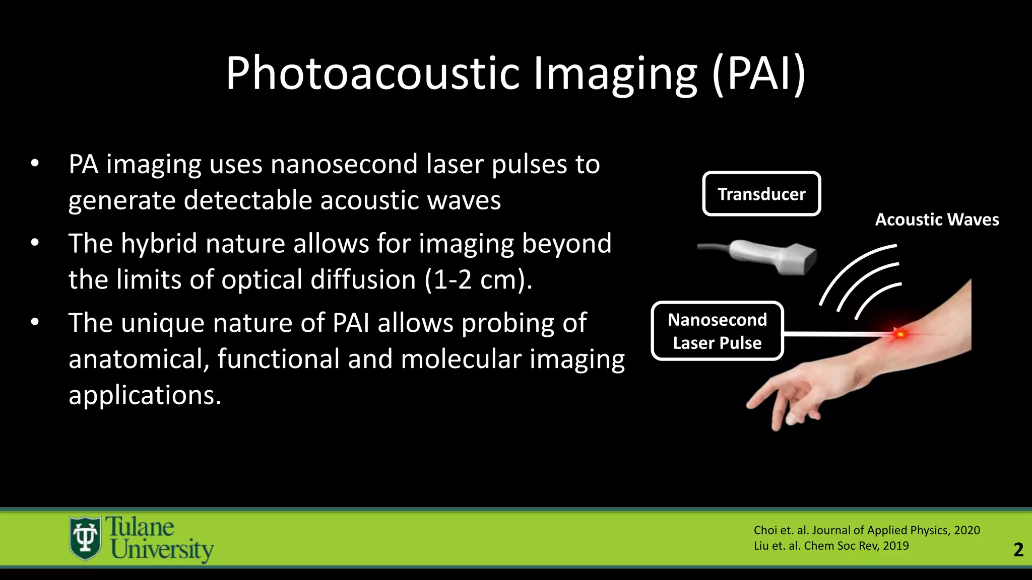
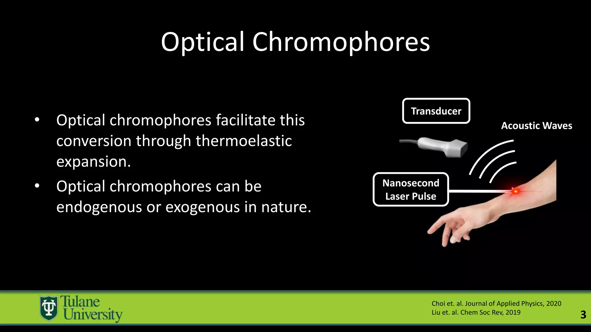
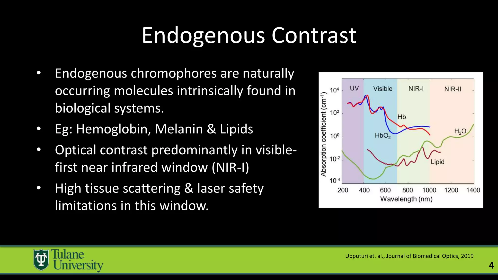
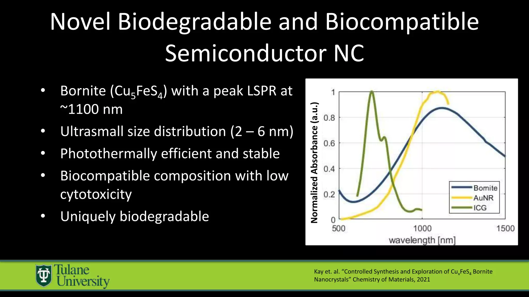
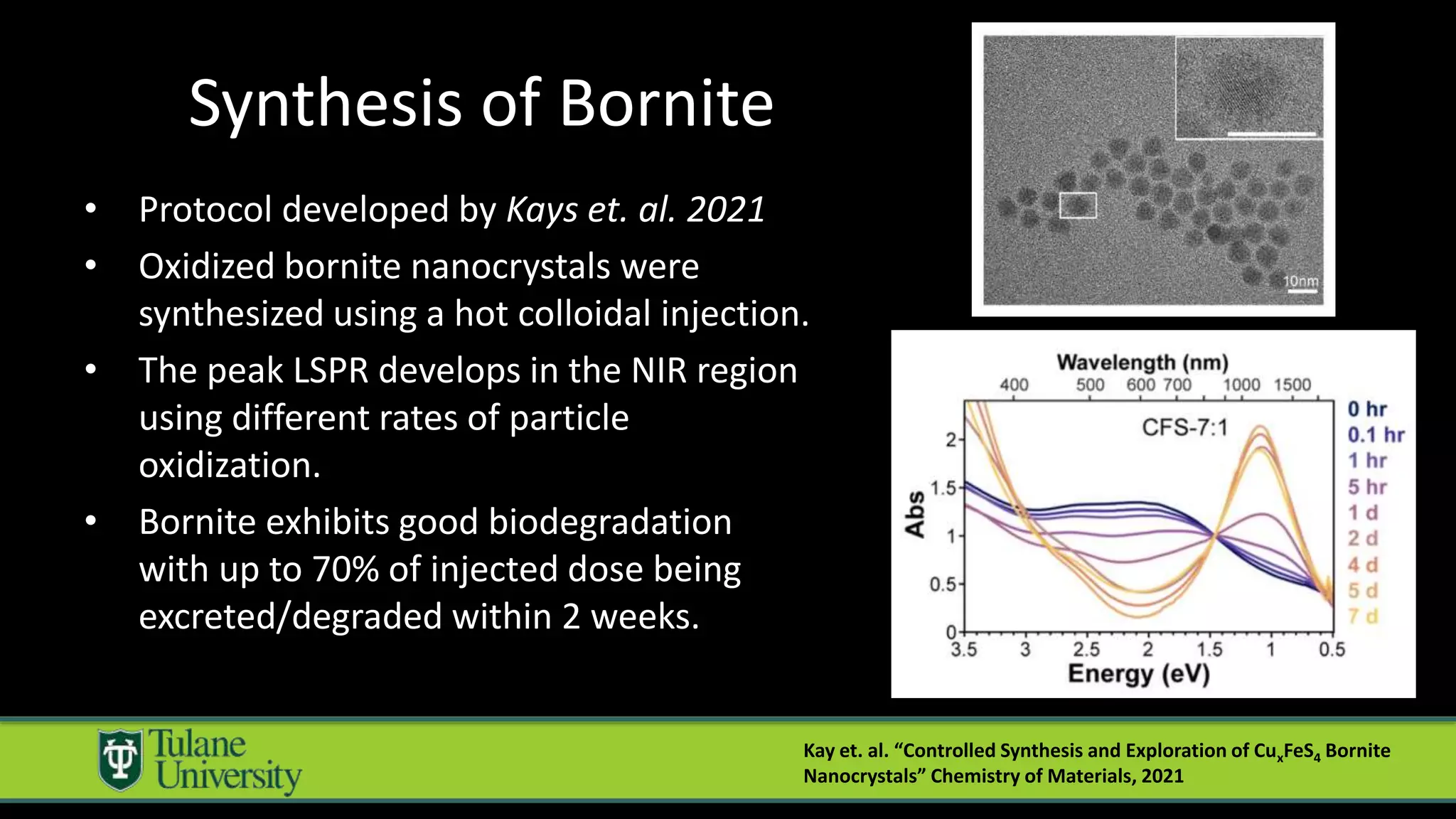
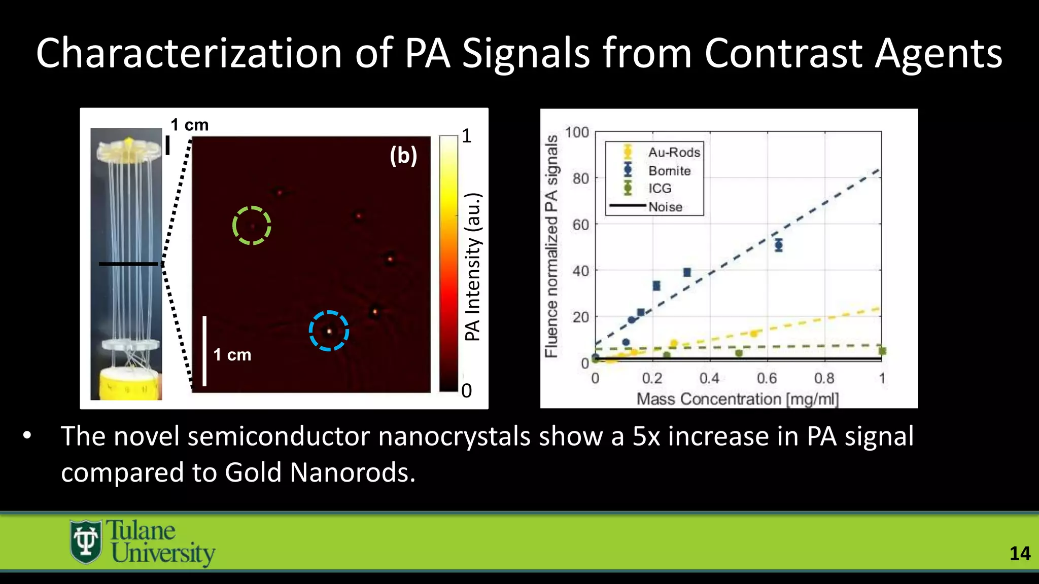
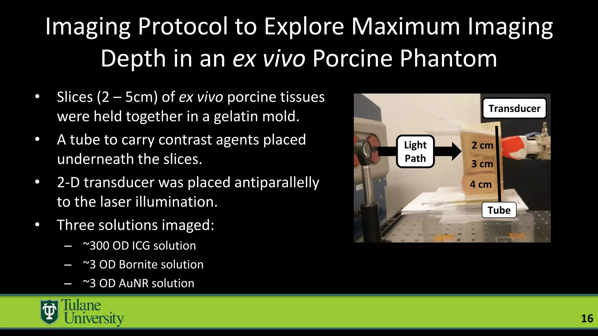
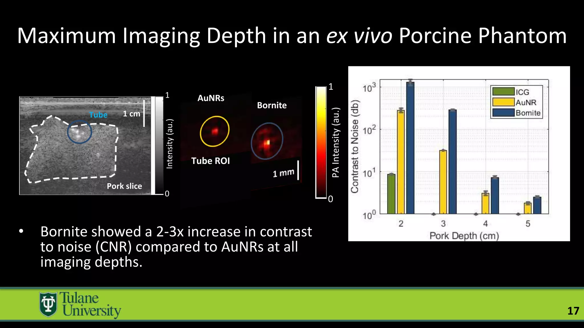
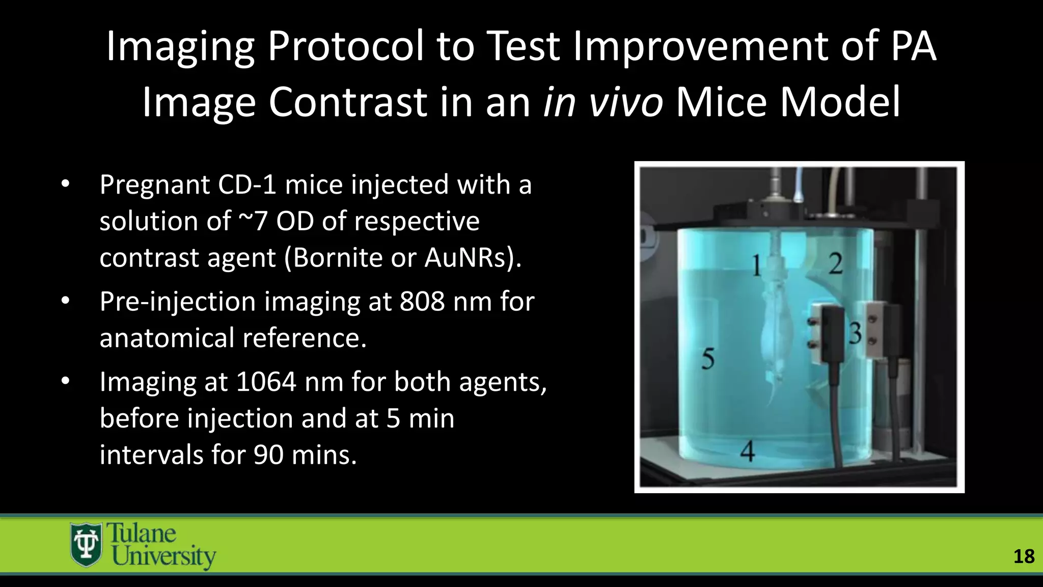
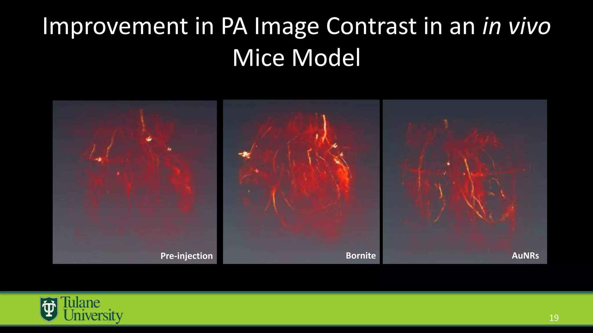
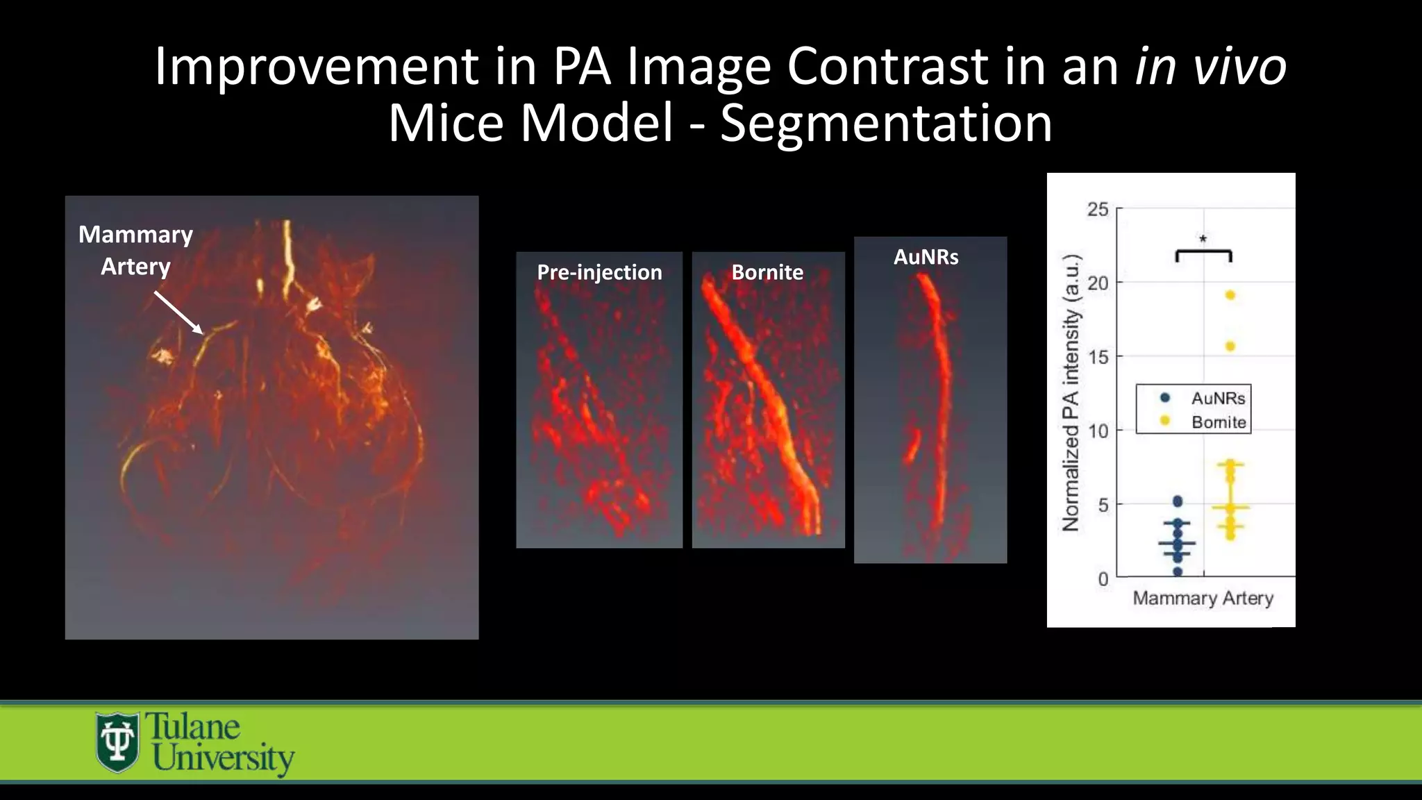
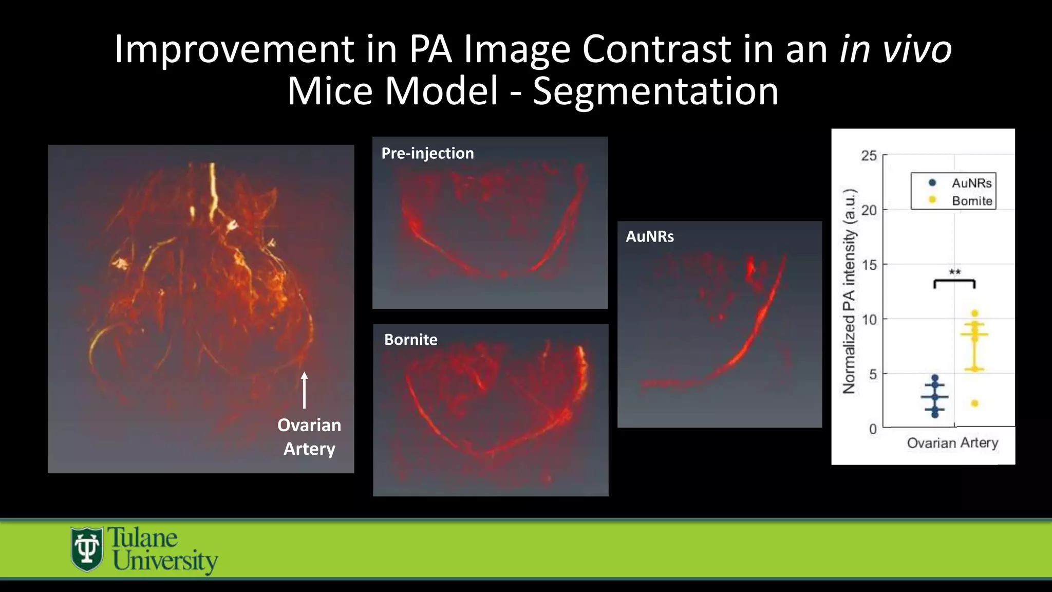
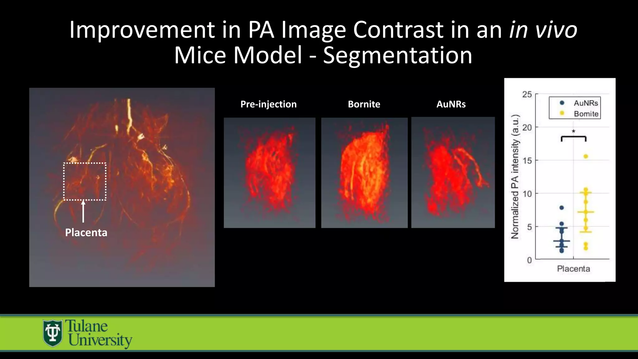
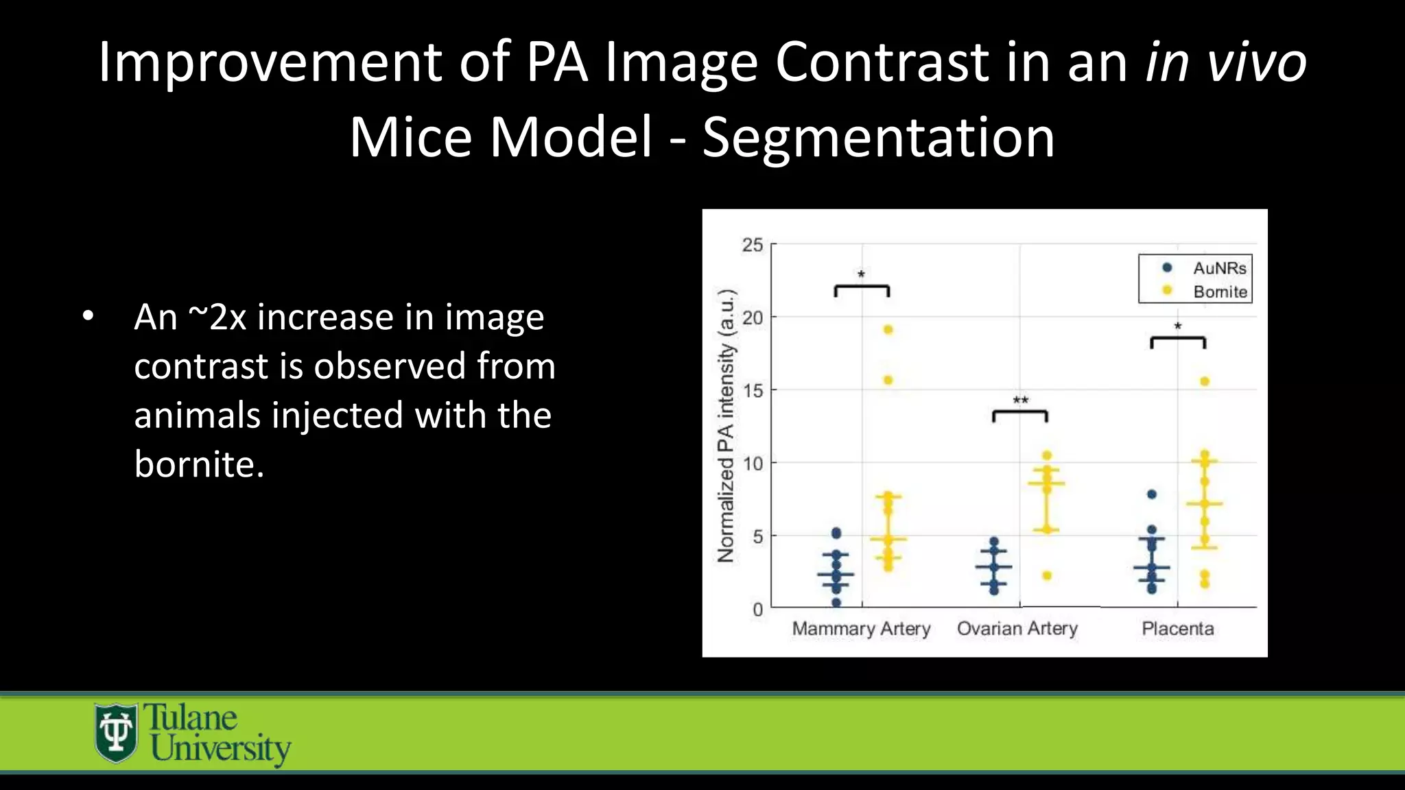


The document introduces a novel biodegradable and biocompatible semiconductor nanocrystal called bornite that could improve photoacoustic imaging contrast for deep tissue applications. Experiments show bornite generates a 5x stronger photoacoustic signal than gold nanorods and indocyanine green. It also allows 2-3x deeper imaging of up to 5cm in tissue phantoms and provides around 2x better contrast in vivo. Bornite could be a safer and more effective photoacoustic contrast agent compared to existing alternatives.


![Photodynamic therapy in endodontics [Autosaved].pptx](https://cdn.slidesharecdn.com/ss_thumbnails/photodynamictherapyinendodonticsautosaved-250928174448-cd9e341b-thumbnail.jpg?width=640&height=640&fit=bounds)


