SlideShare a Scribd company logo
1 of 53
Cell
Division
Mitosis &
Meiosis
From the Virtual Cell Biology Classroom on ScienceProfOnline.com
Image: Cell Division, Wikipedia
CELL DIVISION
• It’s the process by which a cell divides to
form two new cells
• • Three types of cell division or cell
reproduction in an organism
• • Prokaryotes (bacteria)
- Binary fission
- • Divides forming two new identical cells
• Eukaryotes
-Mitosis
• Cell or organism growth • Replacement or
repair of damaged cells
-Meiosis • formation of sex cells, or gametes
WHY DO CELLS DIVIDE?
• Cells divide for growth, development,
repair of worn-out tissues and
reproduction
• To facilitate the exchange of materials
• To control DNA overloading
PROKARYOTIC CELL
DIVISION
• 1. Binary Fission
• Three (3) major steps;
• DNA Replication DNA is copied resulting into two
identical chromosomes
• Chromosome Segregation Chromosomes
separate and move towards ends (poles) of cell
• Cytokinesis (Separation) Cytoplasm divides
forming two (2) cells
• Each new daughter cell is Genetically Identical to
parent cell
EUKARYOTIC CELL DIVISION
• Cell division that results in two daughter cells each
having the same number and kind of chromosomes
as the parent cell
• 1. MITOSIS
• Two (2) main steps:
1. Mitosis
• Fours steps;
[Prophase>Metaphase>Anaphase>Telophase]
2. Cytokinesis
• Cytoplasm divides forming two new daughter cells
• Each daughter cell is Genetically Identical to parent
cell
• Cell division that results in four daughter cells
2. MEIOSIS
• Two (2) major steps:
1. Mitosis Fours steps;
[Prophase>Metaphase>Anaphase>Telophase]
2. Cytokinesis
• Cytoplasm divides forming two new daughter
cells
• Each daughter cell is NOT Genetically Identical to
parent cell
THE CELL CYCLE
• The sequence of events from the time a cell first
arises as a result of cell division until the time when
that cell itself divides.
• Arise – Divide
• This consist of periods of;
• Growth and Development
• DNA Replication
• Preparation For Division
• Cell Division
• Cell after division begins a new cycle
The Cell Cycle
• Consist of two(2) main periods;
I. Interphase
II. Mitotic Phase
Interphase
• Interphase: period of growth and DNA replication
between cell divisions
• Three (3) phases:
• G1 Phase ‒ Cell increases in size
• S Phase ‒ Replication of DNA ‒ Two sister strands
of DNA called chromatids are produced
• G2 Phase ‒ Organelles double ‒ New cytoplasm
forms ‒ All other structures needed for mitosis form
CELL CYCLE
Mitotic Phase
• Mitotic phase is the stage when a cell divides
• Mitosis – the division of a single nucleus into two
genetically identical daughter nuclei • This division
involves two(2) processes;
‒ Division of the nucleus
‒ Separation of the cytoplasm and the new nuclei
into daughter cells
Mitotic Phase
• Divided into two (2) mitotic phases
• 1st Mitotic Phase contain four stages (P-
MAT)
‒ Prophase, metaphase, anaphase and
telophase
• 2nd Mitotic Phase is cytokinesis
Mitosis
1. Prophase
2. Metaphase
3. Anaphase
4. Telophase
•P-MAT
Interphase
• The period when the cell is in a non-dividing state
• A cell spends most of its time in this phase
• During this time it grows, replicates its
chromosomes and prepares for cell division.
• The cell then leaves interphase, undergoes
mitosis, and completes its division.
Early Prophase
• Chromatids condense becoming chromosomes
• Nucleolus disappears
• Centrioles separate and start moving to opposite
ends of the cell
• Spindle begins to form
Late Prophase
• The nuclear membrane fragments and the
microtubules invade the nuclear area
• Centrioles have moved to the opposite poles
• The spindle is completely formed
Metaphase
• The chromosomes are aligned at the metaphase
plate
• Centrioles move at polar ends and projects
spindle fibers to connect each chromosome
Anaphase
• The paired chromosomes (sister chromatids)
separate
• Separated chromatids move to opposite pole
• Partial division of cytoplasm begins
Telophase
• Chromosomes are at the poles
• Chromosomes uncoil-turn chromatin
• Nuclear envelops reforms
• Spindle fiber disappear
Cytokinesis
• Occurs at the end of mitosis
• Animal cells: a cleavage furrow separates the
daughter cells
• Plant cell: a cell plate separates the daughter cells
• Daughter cells are genetically identical
CONTROL OF THE CELL
CYCLE
• Regulatory proteins called cyclins control the cell
cycle at checkpoints:
• G1 Checkpoint—decides whether or not cell will
divide
• S Checkpoint—determines if DNA has been
properly replicated
• Mitotic Spindle Checkpoint—ensures
chromosomes are aligned at mitotic plate
Eukaryotic Cell Cycle
– Cell grows.
– DNA is replicated.
– Mitotic cell division produces
daughter cells identical to the
parent.
– Repeat.
The timing of replication and
cell division is highly
regulated.
Image: Cell cycle by Richard Wheeler From the Virtual Cell Biology Classroom on ScienceProfOnline.com
2 major phases:
• Interphase (3 stages)
– DNA is not condensed
• Mitosis (4 stages + cytokinesis)
– Nuclear division & division of
cytoplasm
– DNA condensed
Eukaryotic Cell Cycle
Interphase
Non-dividing state
with 3 sub-stages:
Gap 1 – cell grows in size
– organelles replicated
Synthesis – replication of DNA
– synthesis of proteins
associated with DNA
Gap 2 – synthesis of proteins
associated with mitosis
Image: Cell cycle by Richard Wheeler; Interphase in Onion Cell
Drawing & Photo, Source Unknown From the Virtual Cell Biology Classroom on ScienceProfOnline.com
Mitosis
Division of somatic cells (non-reproductive
cells) in eukaryotic organisms.
A single cell divides into two identical
daughter cells.
Daughter cells have same number of
chromosomes as does parent cell.
Packing for the move…
When the cell is not dividing…
• DNA molecules are in extended,
uncondensed form = chromatin
• Cell can only replicate and
transcribe DNA when it is in the
extended state.
When the cell is preparing for
division…
• DNA molecules condense to form
chromosomes prior to division.
• each chromosome is a single
molecule of DNA
• easier to sort and organize the
replicated DNA into daughter cells
From the Virtual Cell Biology Classroom on ScienceProfOnline.com
Mitosis
4 sub-phases:
1st – Prophase
2nd – Metaphase
3rd – Anaphase
4th – Telophase
followed by
Cytokinesis
From the Virtual Cell Biology Classroom on ScienceProfOnline.com
Image: Mitosis diagram, Marek Kultys
1. Prophase
Three Major Events
1. chromosomes
condense
2. spindle fibers form
1. (spindle fibers are
specialized microtubules
radiating out from centrioles)
3. chromosomes are captured by spindle
From the Virtual Cell Biology Classroom on ScienceProfOnline.com
chromatin
nucleolus
nucleus
centrioles
condensing
chromosomes
• chromosomes align along the
equator of the cell, with one
chromatid facing each pole
centrioles
spindle fibers
chromosomes
2. Metaphase
Images: Metaphase drawing, Henry Gray's Anatomy of the Human Body;
Metaphase Onion Cell Drawing & Photo, Source Unknown
3. Anaphase
• sister chromatids separate
• spindle fibers attached to
kinetochores shorten and pull
chromatids towards the poles.
• free spindle fibers lengthen and
push the poles of the cell apart
Images: Anaphase drawing, Henry Gray's Anatomy of the Human Body;
Anaphase Onion Cell Drawing & Photo, Source Unknown From the Virtual Cell Biology Classroom on ScienceProfOnline.com
4. Telophase
• spindle fibers disintegrate
• nuclear envelopes form around both groups
of chromosomes
• chromosomes revert to their extended state
• cytokinesis occurs, enclosing each daughter
nucleus into a separate cell
Images: Telophase drawing, Henry Gray's Anatomy of the Human Body;
Telophase Onion Cell Drawing & Photo, Source Unknown From the Virtual Cell Biology Classroom on ScienceProfOnline.com
Cytokinesis – Plant vs. Animal Cell
• Plant cells undergo cytokinesis
by forming a cell plate between
the two daughter nuclei.
• Animal cells undergo
cytokinesis through the
formation of a cleavage furrow. A
ring of microtubules contract,
pinching the cell in half.
From the Virtual Cell Biology Classroom on ScienceProfOnline.com
Images: Telophase drawing, Henry Gray's Anatomy of the Human Body; Ciliate
dividing, TheAlphaWolf; Telophase Onion Cell Photo, Source Unknown
- A single germ cell divides into four unique daughter cells.
- Daughter cells have half the # of chromosomes as parent cell, so
they are considered haploid.
Image: Overview of Meiosis,
National Institutes of Health
What is cell division of gametes called?
Meiosis
From the Virtual Cell Biology Classroom on ScienceProfOnline.com
Diploid organisms receive one of each type of
chromosome from female parent (maternal chromosomes) and
one of each type of chromosome from male parent
(paternal chromosomes)
Refers to the number of sets of
chromosomes in cells.
● Haploid – one copy of each chromosome
– designated as “n”, the number of
chromosomes in one “set”
– gametes
● Diploid – two sets of chromosomes (two of
each chromosome)
– designated as “2n”
– somatic cells
Genetics Terminology: Ploidy
From the Virtual Cell Biology Classroom on ScienceProfOnline.com
Genetics Terminology: Homologues
Chromosomes exist in homologous pairs in
diploid (2n) cells.
Exception: Sex chromosomes (X, Y).
All other chromosomes (autosomes) have homologues.
From the Virtual Cell Biology Classroom on ScienceProfOnline.com
Karyotype
• Q: How many homologous
pairs are in each
karyotype?
• Q: How is the bottom
karyotype different from
the top two?
Image: Karyotype, National Human Genome Research Institute
From the Virtual Cell Biology Classroom on ScienceProfOnline.com
Sexual Reproduction
• Fusion of two gametes to
produce a single zygote.
• Introduces greater genetic
variation, allows genetic
recombination.
• Zygote has gametes from
two different parents
(except in cases of self-
fertilizing organisms).
Rose + Greg = Steven
Images: Rose, Greg, and Steven, Steven Universe From the Virtual Cell Biology Classroom on ScienceProfOnline.com
Sexual reproduction in humans …
• At fertilization, 23 chromosomes
are donated by each parent.
(total = 46 or 23 pairs).
• Gametes (sperm/ova):
– Contain 22 autosomes and 1 sex
chromosome.
– Are haploid (haploid number “n” = 23
in humans).
• Fertilization results in diploid zygote.
– Diploid cell; 2n = 46. (n = 23 in humans)
• Q: Most cells in the body are produced through what type of cell division?
(Remember, only gametes are produced through meiosis)
Image: Superficial human anatomy, Mikael
Häggström& Rainer Zenz; Sperm & egg, Wikipedia From the Virtual Cell Biology Classroom on ScienceProfOnline.com
Meiosis - Sex Cell (Gamete) Formation
In meiosis, there
are 2 divisions
of the nucleus:
meiosis I
&
meiosis II
Image: Overview of Meiosis,
National Institutes of Health From the Virtual Cell Biology Classroom on ScienceProfOnline.com
Image: Meiosis diagram, Marek Kultys
From the Virtual Cell Biology Classroom on ScienceProfOnline.com
Meiosis & Sexual Reproduction
Life Cycle
Image: Animal Life Cycle, Dr. T’s Bio 328 Genetics
Mitosis * *
*
From the Virtual Cell Biology Classroom on ScienceProfOnline.com
Genetic Variation in Diploid Organisms
Fusion of sperm and egg results in unique
offspring…
…but not only because the young are a product
of two individuals with different genetic
makeup.
Meiosis also “shuffles” the genes so that the
an individual’s gametes are genetically
different from one another.
From the Virtual Cell Biology Classroom on ScienceProfOnline.com Image: Meiosis diagram, Marek Kultys
How is this shuffling accomplished?
Genetic shuffling of Meiosis I
In addition to a new combination of chromosomes resulting from
fertilization, there are also events in Meiosis I that shuffle the
genes.
1. Crossing over in Prophase I.
2. Independent assortment in Metaphase I.
From the Virtual Cell Biology Classroom on ScienceProfOnline.com
Crossing Over
• Homologues break at identical
locations, then rejoin opposite
partners.
• This creates new combinations
of the alleles on each
chromosome.
• Occurs randomly several times
on every chromosome.
• Results in mixing of the genes
you inherited from your
parents.
From the Virtual Cell Biology Classroom on ScienceProfOnline.com
Independent Assortment
Variation from genetic
recombination
• Independent assortment of chromosomes
– meiosis introduces genetic variation
– gametes of offspring do not have same
combination of genes as gametes from parents
• random assortment in humans produces
223 (8,388,608) different combinations in gametes
from Dad
from Mom offspring
new gametes
made by offspring
Mitosis vs. Meiosis
• 2n
• Clone
• Same genetic
information in parent
cell and daughter cell.
• Give me another one
just like the other
one!
• 1n
• Daughter cells different
from parent cell and from
each other.
• Daughter cells have ½ the
number of chromosomes
as somatic cell.
• Shuffling the genes
(Mix it up!)
From the Virtual Cell Biology Classroom on ScienceProfOnline.com
Image: Mitosis diagram & Meiosis diagram, Marek Kultys From the Virtual Cell Biology Classroom on ScienceProfOnline.com
Drawing and Labeling Chromosomes
Sister
Chromatid
Replicated
Uncondensed
Chromosome
(chromatin)
Sister
Chromatid
Centromere
Unreplicated
Uncondensed
Chromosome
(chromatin)
From the Virtual Cell Biology Classroom on ScienceProfOnline.com
Drawing & Labeling Homologous Chromosomes
Unreplicated,
Condensed,
Homologous
Chromosomes
Replicated,
Condensed,
Homologous
Chromosomes
From the Virtual Cell Biology Classroom on ScienceProfOnline.com

More Related Content

What's hot

Technical faults in histopathology lab
Technical faults in histopathology labTechnical faults in histopathology lab
Technical faults in histopathology lab
magdy abdelghany
 

What's hot (17)

Cervix cyto
Cervix cytoCervix cyto
Cervix cyto
 
Golgi complex
Golgi complexGolgi complex
Golgi complex
 
Microtome Procedure
Microtome ProcedureMicrotome Procedure
Microtome Procedure
 
Cell biology
Cell biologyCell biology
Cell biology
 
Technical faults in histopathology lab
Technical faults in histopathology labTechnical faults in histopathology lab
Technical faults in histopathology lab
 
Cryostat and frozen section
Cryostat and frozen sectionCryostat and frozen section
Cryostat and frozen section
 
SEMEN ANALYSIS PPT.pptx
SEMEN ANALYSIS PPT.pptxSEMEN ANALYSIS PPT.pptx
SEMEN ANALYSIS PPT.pptx
 
Pathology of the_placenta_-_lecture
Pathology of the_placenta_-_lecturePathology of the_placenta_-_lecture
Pathology of the_placenta_-_lecture
 
Histology of ovary
Histology of ovaryHistology of ovary
Histology of ovary
 
MICROTOME and it's TYPES
MICROTOME and it's TYPES MICROTOME and it's TYPES
MICROTOME and it's TYPES
 
9. automation in urine analysis
9. automation in urine analysis9. automation in urine analysis
9. automation in urine analysis
 
The m phase
The m phaseThe m phase
The m phase
 
Biology 12 - Section 3-2 Cell Organelles
Biology 12 - Section 3-2 Cell OrganellesBiology 12 - Section 3-2 Cell Organelles
Biology 12 - Section 3-2 Cell Organelles
 
Cellular communication (signal transduction)
Cellular communication (signal transduction)Cellular communication (signal transduction)
Cellular communication (signal transduction)
 
Freezing microtome_ cryostat.pptx
Freezing microtome_ cryostat.pptxFreezing microtome_ cryostat.pptx
Freezing microtome_ cryostat.pptx
 
Microtome (2)
Microtome (2)Microtome (2)
Microtome (2)
 
Female Histology
Female HistologyFemale Histology
Female Histology
 

Similar to mitosis and meiosis 02152018.pptx

5. Cell Cycle and Mitosis.ppt
5. Cell Cycle and Mitosis.ppt5. Cell Cycle and Mitosis.ppt
5. Cell Cycle and Mitosis.ppt
HananeKatir1
 
Cell reproduction notes
Cell reproduction notesCell reproduction notes
Cell reproduction notes
mjnepa
 
Cell_Division_Mitosis_Meiosis_ppt.pptx
Cell_Division_Mitosis_Meiosis_ppt.pptxCell_Division_Mitosis_Meiosis_ppt.pptx
Cell_Division_Mitosis_Meiosis_ppt.pptx
CharlesIbus
 
Cell_Division_Mitosis_Meiosis_ppt_{SIS7DDA7B647659}.pptx
Cell_Division_Mitosis_Meiosis_ppt_{SIS7DDA7B647659}.pptxCell_Division_Mitosis_Meiosis_ppt_{SIS7DDA7B647659}.pptx
Cell_Division_Mitosis_Meiosis_ppt_{SIS7DDA7B647659}.pptx
AnnalieLobiano1
 
CELL DIVISION- Decoding Cell Division: The Dance of Life's Continuity
CELL DIVISION- Decoding Cell Division: The Dance of Life's ContinuityCELL DIVISION- Decoding Cell Division: The Dance of Life's Continuity
CELL DIVISION- Decoding Cell Division: The Dance of Life's Continuity
Sagar Pandya
 
5 10 cell division and cell cycle ppt
5 10 cell division and cell cycle ppt5 10 cell division and cell cycle ppt
5 10 cell division and cell cycle ppt
AshwinKshirsagar2
 
W60Aff6ZLR20CNoL269.pptx
W60Aff6ZLR20CNoL269.pptxW60Aff6ZLR20CNoL269.pptx
W60Aff6ZLR20CNoL269.pptx
RCGaur1
 

Similar to mitosis and meiosis 02152018.pptx (20)

5. Cell Cycle and Mitosis.ppt
5. Cell Cycle and Mitosis.ppt5. Cell Cycle and Mitosis.ppt
5. Cell Cycle and Mitosis.ppt
 
Cell division
Cell division Cell division
Cell division
 
Biology theory 4: cell division and cell cycle
Biology theory 4: cell division and cell cycleBiology theory 4: cell division and cell cycle
Biology theory 4: cell division and cell cycle
 
Cell reproduction notes
Cell reproduction notesCell reproduction notes
Cell reproduction notes
 
Cell and Cell division.pptx
Cell and Cell division.pptxCell and Cell division.pptx
Cell and Cell division.pptx
 
12 - The Cell Division Cycle
12 - The Cell Division Cycle12 - The Cell Division Cycle
12 - The Cell Division Cycle
 
Cell_Division_Mitosis_Meiosis_ppt.pptx
Cell_Division_Mitosis_Meiosis_ppt.pptxCell_Division_Mitosis_Meiosis_ppt.pptx
Cell_Division_Mitosis_Meiosis_ppt.pptx
 
Cell_Division_Mitosis_Meiosis_ppt_{SIS7DDA7B647659}.pptx
Cell_Division_Mitosis_Meiosis_ppt_{SIS7DDA7B647659}.pptxCell_Division_Mitosis_Meiosis_ppt_{SIS7DDA7B647659}.pptx
Cell_Division_Mitosis_Meiosis_ppt_{SIS7DDA7B647659}.pptx
 
Cell and Cell division.pptx
Cell and Cell division.pptxCell and Cell division.pptx
Cell and Cell division.pptx
 
Cell reproduction
Cell reproductionCell reproduction
Cell reproduction
 
CELL DIVISION- Decoding Cell Division: The Dance of Life's Continuity
CELL DIVISION- Decoding Cell Division: The Dance of Life's ContinuityCELL DIVISION- Decoding Cell Division: The Dance of Life's Continuity
CELL DIVISION- Decoding Cell Division: The Dance of Life's Continuity
 
MITOSIS
MITOSISMITOSIS
MITOSIS
 
Chapter 5 cell division SPM Biology Form 4
Chapter 5 cell division SPM Biology Form 4Chapter 5 cell division SPM Biology Form 4
Chapter 5 cell division SPM Biology Form 4
 
5 10 cell division and cell cycle ppt
5 10 cell division and cell cycle ppt5 10 cell division and cell cycle ppt
5 10 cell division and cell cycle ppt
 
独中高中生物Chapter 16 cell division
独中高中生物Chapter 16 cell division独中高中生物Chapter 16 cell division
独中高中生物Chapter 16 cell division
 
Cell division, a new way.
Cell division, a new way.Cell division, a new way.
Cell division, a new way.
 
mitosis
mitosismitosis
mitosis
 
2-190118112234.pdf
2-190118112234.pdf2-190118112234.pdf
2-190118112234.pdf
 
W60Aff6ZLR20CNoL269.pptx
W60Aff6ZLR20CNoL269.pptxW60Aff6ZLR20CNoL269.pptx
W60Aff6ZLR20CNoL269.pptx
 
The Cell Cycle and Cell Division
The Cell Cycle and Cell Division The Cell Cycle and Cell Division
The Cell Cycle and Cell Division
 

More from sanarao25

Data Retreival, Protein sequence database, Gene Pridiction 3.pptx
Data Retreival, Protein sequence database, Gene Pridiction 3.pptxData Retreival, Protein sequence database, Gene Pridiction 3.pptx
Data Retreival, Protein sequence database, Gene Pridiction 3.pptx
sanarao25
 

More from sanarao25 (17)

Lecture Environmental Biotechnology, Bioremediation 24-12-2019.ppt
Lecture Environmental Biotechnology, Bioremediation 24-12-2019.pptLecture Environmental Biotechnology, Bioremediation 24-12-2019.ppt
Lecture Environmental Biotechnology, Bioremediation 24-12-2019.ppt
 
Parasitic diseases.pptx
Parasitic diseases.pptxParasitic diseases.pptx
Parasitic diseases.pptx
 
Prenylation.pptx
Prenylation.pptxPrenylation.pptx
Prenylation.pptx
 
Prenylation.pptx
Prenylation.pptxPrenylation.pptx
Prenylation.pptx
 
Microarray data Analysis.pptx
Microarray data Analysis.pptxMicroarray data Analysis.pptx
Microarray data Analysis.pptx
 
Data Retreival, Protein sequence database, Gene Pridiction 3.pptx
Data Retreival, Protein sequence database, Gene Pridiction 3.pptxData Retreival, Protein sequence database, Gene Pridiction 3.pptx
Data Retreival, Protein sequence database, Gene Pridiction 3.pptx
 
COPI and COPII Secretery pathways.pptx
COPI and COPII Secretery pathways.pptxCOPI and COPII Secretery pathways.pptx
COPI and COPII Secretery pathways.pptx
 
citrus SH presentation.ppt
citrus SH presentation.pptcitrus SH presentation.ppt
citrus SH presentation.ppt
 
Molecular markers.ppt
Molecular markers.pptMolecular markers.ppt
Molecular markers.ppt
 
An efficient cucumber (Cucumis sativus L.pdf
An efficient cucumber (Cucumis sativus L.pdfAn efficient cucumber (Cucumis sativus L.pdf
An efficient cucumber (Cucumis sativus L.pdf
 
L 7 Plant protoplasts .ppt
L 7 Plant  protoplasts .pptL 7 Plant  protoplasts .ppt
L 7 Plant protoplasts .ppt
 
helgesonsomatic.ppt
helgesonsomatic.ppthelgesonsomatic.ppt
helgesonsomatic.ppt
 
17465299.ppt
17465299.ppt17465299.ppt
17465299.ppt
 
GAMETOGENESIS.pptx
GAMETOGENESIS.pptxGAMETOGENESIS.pptx
GAMETOGENESIS.pptx
 
23_clark_missing_epistasis.pptx
23_clark_missing_epistasis.pptx23_clark_missing_epistasis.pptx
23_clark_missing_epistasis.pptx
 
Presentation1.pptx
Presentation1.pptxPresentation1.pptx
Presentation1.pptx
 
mitosis and meiosis 02152018.pptx
mitosis and meiosis 02152018.pptxmitosis and meiosis 02152018.pptx
mitosis and meiosis 02152018.pptx
 

Recently uploaded

CYTOGENETIC MAP................ ppt.pptx
CYTOGENETIC MAP................ ppt.pptxCYTOGENETIC MAP................ ppt.pptx
CYTOGENETIC MAP................ ppt.pptx
Cherry
 
PODOCARPUS...........................pptx
PODOCARPUS...........................pptxPODOCARPUS...........................pptx
PODOCARPUS...........................pptx
Cherry
 
Pteris : features, anatomy, morphology and lifecycle
Pteris : features, anatomy, morphology and lifecyclePteris : features, anatomy, morphology and lifecycle
Pteris : features, anatomy, morphology and lifecycle
Cherry
 
Porella : features, morphology, anatomy, reproduction etc.
Porella : features, morphology, anatomy, reproduction etc.Porella : features, morphology, anatomy, reproduction etc.
Porella : features, morphology, anatomy, reproduction etc.
Cherry
 
(May 9, 2024) Enhanced Ultrafast Vector Flow Imaging (VFI) Using Multi-Angle ...
(May 9, 2024) Enhanced Ultrafast Vector Flow Imaging (VFI) Using Multi-Angle ...(May 9, 2024) Enhanced Ultrafast Vector Flow Imaging (VFI) Using Multi-Angle ...
(May 9, 2024) Enhanced Ultrafast Vector Flow Imaging (VFI) Using Multi-Angle ...
Scintica Instrumentation
 
POGONATUM : morphology, anatomy, reproduction etc.
POGONATUM : morphology, anatomy, reproduction etc.POGONATUM : morphology, anatomy, reproduction etc.
POGONATUM : morphology, anatomy, reproduction etc.
Cherry
 
Human genetics..........................pptx
Human genetics..........................pptxHuman genetics..........................pptx
Human genetics..........................pptx
Cherry
 
Lipids: types, structure and important functions.
Lipids: types, structure and important functions.Lipids: types, structure and important functions.
Lipids: types, structure and important functions.
Cherry
 
Reboulia: features, anatomy, morphology etc.
Reboulia: features, anatomy, morphology etc.Reboulia: features, anatomy, morphology etc.
Reboulia: features, anatomy, morphology etc.
Cherry
 
Cyathodium bryophyte: morphology, anatomy, reproduction etc.
Cyathodium bryophyte: morphology, anatomy, reproduction etc.Cyathodium bryophyte: morphology, anatomy, reproduction etc.
Cyathodium bryophyte: morphology, anatomy, reproduction etc.
Cherry
 

Recently uploaded (20)

GBSN - Microbiology (Unit 3)Defense Mechanism of the body
GBSN - Microbiology (Unit 3)Defense Mechanism of the body GBSN - Microbiology (Unit 3)Defense Mechanism of the body
GBSN - Microbiology (Unit 3)Defense Mechanism of the body
 
FS P2 COMBO MSTA LAST PUSH past exam papers.
FS P2 COMBO MSTA LAST PUSH past exam papers.FS P2 COMBO MSTA LAST PUSH past exam papers.
FS P2 COMBO MSTA LAST PUSH past exam papers.
 
CYTOGENETIC MAP................ ppt.pptx
CYTOGENETIC MAP................ ppt.pptxCYTOGENETIC MAP................ ppt.pptx
CYTOGENETIC MAP................ ppt.pptx
 
Use of mutants in understanding seedling development.pptx
Use of mutants in understanding seedling development.pptxUse of mutants in understanding seedling development.pptx
Use of mutants in understanding seedling development.pptx
 
PODOCARPUS...........................pptx
PODOCARPUS...........................pptxPODOCARPUS...........................pptx
PODOCARPUS...........................pptx
 
Pteris : features, anatomy, morphology and lifecycle
Pteris : features, anatomy, morphology and lifecyclePteris : features, anatomy, morphology and lifecycle
Pteris : features, anatomy, morphology and lifecycle
 
Plasmid: types, structure and functions.
Plasmid: types, structure and functions.Plasmid: types, structure and functions.
Plasmid: types, structure and functions.
 
CURRENT SCENARIO OF POULTRY PRODUCTION IN INDIA
CURRENT SCENARIO OF POULTRY PRODUCTION IN INDIACURRENT SCENARIO OF POULTRY PRODUCTION IN INDIA
CURRENT SCENARIO OF POULTRY PRODUCTION IN INDIA
 
Porella : features, morphology, anatomy, reproduction etc.
Porella : features, morphology, anatomy, reproduction etc.Porella : features, morphology, anatomy, reproduction etc.
Porella : features, morphology, anatomy, reproduction etc.
 
Role of AI in seed science Predictive modelling and Beyond.pptx
Role of AI in seed science  Predictive modelling and  Beyond.pptxRole of AI in seed science  Predictive modelling and  Beyond.pptx
Role of AI in seed science Predictive modelling and Beyond.pptx
 
(May 9, 2024) Enhanced Ultrafast Vector Flow Imaging (VFI) Using Multi-Angle ...
(May 9, 2024) Enhanced Ultrafast Vector Flow Imaging (VFI) Using Multi-Angle ...(May 9, 2024) Enhanced Ultrafast Vector Flow Imaging (VFI) Using Multi-Angle ...
(May 9, 2024) Enhanced Ultrafast Vector Flow Imaging (VFI) Using Multi-Angle ...
 
POGONATUM : morphology, anatomy, reproduction etc.
POGONATUM : morphology, anatomy, reproduction etc.POGONATUM : morphology, anatomy, reproduction etc.
POGONATUM : morphology, anatomy, reproduction etc.
 
ABHISHEK ANTIBIOTICS PPT MICROBIOLOGY // USES OF ANTIOBIOTICS TYPES OF ANTIB...
ABHISHEK ANTIBIOTICS PPT MICROBIOLOGY  // USES OF ANTIOBIOTICS TYPES OF ANTIB...ABHISHEK ANTIBIOTICS PPT MICROBIOLOGY  // USES OF ANTIOBIOTICS TYPES OF ANTIB...
ABHISHEK ANTIBIOTICS PPT MICROBIOLOGY // USES OF ANTIOBIOTICS TYPES OF ANTIB...
 
Human genetics..........................pptx
Human genetics..........................pptxHuman genetics..........................pptx
Human genetics..........................pptx
 
Understanding Partial Differential Equations: Types and Solution Methods
Understanding Partial Differential Equations: Types and Solution MethodsUnderstanding Partial Differential Equations: Types and Solution Methods
Understanding Partial Differential Equations: Types and Solution Methods
 
Genome sequencing,shotgun sequencing.pptx
Genome sequencing,shotgun sequencing.pptxGenome sequencing,shotgun sequencing.pptx
Genome sequencing,shotgun sequencing.pptx
 
Lipids: types, structure and important functions.
Lipids: types, structure and important functions.Lipids: types, structure and important functions.
Lipids: types, structure and important functions.
 
module for grade 9 for distance learning
module for grade 9 for distance learningmodule for grade 9 for distance learning
module for grade 9 for distance learning
 
Reboulia: features, anatomy, morphology etc.
Reboulia: features, anatomy, morphology etc.Reboulia: features, anatomy, morphology etc.
Reboulia: features, anatomy, morphology etc.
 
Cyathodium bryophyte: morphology, anatomy, reproduction etc.
Cyathodium bryophyte: morphology, anatomy, reproduction etc.Cyathodium bryophyte: morphology, anatomy, reproduction etc.
Cyathodium bryophyte: morphology, anatomy, reproduction etc.
 

mitosis and meiosis 02152018.pptx

  • 1. Cell Division Mitosis & Meiosis From the Virtual Cell Biology Classroom on ScienceProfOnline.com Image: Cell Division, Wikipedia
  • 2. CELL DIVISION • It’s the process by which a cell divides to form two new cells • • Three types of cell division or cell reproduction in an organism • • Prokaryotes (bacteria) - Binary fission - • Divides forming two new identical cells
  • 3. • Eukaryotes -Mitosis • Cell or organism growth • Replacement or repair of damaged cells -Meiosis • formation of sex cells, or gametes
  • 4. WHY DO CELLS DIVIDE? • Cells divide for growth, development, repair of worn-out tissues and reproduction • To facilitate the exchange of materials • To control DNA overloading
  • 5. PROKARYOTIC CELL DIVISION • 1. Binary Fission • Three (3) major steps; • DNA Replication DNA is copied resulting into two identical chromosomes • Chromosome Segregation Chromosomes separate and move towards ends (poles) of cell • Cytokinesis (Separation) Cytoplasm divides forming two (2) cells • Each new daughter cell is Genetically Identical to parent cell
  • 6.
  • 7. EUKARYOTIC CELL DIVISION • Cell division that results in two daughter cells each having the same number and kind of chromosomes as the parent cell • 1. MITOSIS • Two (2) main steps: 1. Mitosis • Fours steps; [Prophase>Metaphase>Anaphase>Telophase] 2. Cytokinesis • Cytoplasm divides forming two new daughter cells • Each daughter cell is Genetically Identical to parent cell
  • 8.
  • 9. • Cell division that results in four daughter cells 2. MEIOSIS • Two (2) major steps: 1. Mitosis Fours steps; [Prophase>Metaphase>Anaphase>Telophase] 2. Cytokinesis • Cytoplasm divides forming two new daughter cells • Each daughter cell is NOT Genetically Identical to parent cell
  • 10. THE CELL CYCLE • The sequence of events from the time a cell first arises as a result of cell division until the time when that cell itself divides. • Arise – Divide • This consist of periods of; • Growth and Development • DNA Replication • Preparation For Division • Cell Division • Cell after division begins a new cycle
  • 11. The Cell Cycle • Consist of two(2) main periods; I. Interphase II. Mitotic Phase
  • 12. Interphase • Interphase: period of growth and DNA replication between cell divisions • Three (3) phases: • G1 Phase ‒ Cell increases in size • S Phase ‒ Replication of DNA ‒ Two sister strands of DNA called chromatids are produced • G2 Phase ‒ Organelles double ‒ New cytoplasm forms ‒ All other structures needed for mitosis form
  • 14. Mitotic Phase • Mitotic phase is the stage when a cell divides • Mitosis – the division of a single nucleus into two genetically identical daughter nuclei • This division involves two(2) processes; ‒ Division of the nucleus ‒ Separation of the cytoplasm and the new nuclei into daughter cells
  • 15. Mitotic Phase • Divided into two (2) mitotic phases • 1st Mitotic Phase contain four stages (P- MAT) ‒ Prophase, metaphase, anaphase and telophase • 2nd Mitotic Phase is cytokinesis
  • 16. Mitosis 1. Prophase 2. Metaphase 3. Anaphase 4. Telophase •P-MAT
  • 17. Interphase • The period when the cell is in a non-dividing state • A cell spends most of its time in this phase • During this time it grows, replicates its chromosomes and prepares for cell division. • The cell then leaves interphase, undergoes mitosis, and completes its division.
  • 18. Early Prophase • Chromatids condense becoming chromosomes • Nucleolus disappears • Centrioles separate and start moving to opposite ends of the cell • Spindle begins to form
  • 19. Late Prophase • The nuclear membrane fragments and the microtubules invade the nuclear area • Centrioles have moved to the opposite poles • The spindle is completely formed
  • 20. Metaphase • The chromosomes are aligned at the metaphase plate • Centrioles move at polar ends and projects spindle fibers to connect each chromosome
  • 21. Anaphase • The paired chromosomes (sister chromatids) separate • Separated chromatids move to opposite pole • Partial division of cytoplasm begins
  • 22. Telophase • Chromosomes are at the poles • Chromosomes uncoil-turn chromatin • Nuclear envelops reforms • Spindle fiber disappear
  • 23. Cytokinesis • Occurs at the end of mitosis • Animal cells: a cleavage furrow separates the daughter cells • Plant cell: a cell plate separates the daughter cells • Daughter cells are genetically identical
  • 24. CONTROL OF THE CELL CYCLE • Regulatory proteins called cyclins control the cell cycle at checkpoints: • G1 Checkpoint—decides whether or not cell will divide • S Checkpoint—determines if DNA has been properly replicated • Mitotic Spindle Checkpoint—ensures chromosomes are aligned at mitotic plate
  • 25. Eukaryotic Cell Cycle – Cell grows. – DNA is replicated. – Mitotic cell division produces daughter cells identical to the parent. – Repeat. The timing of replication and cell division is highly regulated. Image: Cell cycle by Richard Wheeler From the Virtual Cell Biology Classroom on ScienceProfOnline.com
  • 26. Image: Cell cycle by Richard Wheeler From the Virtual Cell Biology Classroom on ScienceProfOnline.com 2 major phases: • Interphase (3 stages) – DNA is not condensed • Mitosis (4 stages + cytokinesis) – Nuclear division & division of cytoplasm – DNA condensed Eukaryotic Cell Cycle
  • 27. Interphase Non-dividing state with 3 sub-stages: Gap 1 – cell grows in size – organelles replicated Synthesis – replication of DNA – synthesis of proteins associated with DNA Gap 2 – synthesis of proteins associated with mitosis Image: Cell cycle by Richard Wheeler; Interphase in Onion Cell Drawing & Photo, Source Unknown From the Virtual Cell Biology Classroom on ScienceProfOnline.com
  • 28. Mitosis Division of somatic cells (non-reproductive cells) in eukaryotic organisms. A single cell divides into two identical daughter cells. Daughter cells have same number of chromosomes as does parent cell.
  • 29. Packing for the move… When the cell is not dividing… • DNA molecules are in extended, uncondensed form = chromatin • Cell can only replicate and transcribe DNA when it is in the extended state. When the cell is preparing for division… • DNA molecules condense to form chromosomes prior to division. • each chromosome is a single molecule of DNA • easier to sort and organize the replicated DNA into daughter cells From the Virtual Cell Biology Classroom on ScienceProfOnline.com
  • 30. Mitosis 4 sub-phases: 1st – Prophase 2nd – Metaphase 3rd – Anaphase 4th – Telophase followed by Cytokinesis From the Virtual Cell Biology Classroom on ScienceProfOnline.com Image: Mitosis diagram, Marek Kultys
  • 31. 1. Prophase Three Major Events 1. chromosomes condense 2. spindle fibers form 1. (spindle fibers are specialized microtubules radiating out from centrioles) 3. chromosomes are captured by spindle From the Virtual Cell Biology Classroom on ScienceProfOnline.com chromatin nucleolus nucleus centrioles condensing chromosomes
  • 32. • chromosomes align along the equator of the cell, with one chromatid facing each pole centrioles spindle fibers chromosomes 2. Metaphase Images: Metaphase drawing, Henry Gray's Anatomy of the Human Body; Metaphase Onion Cell Drawing & Photo, Source Unknown
  • 33. 3. Anaphase • sister chromatids separate • spindle fibers attached to kinetochores shorten and pull chromatids towards the poles. • free spindle fibers lengthen and push the poles of the cell apart Images: Anaphase drawing, Henry Gray's Anatomy of the Human Body; Anaphase Onion Cell Drawing & Photo, Source Unknown From the Virtual Cell Biology Classroom on ScienceProfOnline.com
  • 34. 4. Telophase • spindle fibers disintegrate • nuclear envelopes form around both groups of chromosomes • chromosomes revert to their extended state • cytokinesis occurs, enclosing each daughter nucleus into a separate cell Images: Telophase drawing, Henry Gray's Anatomy of the Human Body; Telophase Onion Cell Drawing & Photo, Source Unknown From the Virtual Cell Biology Classroom on ScienceProfOnline.com
  • 35. Cytokinesis – Plant vs. Animal Cell • Plant cells undergo cytokinesis by forming a cell plate between the two daughter nuclei. • Animal cells undergo cytokinesis through the formation of a cleavage furrow. A ring of microtubules contract, pinching the cell in half. From the Virtual Cell Biology Classroom on ScienceProfOnline.com Images: Telophase drawing, Henry Gray's Anatomy of the Human Body; Ciliate dividing, TheAlphaWolf; Telophase Onion Cell Photo, Source Unknown
  • 36. - A single germ cell divides into four unique daughter cells. - Daughter cells have half the # of chromosomes as parent cell, so they are considered haploid. Image: Overview of Meiosis, National Institutes of Health What is cell division of gametes called? Meiosis From the Virtual Cell Biology Classroom on ScienceProfOnline.com
  • 37. Diploid organisms receive one of each type of chromosome from female parent (maternal chromosomes) and one of each type of chromosome from male parent (paternal chromosomes) Refers to the number of sets of chromosomes in cells. ● Haploid – one copy of each chromosome – designated as “n”, the number of chromosomes in one “set” – gametes ● Diploid – two sets of chromosomes (two of each chromosome) – designated as “2n” – somatic cells Genetics Terminology: Ploidy From the Virtual Cell Biology Classroom on ScienceProfOnline.com
  • 38. Genetics Terminology: Homologues Chromosomes exist in homologous pairs in diploid (2n) cells. Exception: Sex chromosomes (X, Y). All other chromosomes (autosomes) have homologues. From the Virtual Cell Biology Classroom on ScienceProfOnline.com
  • 39. Karyotype • Q: How many homologous pairs are in each karyotype? • Q: How is the bottom karyotype different from the top two? Image: Karyotype, National Human Genome Research Institute From the Virtual Cell Biology Classroom on ScienceProfOnline.com
  • 40. Sexual Reproduction • Fusion of two gametes to produce a single zygote. • Introduces greater genetic variation, allows genetic recombination. • Zygote has gametes from two different parents (except in cases of self- fertilizing organisms). Rose + Greg = Steven Images: Rose, Greg, and Steven, Steven Universe From the Virtual Cell Biology Classroom on ScienceProfOnline.com
  • 41. Sexual reproduction in humans … • At fertilization, 23 chromosomes are donated by each parent. (total = 46 or 23 pairs). • Gametes (sperm/ova): – Contain 22 autosomes and 1 sex chromosome. – Are haploid (haploid number “n” = 23 in humans). • Fertilization results in diploid zygote. – Diploid cell; 2n = 46. (n = 23 in humans) • Q: Most cells in the body are produced through what type of cell division? (Remember, only gametes are produced through meiosis) Image: Superficial human anatomy, Mikael Häggström& Rainer Zenz; Sperm & egg, Wikipedia From the Virtual Cell Biology Classroom on ScienceProfOnline.com
  • 42. Meiosis - Sex Cell (Gamete) Formation In meiosis, there are 2 divisions of the nucleus: meiosis I & meiosis II Image: Overview of Meiosis, National Institutes of Health From the Virtual Cell Biology Classroom on ScienceProfOnline.com
  • 43. Image: Meiosis diagram, Marek Kultys From the Virtual Cell Biology Classroom on ScienceProfOnline.com
  • 44. Meiosis & Sexual Reproduction Life Cycle Image: Animal Life Cycle, Dr. T’s Bio 328 Genetics Mitosis * * * From the Virtual Cell Biology Classroom on ScienceProfOnline.com
  • 45. Genetic Variation in Diploid Organisms Fusion of sperm and egg results in unique offspring… …but not only because the young are a product of two individuals with different genetic makeup. Meiosis also “shuffles” the genes so that the an individual’s gametes are genetically different from one another. From the Virtual Cell Biology Classroom on ScienceProfOnline.com Image: Meiosis diagram, Marek Kultys How is this shuffling accomplished?
  • 46. Genetic shuffling of Meiosis I In addition to a new combination of chromosomes resulting from fertilization, there are also events in Meiosis I that shuffle the genes. 1. Crossing over in Prophase I. 2. Independent assortment in Metaphase I. From the Virtual Cell Biology Classroom on ScienceProfOnline.com
  • 47. Crossing Over • Homologues break at identical locations, then rejoin opposite partners. • This creates new combinations of the alleles on each chromosome. • Occurs randomly several times on every chromosome. • Results in mixing of the genes you inherited from your parents. From the Virtual Cell Biology Classroom on ScienceProfOnline.com
  • 49. Variation from genetic recombination • Independent assortment of chromosomes – meiosis introduces genetic variation – gametes of offspring do not have same combination of genes as gametes from parents • random assortment in humans produces 223 (8,388,608) different combinations in gametes from Dad from Mom offspring new gametes made by offspring
  • 50. Mitosis vs. Meiosis • 2n • Clone • Same genetic information in parent cell and daughter cell. • Give me another one just like the other one! • 1n • Daughter cells different from parent cell and from each other. • Daughter cells have ½ the number of chromosomes as somatic cell. • Shuffling the genes (Mix it up!) From the Virtual Cell Biology Classroom on ScienceProfOnline.com
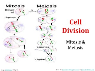
  • 51. Image: Mitosis diagram & Meiosis diagram, Marek Kultys From the Virtual Cell Biology Classroom on ScienceProfOnline.com
  • 52. Drawing and Labeling Chromosomes Sister Chromatid Replicated Uncondensed Chromosome (chromatin) Sister Chromatid Centromere Unreplicated Uncondensed Chromosome (chromatin) From the Virtual Cell Biology Classroom on ScienceProfOnline.com
  • 53. Drawing & Labeling Homologous Chromosomes Unreplicated, Condensed, Homologous Chromosomes Replicated, Condensed, Homologous Chromosomes From the Virtual Cell Biology Classroom on ScienceProfOnline.com