Procuring digital preservation CAN be quick and painless with our new dynamic...
32 an overview of animal diversity
1. 32
An Overview of Animal
Diversity
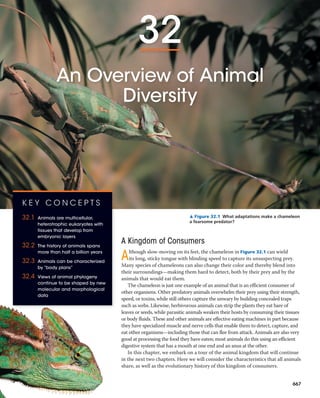
▲ Figure 32.1 What adaptations make a chameleon
a fearsome predator?
A Kingdom of Consumers
Although slow-moving on its feet, the chameleon in Figure 32.1 can wield
its long, sticky tongue with blinding speed to capture its unsuspecting prey.
Many species of chameleons can also change their color and thereby blend into
their surroundings—making them hard to detect, both by their prey and by the
animals that would eat them.
The chameleon is just one example of an animal that is an efficient consumer of
other organisms. Other predatory animals overwhelm their prey using their strength,
speed, or toxins, while still others capture the unwary by building concealed traps
such as webs. Likewise, herbivorous animals can strip the plants they eat bare of
leaves or seeds, while parasitic animals weaken their hosts by consuming their tissues
or body fluids. These and other animals are effective eating machines in part because
they have specialized muscle and nerve cells that enable them to detect, capture, and
eat other organisms—including those that can flee from attack. Animals are also very
good at processing the food they have eaten; most animals do this using an efficient
digestive system that has a mouth at one end and an anus at the other.
In this chapter, we embark on a tour of the animal kingdom that will continue
in the next two chapters. Here we will consider the characteristics that all animals
share, as well as the evolutionary history of this kingdom of consumers.
K E Y C O N C E P T S
32.1 Animals are multicellular,
heterotrophic eukaryotes with
tissues that develop from
embryonic layers
32.2 The history of animals spans
more than half a billion years
32.3 Animals can be characterized
by “body plans”
32.4 Views of animal phylogeny
continue to be shaped by new
molecular and morphological
data
667
2. 668 UNIT FIVE The Evolutionary History of Biological Diversity
ball (Figure 32.2). Following the blastula stage is the process of
gastrulation, during which the layers of embryonic tissues that
will develop into adult body parts are produced. The resulting
developmental stage is called a gastrula.
Although some animals, including humans, develop directly
into adults, the life cycles of most animals include at least one
larval stage. A larva is a sexually immature form of an animal
that is morphologically distinct from the adult, usually eats
different food, and may even have a different habitat than
the adult, as in the case of the aquatic larva of a mosquito or
dragonfly. Animal larvae eventually undergo metamorphosis,
a developmental transformation that turns the animal into a
juvenile that resembles an
adult but is not yet sexually
mature.
Though adult
animals vary widely in
morphology, the genes
that control animal de-
velopment are similar
across a broad range of
taxa. All animals have
developmental genes
that regulate the ex-
pression of other genes,
C O N C E P T 32.1
Animals are multicellular, heterotrophic
eukaryotes with tissues that develop
from embryonic layers
Listing features shared by all animals is challenging, as there
are exceptions to nearly every criterion we might select.
When taken together, however, several characteristics of
animals sufficiently describe the group for our discussion.
Nutritional Mode
Animals differ from both plants and fungi in their mode of
nutrition. Plants are autotrophic eukaryotes capable of gen-
erating organic molecules through photosynthesis. Fungi are
heterotrophs that grow on or near their food and that feed
by absorption (often after they have released enzymes that
digest the food outside their bodies). Unlike plants, animals
cannot construct all of their own organic molecules, and so,
in most cases, they ingest them—either by eating other liv-
ing organisms or by eating nonliving organic material. But
unlike fungi, most animals feed by ingesting their food and
then using enzymes to digest it within their bodies.
Cell Structure and Specialization
Animals are eukaryotes, and like plants and most fungi,
animals are multicellular. In contrast to plants and fungi,
however, animals lack the structural support of cell walls.
Instead, proteins external to the cell membrane provide
structural support to animal cells and connect them to one
another (see Figure 6.28). The most abundant of these pro-
teins is collagen, which is not found in plants or fungi.
The cells of most animals are organized into tissues,
groups of similar cells that act as a functional unit. For ex-
ample, muscle tissue and nervous tissue are responsible for
moving the body and conducting nerve impulses, respectively.
The ability to move and conduct nerve impulses underlies
many of the adaptations that differentiate animals from plants
and fungi (which lack muscle and nerve cells). For this reason,
muscle and nerve cells are central to the animal lifestyle.
Reproduction and Development
Most animals reproduce sexually, and the diploid stage usually
dominates the life cycle. In the haploid stage, sperm and egg
cells are produced directly by meiotic division, unlike what oc-
curs in plants and fungi (see Figure 13.6). In most animal spe-
cies, a small, flagellated sperm fertilizes a larger, nonmotile egg,
forming a diploid zygote. The zygote then undergoes cleavage,
a succession of mitotic cell divisions without cell growth be-
tween the divisions. During the development of most animals,
cleavage leads to the formation of a multicellular stage called
a blastula, which in many animals takes the form of a hollow
Blastula
Gastrulation
Cross section
of gastrula
Eight-cell stage
Cross section
of blastula
Zygote
Cleavage
Cleavage
Blastocoel
Blastopore
Blastocoel
Endoderm
Archenteron
Ectoderm
In most animals,
cleavage produces
a multicellular
stage called a
blastula. The blas-
tula is typically a
hollow ball of cells
that surround a
cavity called
the blastocoel.
1
3
4
The pouch formed
by gastrulation, called the
archenteron, opens to the
outside via the blastopore.
5
The endoderm of the
archenteron develops
into the tissue
lining the animal’s
digestive tract.
6
2
Most animals also undergo
gastrulation, a process in which
one end of the embryo folds
inward, expands, and eventually
fills the blastocoel, producing
layers of embryonic tissues: the
ectoderm (outer layer) and the
endoderm (inner layer).
An eight-cell
embryo is formed
by three rounds
of cell division.
The zygote of
an animal
undergoes a
series of mitotic
cell divisions
called cleavage.
▲ Figure 32.2 Early embryonic development in animals.
3. CHAPTE R 32 An Overview of Animal Diversity 669
and many of these regulatory genes contain sets of DNA
sequences called homeoboxes (see Chapter 21). In particular,
most animals share a unique homeobox-containing family of
genes, known as Hox genes. Hox genes play important roles in
the development of animal embryos, controlling the expres-
sion of many other genes that influence morphology.
Sponges, which are among the simplest extant (living) ani-
mals, lack Hox genes. However, they have other homeobox
genes that influence their shape, such as those that regulate
the formation of water channels in the body wall, a key fea-
ture of sponge morphology (see Figure 33.4). In the ancestors
of more complex animals, the Hox gene family arose via the
duplication of earlier homeobox genes. Over time, the Hox
gene family underwent a series of duplications, yielding a ver-
satile “toolkit” for regulating development. In most animals,
Hox genes regulate the formation of the anterior-posterior
(front-to-back) axis, as well as other aspects of development.
Similar sets of conserved genes govern the development of
both flies and humans, despite their obvious differences and
hundreds of millions of years of divergent evolution.
C O N C E P T 32.2
The history of animals spans more than
half a billion years
To date, biologists have identified 1.3 million extant spe-
cies of animals, and estimates of the actual number run far
higher. This vast diversity encompasses a spectacular range
of morphological variation, from corals to cockroaches to
crocodiles. Various studies suggest that this great diversity
originated during the last billion years. For example, re-
searchers have unearthed 710-million-year-old sediments
containing the fossilized remains of steroids that today are
primarily produced by a particular group of sponges. Hence,
these fossil steroids suggest that animals had arisen by 710
million years ago.
DNA analyses generally agree with this fossil biochemical
evidence; for example, one recent molecular clock study es-
timated that sponges originated about 700 million years ago.
These findings are also consistent with molecular analyses
suggesting that the common ancestor of all extant animal
species lived about 770 million years ago. What was this
common ancestor like, and how did animals arise from their
single-celled ancestors?
Steps in the Origin of Multicellular Animals
One way to gather information about the origin of animals is
to identify protist groups that are closely related to animals.
As shown in Figure 32.3, a combination of morphological
Animals
DNA sequence data indicate that choanoflagellates
and animals are sister groups. In addition, genes for
signaling and adhesion proteins previously known only
from animals have been discovered in choanoflagellates.
3
Similar collar cells have been identified in other animals, including
cnidarians, flatworms, and echinoderms—but they have never been
observed in non-choanoflagellate protists or in plants or fungi.
2
OTHER
EUKARYOTES
Choanoflagellates
Sponges
Other animals
Individual
choanoflagellate
Collar cell
(choanocyte)
Morphologically,
choanoflagellate cells
and the collar cells (or
choanocytes) of
sponges are almost
indistinguishable.
1
▼ Figure 32.3 Three lines of evidence that
choanoflagellates are closely related to animals.
Are the data described in 3 consistent with predictions
that could be made from the evidence in 1
and 2 ? Explain.
C O N C E P T C H E C K 3 2 . 1
1. Summarize the main stages of animal development.
What family of control genes plays a major role?
2. W H AT I F ? What animal characteristics would be
needed by an imaginary plant that could chase, cap-
ture, and digest its prey—yet could also extract nutrients
from soil and conduct photosynthesis?
For suggested answers, see Appendix A.
4. and molecular evidence points to choanoflagellates as the
closest living relatives of animals. Based on such evidence,
researchers have hypothesized that the common ancestor of
choanoflagellates and living animals may have been a sus-
pension feeder similar to present-day choanoflagellates.
Scientists exploring how animals may have arisen from
their single-celled ancestors have noted that the origin of
multicellularity requires the evolution of new ways for cells to
adhere (attach) and signal (communicate) to each other. In an
effort to learn more about such mechanisms, Dr. Nicole King
(featured in the interview before Chapter 26) and colleagues
compared the genome of the unicellular choanoflagellate
Monosiga brevicollis with those of representative animals.
This analysis uncovered 78 protein domains in M. brevicollis
that were otherwise only known to occur in animals. (A
domain is a key structural or functional region of a protein.)
For example, M. brevicollis has genes that encode domains of
certain proteins (known as cadherins) that play key roles in
how animal cells attach to one another, as well as genes that
encode protein domains that animals (and only animals) use
in cell-signaling pathways.
Let’s take a closer look at the cadherin attachment pro-
teins we just mentioned. DNA sequence analyses show
that animal cadherin proteins are composed primarily of
domains that are also found in a cadherin-like protein of
choanoflagellates (Figure 32.4). However, animal cadherin
proteins also contain a highly conserved region not found
in the choanoflagellate protein (the “CCD” domain labeled
in Figure 32.4). These data suggest that the cadherin attach-
ment protein originated by the rearrangement of protein
domains found in choanoflagellates plus the incorporation
of a novel domain, the conserved CCD region. Overall, com-
parisons of choanoflagellate and animal genomes suggest
that key steps in the transition to multicellularity in animals
involved new ways of using proteins or parts of proteins that
were encoded by genes found in choanoflagellates.
Next, we’ll survey the fossil evidence for how animals
evolved from their distant common ancestor over four geo-
logic eras (see Table 25.1 to review the geologic time scale).
Neoproterozoic Era (1 Billion–542 Million
Years Ago)
Although data from fossil steroids and molecular clocks indi-
cate an earlier origin, the first generally accepted macroscopic
fossils of animals date from about 560 million years ago.
These fossils are members of an early group of soft-bodied
multicellular eukaryotes, known collectively as the Ediacaran
biota. The name comes from the Ediacara Hills of Austra-
lia, where fossils of these organisms were first discovered
(Figure 32.5). Similar fossils have since been found on other
continents. Among the oldest Ediacaran fossils that resemble
animals, some are thought to be molluscs (snails and their rel-
atives), while others may be related to sponges and cnidarians
(sea anemones and their relatives). Still others have proved
Choanoflagellate
Hydra
“CCD” domain (only
found in animals)
Fruit fly
Mouse
◀ Figure 32.4 Cadherin proteins in choanoflagellates
and animals. The ancestral cadherin-like protein of choanofla-
gellates has seven kinds of domains (regions), each represented
here by a particular symbol. With the exception of the “CCD”
domain, which is found only in animals, the domains of animal
cadherin proteins are present in the choanoflagellate cadherin-
like protein. The cadherin protein domains shown here were
identified from whole-genome sequence data; evolutionary re-
lationships are based on morphological and DNA sequence data.
1.5 cm 0.4 cm
(a) Mawsonites spriggi (b) Spriggina floundersi
▲ Figure 32.5 Ediacaran fossil animals. Fossils dating to about
560 million years ago include those resembling animals (a) with sim-
ple, radial forms and (b) with many body segments.
670 UNIT FIVE The Evolutionary History of Biological Diversity
5. CHAPTE R 32 An Overview of Animal Diversity 671
with hard, mineralized skeletons, look very different from
most living animals (Figure 32.7). Even so, paleontologists
have established that these Cambrian fossils are members
of extant animal phyla, or at least are close relatives. In
particular, most of the fossils from the Cambrian explosion
are of bilaterians, an enormous clade whose members (un-
like sponges and cnidarians) have a two-sided or bilaterally
symmetric form and a complete digestive tract, an efficient
digestive system that has a mouth at one end and an anus at
the other. As we’ll discuss later in the chapter, bilaterians in-
clude molluscs, arthropods, chordates, and most other living
animal phyla.
As the diversity of animal phyla increased during the
Cambrian, the diversity of Ediacaran life-forms declined.
What caused these trends? Fossil evidence suggests that
during the Cambrian period, predators acquired novel adap-
tations, such as forms of locomotion that helped them catch
prey, while prey species acquired new defenses, such as pro-
tective shells. As new predator-prey relationships emerged,
natural selection may have led to the decline of the soft-bod-
ied Ediacaran species and the rise of various bilaterian phyla.
Another hypothesis focuses on an increase in atmospheric
oxygen that preceded the Cambrian explosion. More plenti-
ful oxygen would have enabled animals with higher meta-
bolic rates and larger body sizes to thrive, while potentially
harming other species. A third hypothesis proposes that
genetic changes affecting development, such as the origin of
Hox genes and the addition of new microRNAs (small RNAs
involved in gene regulation), facilitated the evolution of new
0.1 mm
Bore hole
▲ Figure 32.6 Early evidence of predation. This 550-million-
year-old fossil of the animal Cloudina shows evidence of having been
attacked by a predator that bored through its shell.
1 cm
Hallucigenia fossil
(530 mya)
▲ Figure 32.7 A Cambrian seascape. This artist’s reconstruction depicts a diverse array of organisms
found in fossils from the Burgess Shale site in British Columbia, Canada. The animals include Pikaia (eel-like
chordate at top left), Marella (small arthropod swimming at left), Anomalocaris (large animal with grasping
limbs and a circular mouth), and Hallucigenia (animals with toothpick-like spikes on the seafloor and in inset).
Paleozoic era—a phenomenon
referred to as the Cambrian
explosion (see Chapter 25).
In strata formed before the
Cambrian explosion, only a
few animal phyla have been
observed. But in strata that
are 535–525 million years old,
paleontologists have found
the oldest fossils of about half
of all extant animal phyla, in-
cluding the first arthropods,
chordates, and echinoderms.
Many of these fossils, which
include the first large animals
difficult to classify, as they do not seem to be closely related
to any living animal or algal groups. In addition to these mac-
roscopic fossils, Neoproterozoic rocks have also yielded what
may be microscopic fossils of early animal embryos. Although
these microfossils appear to exhibit the basic structural or-
ganization of present-day animal embryos, debate continues
about whether these fossils are indeed of animals.
The fossil record from the Ediacaran period (635–542
million years ago) also provides early evidence of predation.
Consider Cloudina, a small animal whose body was pro-
tected by a shell resembling a series of nested cones
(Figure 32.6). Some Cloudina fossils show signs of at-
tack: round “bore holes” that resemble those formed today
by predators that drill through the shells of their prey to
gain access to the soft-bodied organisms lying within. Like
Cloudina, some other small Ediacaran animals had shells or
other defensive structures that may have been selected for by
predators. Overall, the fossil evidence indicates that the Edia-
caran was a time of increasing animal diversity—a trend that
continued in the Paleozoic.
Paleozoic Era (542–251 Million Years Ago)
Another wave of animal diversification occurred 535–
525 million years ago, during the Cambrian period of the
6. 672 UNIT FIVE The Evolutionary History of Biological Diversity
body forms. In the Scientific Skills Exercise, you can inves-
tigate whether there is a correlation between microRNAs
(miRNAs; see Figure 18.14) and body complexity in various
animal phyla. These various hypotheses are not mutually
exclusive; predator-prey relationships, atmospheric changes,
and changes in development may each have played a role.
The Cambrian period was followed by the Ordovician,
Silurian, and Devonian periods, when animal diversity con-
tinued to increase, although punctuated by episodes of mass
extinction (see Figure 25.17). Vertebrates (fishes) emerged
as the top predators of the marine food web. By 450 mil-
lion years ago, groups that diversified during the Cambrian
period began to make an impact on land. Arthropods were
the first animals to adapt to terrestrial habitats, as indicated
by fragments of arthropod remains and by well-preserved
fossils from several continents of millipedes, centipedes,
and spiders. Another clue is seen in fossilized fern galls—
enlarged cavities that fern plants form in response to
stimulation by resident insects, which then use the galls for
protection. Fossils indicate that fern galls date back at least
302 million years, suggesting that insects and plants were
influencing each other’s evolution by that time.
Vertebrates colonized land around 365 million years
ago and diversified into numerous terrestrial groups. Two
of these survive today: the amphibians (such as frogs and
salamanders) and the amniotes (reptiles, including birds,
and mammals). We will explore these groups, known col-
lectively as the tetrapods, in more detail in Chapter 34.
S C I E N T I F I C S K I L L S E X E R C I S E
Is Animal Complexity Corre-
lated with miRNA Diversity? Ani-
mal phyla vary greatly in morphology,
from simple sponges that lack tissues
and symmetry to complex vertebrates.
Members of different animal phyla
have similar developmental genes, but
the number of miRNAs varies consid-
erably. In this exercise, you will explore
whether miRNA diversity is correlated
to morphological complexity.
How the Study Was Done In the
analysis, miRNA diversity is repre-
sented by the average number of
miRNAs in a phylum (x), while mor-
phological complexity is represented
by the average number of cell types
Calculating and Interpreting Correlation Coefficients
Data from the Study
Animal Phylum i
No. of
miRNAs (xi) (xi – x) (xi – x)2
No. of Cell
Types (yi) (yi – y) (yi – y)2
(xi – x)(yi – y)
Porifera 1 5.8 25
Platyhelminthes 2 35 30
Cnidaria 3 2.5 34
Nematoda 4 26 38
Echinodermata 5 38.6 45
Cephalochordata 6 33 68
Arthropoda 7 59.1 73
Urochordata 8 25 77
Mollusca 9 50.8 83
Annelida 10 58 94
Vertebrata 11 147.5 172.5
x = a = y = a = a =
sx = sy =
miRNAs (x) and the mean number of cell types (y) and enter them in
the data table (for y, replace each x in the formula with a y). (b) Next,
calculate (xi - x) and (yi - y) for each observation, recording your
results in the appropriate column. Square each of those results to
complete the (xi - x)2
and (yi - y)2
columns; sum the results for those
columns. (c) The standard deviation, sx, which describes the varia-
tion found in the data, is calculated using the following formula:
sx =
A
1
n-1 a (xi-x)2
(d) Calculate sx and sy by substituting the results in (b) into the for-
mula for the standard deviation.
3. Next, calculate the correlation coefficient r for the variables x and y.
(a) First, use the results in 3(b) to complete the (xi - x)(yi - y) column;
sum the results in that column. (b) Now use the values for sx and sy
from 3(c) along with the results from 4(a) in the formula for r.
4. Do these data indicate that miRNA diversity and animal complexity are
negatively correlated, positively correlated, or uncorrelated? Explain.
5. What does your analysis suggest about the role of miRNA diversity in
the evolution of animal complexity?
A version of this Scientific Skills Exercise can be assigned in
MasteringBiology.
Data from Bradley Deline, University of West Georgia, and Kevin Peterson, Dartmouth
College, 2013.
(y). The researchers examined the relationship between these two vari-
ables by calculating the correlation coefficient (r). The correlation coef-
ficient indicates the extent and direction of a linear relationship between
two variables (x and y) and ranges in value between -1 and 1. When
r < 0, y and x are negatively correlated, meaning that values of y become
smaller as values of x become larger. When r > 0, y and x are positively
correlated (y becomes larger as x becomes larger). When r = 0, the vari-
ables are not correlated.
The formula for the correlation coefficient r is:
r =
1
n - 1 a (xi - x)(yi - y)
sxsy
In this formula, n is the number of observations, xi is the value of the
ith
observation of variable x, and yi is the value of the ith
observation of
variable y. x and y are the means of variables x and y, and sx and sy are
the standard deviations of variables x and y. The “a” symbol indicates
that the n values of the product (xi - x) (yi - y) are to be added together.
Interpret the Data
1. First, practice reading the data table. For the eighth observation
(i = 8), what are xi and yi? For which phylum are these data?
2. Next, we’ll calculate the mean and standard deviation for each vari-
able. (a) The mean (x) is the sum of the data values divided by n, the
number of observations: x =
a xi
n
. Calculate the mean number of
7. CHAPTE R 32 An Overview of Animal Diversity 673
C O N C E P T 32.3
Animals can be characterized by
“body plans”
Animal species vary tremendously in morphology, but
their great diversity in form can be described by a relatively
small number of major “body plans.” A body plan is a
particular set of morphological and developmental traits,
Radial symmetry.
A radial animal, such as a sea anemone
(phylum Cnidaria), does not have a left side
and a right side. Any imaginary slice
through the central axis divides the animal
into mirror images.
Bilateral symmetry.
A bilateral animal, such as a lobster
(phylum Arthropoda), has a left side
and a right side. Only one imaginary
cut divides the animal into
mirror-image halves.
(a)
(b)
▲ Figure 32.8 Body symmetry. The flowerpot and shovel are
included to help you remember the radial-bilateral distinction.
C O N C E P T C H E C K 3 2 . 2
1. Put the following milestones in animal evolution in order
from oldest to most recent: (a) origin of mammals, (b)
earliest evidence of terrestrial arthropods, (c) Ediacaran
fauna, (d) extinction of large, nonflying dinosaurs.
2. W H AT I F ? Suppose the most recent common ances-
tor of extant fungi and animals lived 1 billion years ago.
If the first fungi lived 990 million years ago, would extant
animals also have been alive at that time? Explain.
3. M A K E C O N N E C T I O N S Evaluate whether the origin of
cell-to-cell attachment proteins in animals illustrates de-
scent with modification. (See Concept 22.2.)
For suggested answers, see Appendix A.
Mesozoic Era (251–65.5 Million Years Ago)
The animal phyla that had evolved during the Paleozoic now
began to spread into new habitats. In the oceans, the first
coral reefs formed, providing other marine animals with new
places to live. Some reptiles returned to the water, leaving
plesiosaurs (see Figure 25.5) and other large aquatic preda-
tors as their descendants. On land, descent with modification
in some tetrapods led to the origin of wings and other flight
equipment in pterosaurs and birds. Large and small dinosaurs
emerged, both as predators and herbivores. At the same time,
the first mammals—tiny nocturnal insect-eaters—appeared
on the scene. In addition, as you read in Chapter 30, flowering
plants (angiosperms) and insects both underwent dramatic
diversifications during the late Mesozoic.
Cenozoic Era (65.5 Million Years Ago
to the Present)
Mass extinctions of both terrestrial and marine animals
ushered in a new era, the Cenozoic. Among the groups of
species that disappeared were the large, nonflying dinosaurs
and the marine reptiles. The fossil record of the early Ceno-
zoic documents the rise of large mammalian herbivores and
predators as mammals began to exploit the vacated ecologi-
cal niches. The global climate gradually cooled throughout
the Cenozoic, triggering significant shifts in many animal
lineages. Among primates, for example, some species in
Africa adapted to the open woodlands and savannas that
replaced many of the former dense forests. The ancestors of
our own species were among those grassland apes.
integrated into a functional whole—the living animal.
The term plan here does not imply that animal forms are
the result of conscious planning or invention. But body
plans do provide a succinct way to compare and con-
trast key animal features. They also are of interest in the
study of evo-devo, the interface between evolution and
development.
Like all features of organisms, animal body plans have
evolved over time. In some cases, including key stages in
gastrulation, novel body plans emerged early in the history
of animal life and have not changed since. As we’ll discuss,
however, other aspects of animal body plans have changed
multiple times over the course of evolution. As we explore
the major features of animal body plans, bear in mind that
similar body forms may have evolved independently in dif-
ferent lineages. In addition, body features can be lost over
the course of evolution, causing some closely related species
to look very different from one another.
Symmetry
A basic feature of animal bodies is their type of symmetry—or
absence of symmetry. (Many sponges, for example, lack sym-
metry altogether.) Some animals exhibit radial symmetry,
the type of symmetry found in a flowerpot (Figure 32.8a). Sea
anemones, for example, have a top side (where the mouth is
located) and a bottom side. But they have no front and back
ends and no left and right sides.
The two-sided symmetry of a shovel is an example of
bilateral symmetry (Figure 32.8b). A bilateral animal has
two axes of orientation: front to back and top to bottom.
Such animals have a dorsal (top) side and a ventral (bottom)
side, a left side and a right side, and an anterior (front) end
8. 674 UNIT FIVE The Evolutionary History of Biological Diversity
form structures that suspend the internal organs. Animals
with a true coelom are known as coelomates (Figure 32.9a).
Some triploblastic animals have a body cavity that is
formed from mesoderm and endoderm (Figure 32.9b).
Such a cavity is called a “pseudocoelom” (from the Greek
pseudo, false), and the animals that have one are called
pseudocoelomates. Despite its name, however, a pseudo-
coelom is not false; it is a fully functional body cavity.
Finally, some triploblastic animals lack a body cavity alto-
gether (Figure 32.9c). They are known collectively as
acoelomates (from the Greek a-, without).
and a posterior (back) end. Many animals with a bilaterally
symmetrical body plan (such as arthropods and mammals)
have sensory equipment concentrated at their anterior end,
including a central nervous system (“brain”) in the head.
The symmetry of an animal generally fits its lifestyle. Many
radial animals are sessile (living attached to a substrate) or
planktonic (drifting or weakly swimming, such as jellies, com-
monly called jellyfishes). Their symmetry equips them to
meet the environment equally well from all sides. In contrast,
bilateral animals typically move actively from place to place.
Most bilateral animals have a central nervous system that
enables them to coordinate the complex movements involved
in crawling, burrowing, flying, or swimming. Fossil evidence
indicates that these two fundamentally different kinds of sym-
metry have existed for at least 550 million years.
Tissues
Animal body plans also vary with regard to tissue organiza-
tion. Recall that tissues are collections of specialized cells
that act as a functional unit; in animals, true tissues are iso-
lated from other tissues by membranous layers. Sponges and
a few other groups lack true tissues. In all other animals, the
embryo becomes layered during gastrulation. As develop-
ment progresses, these layers, called germ layers, form the
various tissues and organs of the body. Ectoderm, the germ
layer covering the surface of the embryo, gives rise to the
outer covering of the animal and, in some phyla, to the cen-
tral nervous system. Endoderm, the innermost germ layer,
lines the pouch that forms during gastrulation (the archen-
teron) and gives rise to the lining of the digestive tract (or
cavity) and organs such as the liver and lungs of vertebrates.
Cnidarians and a few other animal groups that have only
these two germ layers are said to be diploblastic. All bilat-
erally symmetrical animals have a third germ layer, called
the mesoderm, which fills much of the space between the
ectoderm and endoderm. Thus, animals with bilateral sym-
metry are also said to be triploblastic (having three germ
layers). In triploblasts, the mesoderm forms the muscles and
most other organs between the digestive tract and the outer
covering of the animal. Triploblasts include a broad range
of animals, from flatworms to arthropods to vertebrates.
(Although some diploblasts actually do have a third germ
layer, it is not nearly as well developed as the mesoderm of
animals considered to be triploblastic.)
Body Cavities
Most triploblastic animals have a body cavity, a fluid- or
air-filled space located between the digestive tract and the
outer body wall. This body cavity is also called a coelom
(from the Greek koilos, hollow). A so-called “true” coelom
forms from tissue derived from mesoderm. The inner and
outer layers of tissue that surround the cavity connect and
(a) Coelomate
(b) Pseudocoelomate
(c) Acoelomate
Tissue layer
lining coelom
and suspending
internal organs
(from mesoderm)
Coelom
Body covering
(from ectoderm) Tissue-
filled region
(from
mesoderm)
Wall of digestive cavity
(from endoderm)
Body covering
(from ectoderm)
Body covering
(from ectoderm)
Muscle layer
(from
mesoderm)
Digestive tract
(from endoderm)
Digestive tract
(from endoderm)
Pseudocoelom
Acoelomates, such as planarians, lack a body cavity between the
digestive cavity and outer body wall.
Pseudocoelomates, such as roundworms, have a body cavity lined
by tissue derived from mesoderm and by tissue derived from
endoderm.
Coelomates, such as earthworms, have a true coelom, a body
cavity completely lined by tissue derived from mesoderm.
Ectoderm Mesoderm Endoderm
Key
▼ Figure 32.9 Body cavities of triploblastic animals. The
organ systems develop from the three embryonic germ layers.
9. CHAPTE R 32 An Overview of Animal Diversity 675
modes: protostome development or deuterostome
development. These modes can generally be distinguished
by differences in cleavage, coelom formation, and fate of the
blastopore.
Cleavage
Many animals with protostome development undergo spiral
cleavage, in which the planes of cell division are diagonal to
the vertical axis of the embryo; as seen in the eight-cell stage
of the embryo, smaller cells are centered over the grooves
between larger, underlying cells (Figure 32.10a, left). Fur-
thermore, the so-called determinate cleavage of some
animals with protostome development rigidly casts (“deter-
mines”) the developmental fate of each embryonic cell very
early. A cell isolated from a snail at the four-cell stage, for
example, cannot develop into a whole animal. Instead, after
repeated divisions, such a cell will form an inviable embryo
that lacks many parts.
In contrast to the spiral cleavage pattern, deuterostome
development is predominantly characterized by radial
cleavage. The cleavage planes are either parallel or per-
pendicular to the vertical axis of the embryo; as seen at the
eight-cell stage, the tiers of cells are aligned, one directly
above the other (see Figure 32.10a, right). Most animals
with deuterostome development also have indetermi-
nate cleavage, meaning that each cell produced by early
A body cavity has many functions. Its fluid cushions the
suspended organs, helping to prevent internal injury. In
soft-bodied coelomates, such as earthworms, the coelom
contains noncompressible fluid that acts like a skeleton
against which muscles can work. The cavity also enables
the internal organs to grow and move independently of the
outer body wall. If it were not for your coelom, for example,
every beat of your heart or ripple of your intestine would
warp your body’s surface.
Terms such as coelomates and pseudocoelomates refer to
organisms that have a similar body plan and hence belong
to the same grade (a group whose members share key bio-
logical features). However, phylogenetic studies show that
true coeloms and pseudocoeloms have been independently
gained or lost multiple times in the course of animal evolu-
tion. As shown by this example, a grade is not necessarily
equivalent to a clade (a group that includes an ancestral spe-
cies and all of its descendants). Thus, while terms such as
coelomate or pseudocoelomate can be helpful in describing
an organism’s features, these terms must be interpreted with
caution when seeking to understand evolutionary history.
Protostome and Deuterostome Development
Based on certain aspects of early development, many ani-
mals can be described as having one of two developmental
Protostome development
(examples: molluscs,
annelids)
Deuterostome development
(examples: echinoderms,
chordates)
Eight-cell stage
Spiral and determinate Radial and indeterminate
Coelom
Coelom
Archenteron
Digestive tube
Anus Mouth
Mouth Anus
BlastoporeMesoderm
Solid masses of mesoderm
split and form coelom.
Folds of archenteron
form coelom.
Mouth develops from blastopore. Anus develops from blastopore.
(a) Cleavage. In general,
protostome development
begins with spiral, determinate
cleavage. Deuterostome
development is characterized
by radial, indeterminate
cleavage.
(b) Coelom formation. Coelom
formation begins in the
gastrula stage. In protostome
development, the coelom
forms from splits in the
mesoderm. In deuterostome
development, the coelom
forms from mesodermal
outpocketings of the
archenteron.
(c) Fate of the blastopore. In
protostome development,
the mouth forms from the
blastopore. In deuterostome
development, the mouth
forms from a secondary
opening.
Eight-cell stage
MesodermBlastopore
Ectoderm
Mesoderm
Endoderm
Key
◀ Figure 32.10 A comparison
of protostome and deutero-
stome development. These are
useful general distinctions, though
there are many variations and ex-
ceptions to these patterns.
M A K E C O N N E C T I O N S Re-
view Figure 20.21. As an early
embryo, which would more likely
have stem cells capable of giving rise
to cells of any type: an animal with
protostome development or one
with deuterostome development?
Explain.
10. 676 UNIT FIVE The Evolutionary History of Biological Diversity
alive today were established. Next, we’ll examine relation-
ships among these taxa along with some remaining ques-
tions that are currently being addressed using genomic data.
The Diversification of Animals
Zoologists currently recognize about three dozen phyla of
extant animals, 15 of which are shown in Figure 32.11. Re-
searchers infer evolutionary relationships among these phyla
by analyzing whole genomes, as well as morphological traits,
ribosomal RNA (rRNA) genes, Hox genes, protein-coding
nuclear genes, and mitochondrial genes. Notice how the fol-
lowing points are reflected in Figure 32.11.
1. All animals share a common ancestor. Current evi-
dence indicates that animals are monophyletic, forming
a clade called Metazoa. All extant and extinct animal
lineages have descended from a common ancestor.
2. Sponges are basal animals. Among the extant taxa,
sponges (phylum Porifera) branch from the base of the
animal tree. Recent morphological and molecular analyses
indicate that sponges are monophyletic, as shown here.
3. Eumetazoa is a clade of animals with true tissues. All
animals except for sponges and a few others belong to
a clade of eumetazoans (“true animals”). True tissues
evolved in the common ancestor of living eumetazo-
ans. Basal eumetazoans, which include the phyla Cte-
nophora (comb jellies) and Cnidaria, are diploblastic
and generally have radial symmetry.
4. Most animal phyla belong to the clade Bilateria. Bi-
lateral symmetry and the presence of three prominent
germ layers are shared derived characters that help de-
fine the clade Bilateria. This clade contains the majority
of animal phyla, and its members are known as bilat-
erians. The Cambrian explosion was primarily a rapid
diversification of bilaterians.
5. There are three major clades of bilaterian animals.
Bilaterians have diversified into three main lineages,
Deuterostomia, Lophotrochozoa, and Ecdysozoa. With
one exception, the phyla in these clades consist entirely
of invertebrates, animals that lack a backbone; Chor-
data is the only phylum that includes vertebrates, ani-
mals with a backbone.
As seen in Figure 32.11, hemichordates (acorn worms),
echinoderms (sea stars and relatives), and chordates are
members of the bilaterian clade Deuterostomia; thus, the
term deuterostome refers not only to a mode of animal de-
velopment, but also to the members of this clade. (The dual
meaning of this term can be confusing since some organ-
isms with a deuterostome developmental pattern are not
members of clade Deuterostomia.) Hemichordates share
some characteristics with chordates, such as gill slits and a
dorsal nerve cord; echinoderms lack these characteristics.
These shared traits may have been present in the common
cleavage divisions retains the capacity to develop into a
complete embryo. For example, if the cells of a sea urchin
embryo are separated at the four-cell stage, each can form
a complete larva. Similarly, it is the indeterminate cleavage
of the human zygote that makes identical twins possible.
Coelom Formation
During gastrulation, an embryo’s developing digestive tube
initially forms as a blind pouch, the archenteron, which be-
comes the gut (Figure 32.10b). As the archenteron forms in
protostome development, initially solid masses of mesoderm
split and form the coelom. In contrast, in deuterostome de-
velopment, the mesoderm buds from the wall of the archen-
teron, and its cavity becomes the coelom.
Fate of the Blastopore
Protostome and deuterostome development often differ
in the fate of the blastopore, the indentation that dur-
ing gastrulation leads to the formation of the archenteron
(Figure 32.10c). After the archenteron develops, in most
animals a second opening forms at the opposite end of the
gastrula. In many species, the blastopore and this second
opening become the two openings of the digestive tube:
the mouth and the anus. In protostome development, the
mouth generally develops from the first opening, the blas-
topore, and it is for this characteristic that the term pro-
tostome derives (from the Greek protos, first, and stoma,
mouth). In deuterostome development (from the Greek
deuteros, second), the mouth is derived from the secondary
opening, and the blastopore usually forms the anus.
C O N C E P T C H E C K 3 2 . 3
1. Distinguish the terms grade and clade.
2. Compare three aspects of the early development of a
snail (a mollusc) and a human (a chordate).
3. W H AT I F ? Evaluate this claim: Ignoring the details
of their specific anatomy, worms, humans, and most
other triploblasts have a shape analogous to that of a
doughnut.
For suggested answers, see Appendix A.
C O N C E P T 32.4
Views of animal phylogeny continue
to be shaped by new molecular and
morphological data
As animals with diverse body plans radiated during the early
Cambrian, some lineages arose, thrived for a period of time,
and then became extinct, leaving no descendants. However,
by 500 million years ago, most animal phyla with members
11. CHAPTE R 32 An Overview of Animal Diversity 677
ANCESTRAL
PROTIST
Porifera
Ctenophora
Cnidaria
Hemichordata
Echinodermata
Chordata
770 million
years ago
680 million
years ago
670 million
years ago
Platyhelminthes
Mollusca
Brachiopoda
Rotifera
Ectoprocta
Annelida
Nematoda
Arthropoda
Metazoa
Eumetazoa
Bilateria
Deuterostomia
LophotrochozoaEcdysozoa
Acoela
▲ Figure 32.11 A phylogeny of living
animals. This phylogeny shows a leading hy-
pothesis about the relationships among selected
animal phyla. The bilaterians are divided into
three main lineages: deuterostomes, lophotro-
chozoans, and ecdysozoans. The dates of origin
identified here are based on the results of a re-
cent molecular clock study.
? Which phylum is the sister group of Bilateria
in this tree?
Apical tuft
of cilia
Lophophore
Anus
Mouth
(a)
(b)
Lophophore feeding
structures of an ectoproct
Structure of a trochophore larva
▲ Figure 32.12 Morphological char-
acteristics of lophotrochozoans.
ancestor of the deuterostome clade (and lost in the echi-
noderm lineage). As mentioned above, phylum Chordata,
the only phylum with vertebrate members, also includes
invertebrates.
Bilaterians also diversified in two major clades that are
composed entirely of invertebrates: the ecdysozoans and the
lophotrochozoans. The clade name Ecdysozoa refers to a
characteristic shared by nematodes, arthropods, and some
of the other ecdysozoan phyla that are not included in our
survey. These animals secrete external skeletons (exoskel-
etons); the stiff covering of a cricket and the flexible cuticle
of a nematode are examples. As the animal grows, it molts,
squirming out of its old exoskeleton and secreting a larger
one. The process of shedding the old exoskeleton is called
ecdysis. Though named for this characteristic, the clade was
proposed mainly on the basis of molecular data that support
the common ancestry of its members. Furthermore, some
taxa excluded from this clade by their molecular data, such
as certain species of leeches, do in fact molt.
The name Lophotrochozoa refers to two different fea-
tures observed in some animals belonging to this clade.
Some lophotrochozoans, such as ectoprocts, develop a
unique structure called a lophophore (from the Greek
lophos, crest, and pherein, to carry), a crown of ciliated
tentacles that function in feeding (Figure 32.12a). Indi-
viduals in other phyla, including molluscs and annelids,
go through a distinctive developmental stage called the
trochophore larva (Figure 32.12b)—hence the name
lophotrochozoan.
Future Directions in Animal Systematics
While many scientists think that current evidence supports
the evolutionary relationships shown in Figure 32.11, as-
pects of this phylogeny continue to be debated. Although it
can be frustrating that the phylogenies in textbooks cannot
be memorized as set-in-stone truths, the uncertainty inher-
ent in these diagrams is a healthy reminder that science
12. 32 Chapter Review
C O N C E P T 32.2
The history of animals spans more than half a billion years
(pp. 669–673)
Fossil biochemical evidence and molecular clock analyses indi-
cate that animals arose over 700 million years ago.
Genomic analyses suggest that key steps in the origin of animals
involved new ways of using proteins that were encoded by genes
found in choanoflagellates.
560 mya:
Ediacaran animals
Diversification
of mammals
535–525 mya:
Cambrian explosion
Origin and
diversification
of dinosaurs365 mya:
Early land
animals
Era
Neoproterozoic Paleozoic Ceno-
zoic
065.5251542
Millions of years ago (mya)
1,000
Mesozoic
? What caused the Cambrian explosion? Describe current hypotheses.
SUMMARY OF KEY CONCEPTS
C O N C E P T 32.1
Animals are multicellular, heterotrophic eukaryotes with
tissues that develop from embryonic layers (pp. 668–669)
Animals are heterotrophs that ingest their food.
Animals are multicellular eukaryotes. Their cells are supported
and connected to one another by collagen and other structural
proteins located outside the cell membrane. Nervous tissue and
muscle tissue are key animal features.
In most animals, gastrulation follows the formation of the
blastula and leads to the formation of embryonic tissue layers.
Most animals have Hox genes that regulate the development
of body form. Although Hox genes have been highly conserved
over the course of evolution, they can produce a wide diversity
of animal morphology.
? Describe key ways that animals differ from plants and fungi.
678 UNIT FIVE The Evolutionary History of Biological Diversity
is an ongoing, dynamic process of inquiry. We’ll conclude
with three questions that are the focus of ongoing research.
1. Are sponges monophyletic? Traditionally, sponges
were placed in a single phylum, Porifera. This view
began to change in the 1990s, when molecular studies
indicated that sponges were paraphyletic; as a result,
sponges were placed into several different phyla that
branched near the base of the animal tree. Since 2009,
however, several morphological and molecular studies
have concluded that sponges are a monophyletic group
after all, as traditionally thought and as shown in
Figure 32.11. Researchers are currently sequencing
the entire genomes of various sponges to investigate
whether sponges are indeed monophyletic.
2. Are ctenophores basal metazoans? Many research-
ers have concluded that sponges are basal metazoans
(see Figure 32.11). However, several recent studies have
placed the comb jellies (phylum Ctenophora) at the base
of the animal tree. Data that are consistent with plac-
ing sponges at the base of the animal tree include fossil
steroid evidence, molecular clock analyses, the mor-
phological similarity of sponge collar cells to the cells
of choanoflagellates (see Figure 32.3), and the fact that
sponges are one of the few animal groups that lack true
tissues (as might be expected for basal animals). Cteno-
phores, on the other hand, have true tissues and their
cells do not resemble the cells of choanoflagellates. At
present, the idea that ctenophores are basal metazoans
remains an intriguing but controversial hypothesis.
3. Are acoelomate flatworms basal bilaterians? A series
of recent molecular papers have indicated that acoelo-
mate flatworms (phylum Acoela) are basal bilaterians,
as shown in Figure 32.11. A different conclusion was
supported by a 2011 analysis, which placed acoelomates
within Deuterostomia. Researchers are currently se-
quencing the genomes of several acoelomates and species
from closely related groups to provide a more definitive
test of the hypothesis that acoelomate flatworms are
basal bilaterians. If further evidence supports this hy-
pothesis, this would suggest that the bilaterians may have
descended from a common ancestor that resembled liv-
ing acoelomate flatworms—that is, from an ancestor that
had a simple nervous system, a saclike gut with a single
opening (the “mouth”), and no excretory system.
C O N C E P T C H E C K 3 2 . 4
1. Describe the evidence that cnidarians share a more
recent common ancestor with other animals than with
sponges.
2. W H AT I F ? Suppose ctenophores are basal metazo-
ans and sponges are the sister group of all remaining
animals. Under this hypothesis, redraw Figure 32.11 and
discuss whether animals with true tissues would form a
clade.
3. M A K E C O N N E C T I O N S Based on the phylogeny in
Figure 32.11 and the information in Figure 25.11, evaluate
this statement:“The Cambrian explosion actually consists
of three explosions, not one.”
For suggested answers, see Appendix A.
13. LEVEL 3: SYNTHESIS/EVALUATION
5. EVOLUTION CONNECTION
A professor begins a lecture on animal phylogeny (as shown
in Figure 32.11) by saying, “We are all worms.” In this context,
what did she mean?
6. SCIENTIFIC INQUIRY
I N T E R P R E T T H E DATA Redraw the bilaterian portion of
Figure 32.11 for the nine phyla in the table below. Consider
these blastopore fates: protostomy (mouth develops from
the blastopore), deuterostomy (anus develops from the blas-
topore), or neither (the blastopore closes and the mouth
develops elsewhere). Depending on the blastopore fate of its
members, label each branch that leads to a phylum with P, D,
N, or a combination of these letters. What is the ancestral blas-
topore fate? How many times has blastopore fate changed over
the course of evolution? Explain.
Blastopore Fate Phyla
Protostomy (P) Platyhelminthes, Rotifera, Nematoda; most
Mollusca, most Annelida; few Arthropoda
Deuterostomy (D) Echinodermata, Chordata; most Arthropoda;
few Mollusca, few Annelida
Neither (N) Acoela
Source: A. Hejnol and M. Martindale, The mouth, the anus, and the
blastopore—open questions about questionable openings. In Animal Evolu-
tion: Genomes, Fossils and Trees, eds. D. T. J. Littlewood and M. J. Telford,
Oxford University Press, pp. 33–40 (2009).
7. WRITE ABOUT A THEME: INTERACTIONS
Animal life changed greatly during the Cambrian explosion,
with some groups expanding in diversity and others declining.
Write a short essay (100–150 words) interpreting these events
as feedback regulation at the level of the biological community.
8. SYNTHESIZE YOUR KNOWLEDGE
C O N C E P T 32.3
Animals can be characterized by“body plans” (pp. 673–676)
Animals may lack symmetry or may have radial or bilateral sym-
metry. Bilaterally symmetrical animals have dorsal and ventral
sides, as well as anterior and posterior ends.
Eumetazoan embryos may be diploblastic (two germ layers) or
triploblastic (three germ layers). Triploblastic animals with a
body cavity may have a pseudocoelom or a true coelom.
Protostome and deuterostome development often differ in pat-
terns of cleavage, coelom formation, and blastopore fate.
? Describe how body plans provide useful information yet should be
interpreted cautiously as evidence of evolutionary relationships.
C O N C E P T 32.4
Views of animal phylogeny continue to be shaped by new
molecular and morphological data (pp. 676–678)
This phylogenetic tree shows key steps in animal evolution:
CHAPTE R 32 An Overview of Animal Diversity 679
Porifera
(basal animals)
Metazoa
Eumetazoa
Bilateria(mostanimals)
Ctenophora
Cnidaria
Acoela (basal
bilaterians)
Deuterostomia
Lophotrochozoa
Ecdysozoa
True
tissues
Bilateral
symmetry
Three germ
layers
? Consider clades Bilateria, Lophotrochozoa, Metazoa, Chordata,
Ecdysozoa, Eumetazoa, and Deuterostomia. List the clades to which hu-
mans belong in order from the most to the least inclusive clade.
TEST YOUR UNDERSTANDING
LEVEL 1: KNOWLEDGE/COMPREHENSION
1. Among the characteristics unique to animals is
a. gastrulation.
b. multicellularity.
Students Go to MasteringBiology for assignments, the eText, and the
Study Area with practice tests, animations, and activities.
Instructors Go to MasteringBiology for automatically graded tutorials and
questions that you can assign to your students, plus Instructor Resources.
This organism is an animal. What can you infer about its body
structure and lifestyle (that might not be obvious from its ap-
pearance)? This animal has a deuterostome developmental pat-
tern and a lophophore. To which major clades does this animal
belong? Explain your selection, and describe when these clades
originated and how they are related to one another.
For selected answers, see Appendix A.
c. sexual reproduction.
d. flagellated sperm.
c. mesoderm.
d. true tissues.
2. The distinction between sponges and other animal phyla is
based mainly on the absence versus the presence of
a. a body cavity.
b. a complete digestive tract.
3. Which of the following was probably the least important factor
in bringing about the Cambrian explosion?
a. the emergence of predator-prey relationships
b. an increase in the concentration of atmospheric oxygen
c. the movement of animals onto land
d. the origin of Hox genes
LEVEL 2: APPLICATION/ANALYSIS
4. Based on the tree in Figure 32.11, which statement is false?
a. The animal kingdom is monophyletic.
b. Acoelomate flatworms are more closely related to echino-
derms than to annelids.
c. Sponges are basal animals.
d. Bilaterians form a clade.