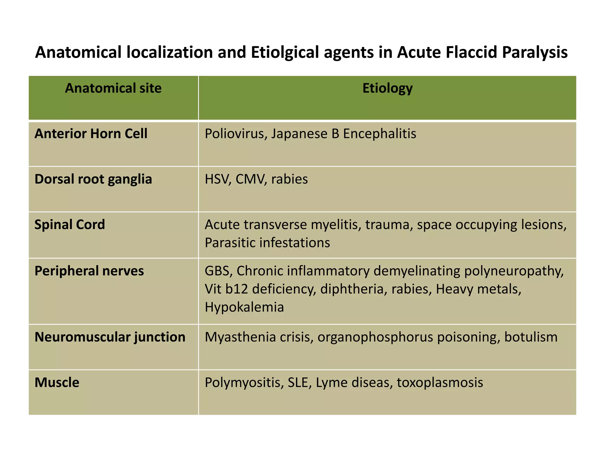
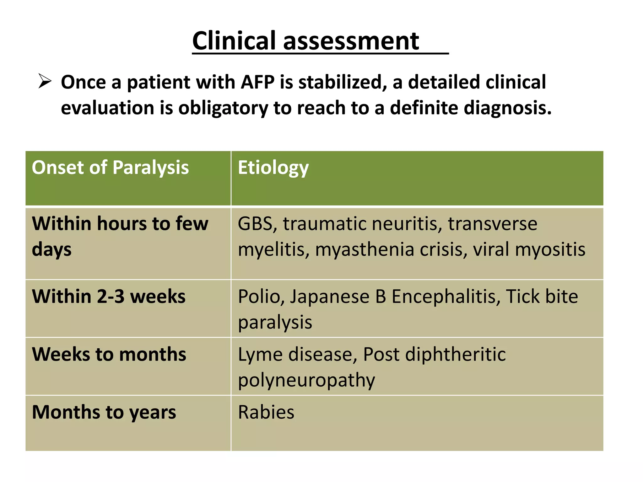
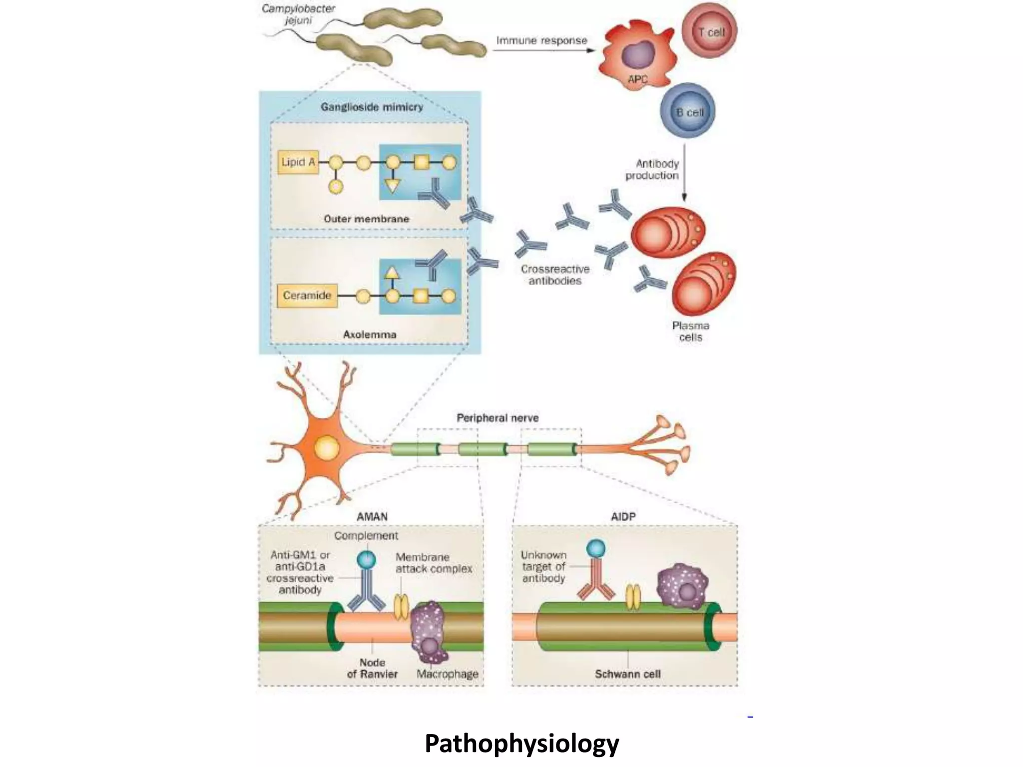
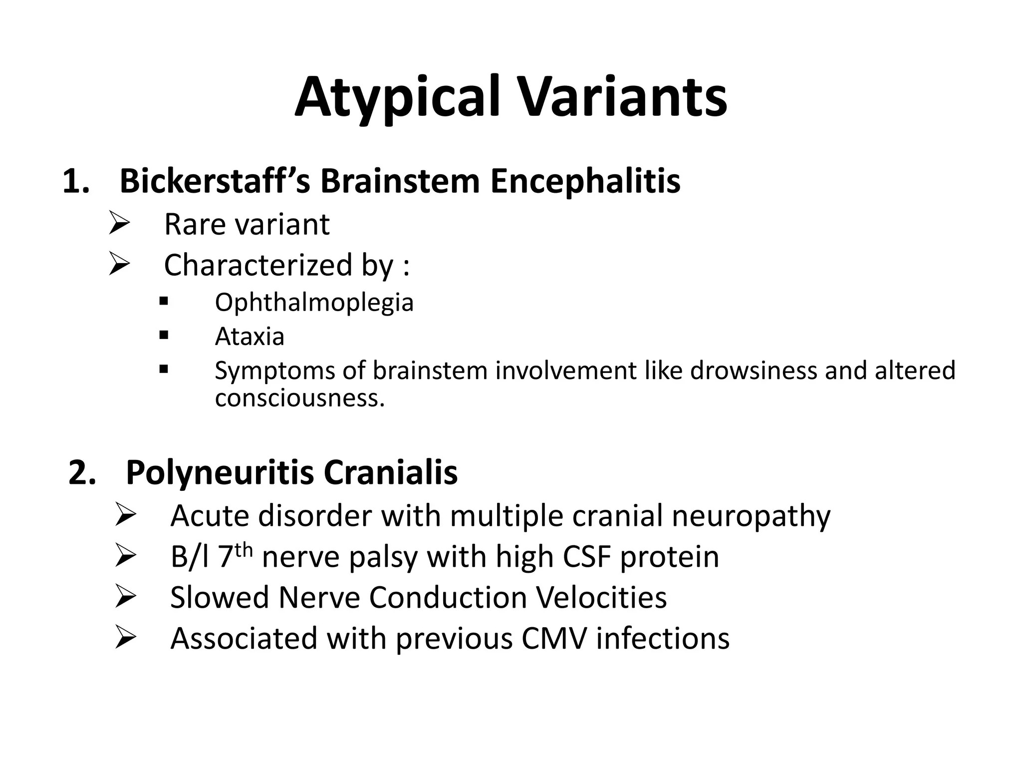
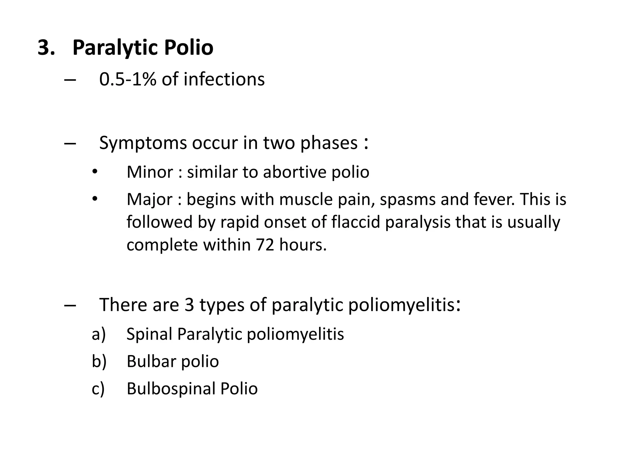
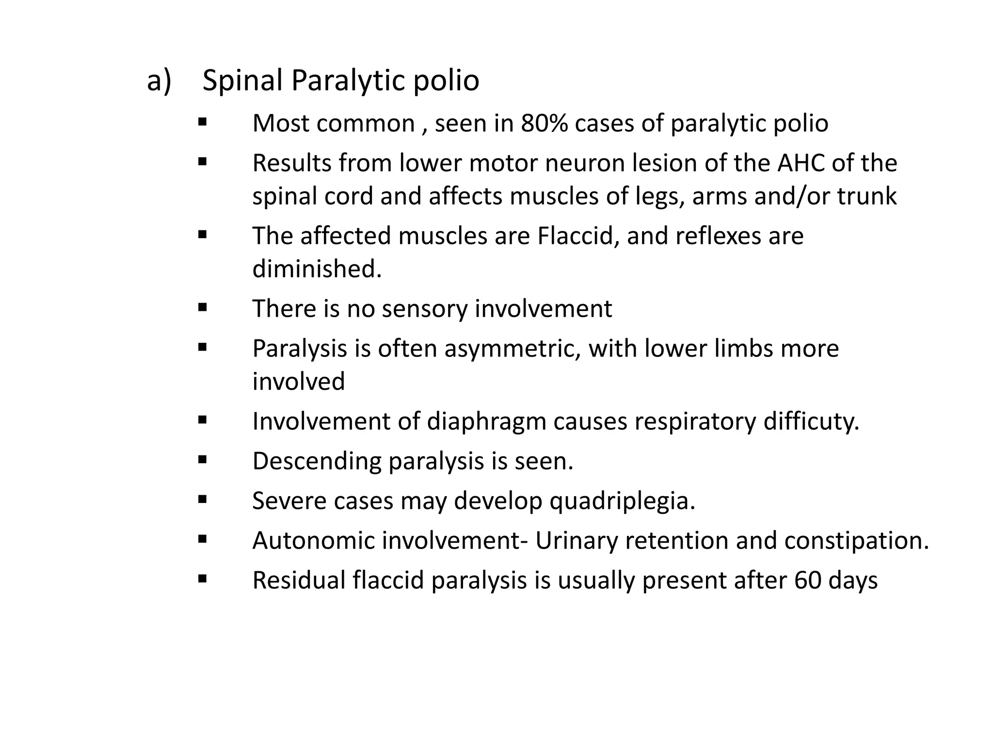
Acute Flaccid Paralysis
( GBS and Poliomyelitis )
Dr. Raghav Kakar
Introduction
• Acute flaccid paralysis (AFP) is defined as onset of
paralysis ( <4 weeks ) in a child (age < 15 years) for which
no obvious cause has been found; or, a paralysis in a
person of any age in whom polio is suspected.
• The Paralysis is associated with loss of muscle tone and loss of
reflexes.
• Reporting of all AFP cases less than 15 years of age is
mandatory in India.
• All AFP cases should be investigated within 48 hours.
Anatomical localization and Etiolgical agents in Acute Flaccid Paralysis
Anatomical site Etiology
Anterior Horn Cell Poliovirus, Japanese B Encephalitis
Dorsal root ganglia HSV, CMV, rabies
Spinal Cord Acute transverse myelitis, trauma, space occupying lesions,
Parasitic infestations
Peripheral nerves GBS, Chronic inflammatory demyelinating polyneuropathy,
Vit b12 deficiency, diphtheria, rabies, Heavy metals,
Hypokalemia
Neuromuscular junction Myasthenia crisis, organophosphorus poisoning, botulism
Muscle Polymyositis, SLE, Lyme diseas, toxoplasmosis
Initial approach to a child with AFP
A child with AFP is a medical emergency. The key steps in
management are as follows:
1. Respiratory care : It is essential to check for respiratory
involvement in the form of rapid breathing, shallow or
paradoxical respiratory efforts and use of accessory muscles.
Early support and intubation is critical in final outcome of
these patients.
2. Bulbar weakness detection and management:
Patient should be monitored for pooling of secretion,
ineffective or weak cough, nasal regurgitation of feeds.
3. Managing Cardiovascular instability :
Under ideal circumstances, ECG electrodes should be
attached to all AFP patients.
4. Rule out dyselectrolytemia and snake envenomation :
As they have treatment implications, these should be ruled
out.
Onset of Paralysis Etiology
Within hours to few
days
GBS, traumatic neuritis, transverse
myelitis, myasthenia crisis, viral myositis
Within 2-3 weeks Polio, Japanese B Encephalitis, Tick bite
paralysis
Weeks to months Lyme disease, Post diphtheritic
polyneuropathy
Months to years Rabies
 Once a patient with AFP is stabilized, a detailed clinical
evaluation is obligatory to reach to a definite diagnosis.
Clinical assessment
Progression of paralysis Disease
Maximum Deficit at onset Traumatic neuropathy
Hours to few days Poliomyelitis, Japanese B encephalitis
Up to 2-4 weeks GBS, Varicella zoster, Tick bite paralysis
Few weeks to months Lyme disease and polymyositis
Topography Disease
Assymetric involvement Poliomyelitis, Japanese B encephalitis,
Traumatic neuropathy
Symmetric involvement GBS, Transverse myelitis, rabies, botulism.
Clinical Feature Disease
Sensory features Neuropathy secondary to neurotropic viruses (Rabies,
varicella zoster)
GBS
Transverse myelitis
Lyme disease
Traumatic neuropathy
DTR-Areflexia Diseases of AHC, peripheral nerves and in spinal shock
phase of cord diseases
Bladder Bowel involvement Transverse myelitis
Fever at onset of paralysis Polio and non polio enteroviruses, other neurotropic
viruses and occasionaly with transverse myelitis, viral
myositis
Investigations
• Serum Potassium
• MRI Spine with contrast
• NCS, NST and EMG
• CSF Examination
• Serum Creatinine phosphokinase
• Urine for porphobilinogen
• Lyme’s serology
Guillain-Barre Syndrome
Introduction
• It is an acute inflammatory demyelinating
polyradiculoneuropathy.
• It is an acute diffuse post-infective disease causing
generalized paralysis and areflexia.
• The average age of incidence is 4-8 years, and rare below
2 years
• There are two major varieties:
1. AMAN : Acute Motor Axonal Neuropathy
2. AIDP : Acute Inflammatory Demyelinating Polyneuropathy
Antecedent causes
 70% cases –post infectious .
 Viral infections : EBV, Measles, Mumps, CMV, HIV , Hep B.
 20-30% cases –Campylobacter jejuni
 Mycoplasma pneumoniae
 Immunosuppression.
 Can be seen in patients with lymphoma, SLE.
Pathophysiology
• It is immune mediated with involvement of both humoral and
cell mediated mechanism.
• Triggers are the common enviornmental pathogens ( C.jejuni-
commonest, EBV, CMV).
• These triggers activate CD 4+ T-cells against variety of
endogeneously produced antigens like GQ1B, GM1 etc
• Resembelance of the triggering factor to the antigen on
peripheral nerves leads to an overzealous autoimmune
response mounted by T-cells and Macrophages
• Activated T-cells stimulate the B-cells to produce specific
antigangliosidic antibodies.
• Cytokines and chemokines released by activated T-cells may
increase capillary permeability and results in more myelin or
axonal injury.
Pathophysiology
Clinical Features
• A progressive ascending, symmetrical paralysis coming on
over hours, days to a few weeks is the hallmark of GBS.
• Pain and refusal to walk are the presenting symptoms.
• Frequent radicular involvement is manifested by the
positive straight leg raising sign. (Lasègue's sign)
• Cranial nerve involvement in 50% cases.
• Autonomic manifestations seen in one third patients
• Motor weakness:
– Begins in lower limbs and progressively involves trunk,
upper limbs, diaphragm, respiratory, pharyngeal and
laryngeal muscles.
– May also involve lower cranial nerves
– Proximal muscles involved more in limbs.
– Progression of paralysis for 4-10 days.
– Areflexia
• Higher functions:
– Consciousness is normal
• Cranial Nerves:
– 7th nerve bilateral involvement is frequently seen.
• Sensory symptoms
– No objective sensory loss.
– There may be distal impairment of vibration and position
sense in some.
Acute Motor Axonal Neuropathy
• Children ,young adults
• Seasonal variation
• Rapid in evolution
• Anti GD1a and Anti GM1 antibodies
• First attack on motor nodes of Ranvier
• Macrophage activation
• Axonal damage is variable
Acute Inflammatory Demyelinating
Polyneuropathy
• Adults > children
• Lack of seasonal variation
• Rapid recovery
• Less severe
• Antigen involved in AIDP still remains unknown
• Demyelinating
• First attack on Schwann cell surface
• Widespread myelin damage
Miller Fischer Syndrome
• Adults,children
• Uncommon
• Anti GQ 1 b anitbodies >90%
(Not seen in other forms of
GBS)
• Cause conduction block
• Pupillary paralysis
• Distal Paresthesia seen.
• Only 5% GBS
• 25-50% show limb weakness (MFS-
GBS overlap syndrome)
Atypical Variants
1. Bickerstaff’s Brainstem Encephalitis
 Rare variant
 Characterized by :
 Ophthalmoplegia
 Ataxia
 Symptoms of brainstem involvement like drowsiness and altered
consciousness.
2. Polyneuritis Cranialis
 Acute disorder with multiple cranial neuropathy
 B/l 7th nerve palsy with high CSF protein
 Slowed Nerve Conduction Velocities
 Associated with previous CMV infections
3. Acute Motor and Sensory Axonal Neuropathy
 Similar to AMAN with sensory and bulbar involvement
and severe course.
4. Pharyngo-cervical-Brachial variant
 Paralysis involves bulbar, neck and upper limb muscles
5. Locked-in state
 Patient may look comatose due to severe quadriplegia
and pan-cranial neuropathy
 Patient doesnt respond to stimuli
6. Acute Pandysautonomia pure sensory variant
 Predominant autonomic or sensory involvement.
Lab Features
CSF :
• Normal pressure
• Xanthochromic appearance
• Raised CSF protein 1-10 g/l (100-1000mg/dl)
• ALBUMINOCYTOLOGICAL DISSOSCIATION
– Cell count normal, no pleocytosis
– Proteins : elevated, twice the normal limit
• Glucose : Normal
• Culture : Sterile
Nerve conduction velocity studies:
• s/o either axonal or demyelinating neuropathy.
• Axonal neuropathy has worst prognosis
Electromyography:
• Evidence of acute denervation is present
Sural Nerve Biopsy may show:
• Segmental demyelination
• Focal inflammation
• Wallerian degeneration
Serum Creatinine kinase may be normal to elevated
Asbury criteria for diagnosis of GBS
REQUIRED :
1. Areflexia
2. Include in differential and rule out other causes
3. Duration < 4 weeks
4. Progressive weakness of 2 or more limbs due to neuropathy
Features supportive of diagnosis:
1. Afebrile
2. Involvement:
– Mild sensory involvement
– Facial or other cranial nerve involvement
3. Demyelinating electrophysiologic evidence
4. Protein in CSF High + Low WBC (Albumino-cytologic dissociation)
5. Symmetrical distribution relatively
Features casting doubt on the diagnosis
1. Asymmetrical weakness
2. Persistent bladder and bowel dysfunction
3. Bladder or bowel dysfunction at onset
4. >50 mononuclear leukocytes/mm3 or presence of
polymorphonuclear leukocytes in CSF
5. Distinct sensory level.
Features that rule out the diagnosis
1. Hexacarbon abuse
2. Abnormal porphyrin metabolism
3. Recent diphtheria infection
4. Lead intoxication
Treatment
• Supportive care : Ventilation, physiotherpary
• IVIG :
– best given within 3-4 days of onset of weakness.
– Indications:
• Acute GBS of less than 2 weeks
• Inability to walk unaided
• Bulbar weakness
– Dose : 0.4mg/kg/day for 5 days or 1g/kg/day for 2
days
• Plasmapharesis
– In cases of rapid progression
– Not much benificial after 3 weeks
– Dose : 200-250 ml/kg/bodyweight of plasma is
removed on alternate days in 4-6 sittings for 8-12
days
– Complications:
• CVS complication
• Dysautonomia
• Hepatitis and AIDS
• Prevention of secondary infections
• Plasmapheresis f/b IVIG is not superior to IVIG or PE
alone
• Oral steroids (methyl prednisolone) may delay recovery,
however a short course is very useful in management of
radicular pain
• Some patients show worsening after initial treatment---
the so called Treatment-related clinical fluctuations.
– If these fluctuations occur more than 3 times or if the recovery
has not occurred 9 weeks into illness, one must consider Acute
variant of Chronic Inflammatory Demyelinating Polyneuropathy
(A-CIDP)
Treatment-related clinical fluctuations :
– Worsening of at least one grade on the GBS disability scale after
initial stabilization or improvement within first 8 weeks after
treatment.
– In a recent study, more TRF were observed in the 2 day IVIg
treatment group (1g/kg for 2 days) than in the 5 day treatment
group ( 0.4g/kg for 5 days )
– TRFs provide evidence that the treatment in a specific patient is
effective, although not lasting long enough, and that the patient
will probably respond again after repeating the same treatment.
– It is rational to retreat the patient with IVIg or PE.
Treatments other than PE and IVIg
– Brain derived neurotrophic factor.
– Interferon Beta-1a
According to cochrane review, the numbers in the study of
above mentioned modalities were too small and larger
sequential RCTs might be more promising
– Tripterygium polyglycoside—a chinese herbal medicine
It showed a significant improvement in disabillity scale 8
weeks after onset of symptoms.
Prognosis
• The clinical course is usually benign, and recovery begins
within 2-3 weeks.
• Some patient might be left with residual weakness.
• Tendon reflexes are usually last to recover.
• Improvement usually follows a gradient opposite the direction
of involvement.
• 3 clinical features are predictive of poor outcome:
– Cranial nerve involvement
– Intubation and
– Max disability at the time of presentation
• Easy fatigue is one of the most common chronic symptom.
• Axonal form of GBS has slower recovery, some requiring upto
years.
• 10% may have persistent neurological symptoms
• 5% may show recurrence.
Poliomyelitis
Introduction
• Greek poliós= "grey", myelós= marrow, and the suffix
- itis= inflammation
• First described by British physician Micheal Underwood in
1799 referring to it as "debility of the lower extremities.“
• A viral infection most often recognized by acute onset of
flaccid paralysis.
• Primarily an infection of human alimentary tract, but may
infect CNS in very small no. (i.e <1%)
• Infection results in a spectrum of clinical manifestations.
Problem statement
World
• A worldwide problem in pre vaccinationera
• With the wide use of polio vaccine from 1954, it is being
eliminated from most of the developed countries
• In 1988 WHA resolved to eradicate the disease globally, GPEI
was estabilished.
• Polio cases have decreased by over 99% since 1988, from an
estimated 3,50,000 cases to only less than 100 in 2014
globally
• In 2016, only 3 countries in the world remain polio endemic.
• A total of 35 cases of paralytic poliomyelitis due to WPV Type
1 were reported in 2016 from these 3 countries
• Failure to eradicate polio from these 3 countries could result
in as many as 200,000 new cases every year within 10 yrs, all
over the world.
India
• In India, Vaccination against polio started in 1978 with
extended programme on immunization.
• Pulse Polio Immunization launched in 1995.
• In 2009, India had half the number of polio cases in the world.
• By 2011, in less than two years’ time, India broughtpolio infections
to the zero level.
• India’s last reported polio case was a 2-year-old girl in the Howrah
district of West Bengal, on 13 January 2011.
• India removed from list of polio-endemic countries in 2012.
• South East Asia region was declared polio free on March 27th
2014.
Epidemiology
• Poliovirus: belongs to “Picorna” viruses which are small RNA-
containing viruses. Non Enveloped.
• Three serotypes- 1, 2 & 3 giving no cross immunity
• Human is the only reservoir. It multiplies in the intestine and
spreads via faeco-oral route.
• The maximum excretion of virus occurs just before the onset
of paralysis and during the first 2 weeks after the onset of
paralysis.
• However, virus is intermittently excreated for upto 2 months
after infection
Etiopathogenesis
• Three antigenically distinct serotypes. Type1, 2 and 3
• Polio virus can remain active for several days at room
temperature and can be stored indefinitely at -20 degree C
• Inactivated by Heat, Chlorine and UV rays.
• Most frequent cause of epidemic polio is Type 1 followed by
type 3.
• The primary site of replication is small intestine and regional
lymph nodes.
• Poliovirus accesses the CNS via peripheral nerves and
primarily infects motor neuron cells in the spinal cord (the
anterior horn cells) and the medulla oblongata.
• Infants born to mothers with few antibodies are protected for
a few weeks.
• Active immunity after natural infection ( including inapparent
and mild infection ) is probably lifetime but protects against
the infecting serotype only.
HOST FACTORS
Age:
– All age groups; children(6 MONTHS TO 3 YEARS most
susceptible)
– more than 95% reported in infancy and childhood with over
50% ofthem in infancy.
Sex:
– no sex ratio differences, but in some countries, males are
infectedmore frequently than females in a ratio 3:1.
Risk factors:
– Fatigue, trauma, im injections, tonsillectomy, immunizing agents
like alum containing DPT vaccine and excessive muscular
exercise…
Clinical manifestations
1. Abortive polio :
– Occurs in 4-8% of infections
– Low grade fever, sore throat
– Vomiting, abdominal pain
– Loss of weigh, malaise
– Recovery is complete and rapid.
– There is no paralysis
2. Non Paralytic Polio :
– 1-2% population
– Non specific illness
– Pre paralytic polio
• Back stiffness,
• Muscle tenderness,
• Head lag and Nuchal rigidity
• Nuchal- Spinal signs present
• Sensory system normal
• Child fully conscious
• IM injection, physical activities should be avoided
• Reflexes : In early stages DTR may be exaggerated. Depression of
DTRs is an indication of impending paralysis
– Recovery within 2-10 days
3. Paralytic Polio
– 0.5-1% of infections
– Symptoms occur in two phases :
• Minor : similar to abortive polio
• Major : begins with muscle pain, spasms and fever. This is
followed by rapid onset of flaccid paralysis that is usually
complete within 72 hours.
– There are 3 types of paralytic poliomyelitis:
a) Spinal Paralytic poliomyelitis
b) Bulbar polio
c) Bulbospinal Polio
a) Spinal Paralytic polio
 Most common , seen in 80% cases of paralytic polio
 Results from lower motor neuron lesion of the AHC of the
spinal cord and affects muscles of legs, arms and/or trunk
 The affected muscles are Flaccid, and reflexes are
diminished.
 There is no sensory involvement
 Paralysis is often asymmetric, with lower limbs more
involved
 Involvement of diaphragm causes respiratory difficuty.
 Descending paralysis is seen.
 Severe cases may develop quadriplegia.
 Autonomic involvement- Urinary retention and constipation.
 Residual flaccid paralysis is usually present after 60 days
b) Bulbar Polio
 2% of paralytic polio cases
 Due to weakness of motor part of cranial nerve.
 9th and 10th CN involvement :Inability to swallow, Nasal
twang, Nasal regurgitation, pooling of saliva, hoarseness of
voice
 Irregular and shaloow respiration
 Cardiac arrhythmias
 Pulse is rapid, weak and thready.
 Patient becomes restless, confused, delirious and comatose.
c) Bulbospinal Polio
 20% of paralytic cases
 Combination of both spinal and bulbar component.
 Residual Paralysis
 Acute phase of illness subsides by 4 weeks and the
recovery begins in paralysed muscles
 The extent of recovery is variable depending upon extent
of damage caused to neurons.
 Max recovery takes place in the first 6 months but slow
recovery continues till 18-24 months.
 After 2 years, no more recovery is expected and the child
is said to have “Post Polio Residual Paralysis” which
remains as such through out the life
Post Polio Syndrome
 Clinical deterioration occurs in survivors after several
decades.
 H/o recent muscle weakness, poor endurance, fatigue and
joint pain.
 Attributed to combination of ageing process and distal
muscle degeneration due to increased metabolic demands
Complications
• Respiratory complications: pneumonia, pulmonary edema
• Cardiovascular complications: myocarditis, cor pulmonale.
• Late complications: soft tissue and bone deformities,
osteoporosis, and chronic distension of the colon.
• Case fatality: varies from 1% to 10% according to the form of
disease (higher in bulbar), complications and age ( fatality
increases with age).
Diagnosis and Lab testing
• Laboratory studies critical to rule out or confirm the diagnosis
of paralytic poliomyelitis.
• Virus isolation
– The likelihood of poliovirus isolation is highest from stool
specimens,
– Intermediate from pharyngeal swabs, and very low from blood or
spinal fluid.
• Serologic testing
– A four-fold titre rise between the acute and convalescent
– specimens suggests poliovirus infection.
Treatment
• The aim of the treatment is to promote recovery and to
minimize residual paralysis and disability.
• Children with bulbospinal polio and respiratory paralysis
would require hospitalization
• Children with isolated limb paralysis should be advised
complete bed rest
• Proper positioning
– The child should be made to lie on a firm bed and maintain limbs in
neutral position.
– Trunk and hip should be straight with slight flexion at the knees and
feet at right angle at ankle.
– External rotation should be prevented.
• Pain Relief
– Warm moist fomentation should be given.
– Massage and IM injections should be avoided during the acute phase
of illness
• Bowel and Bladder
– Laxatives should be avoided
– Intermittent catheterization may be done.
• Physiotherapy to be started once the acute phase subsides.
• Contractures and fixed deformities may require orthopedic
surgery, to be done after 2 years.
Prognosis
• The outcome of inapperent and abortive polio is relatively
good.
• It is bad for older children and those with sudden onset of
illness with high fever.
• In severe bulbar polio, mortality may be as highg as 60%,
whereas in less severe bulbar or spinal polio, mortality varies
from 5-10%
• Type-1 has the greatest propensity for natural poliomyelitis,
whereas Type-3 has a predilection for VDPV.
ACTIVE IMMUNIZATION
 Oral (Sabin) Polio Vaccine
• 1961 by Albert Sabin, Live attenuated vaccine.
• Provide both humoral and Mucosal (Gut) immunity .
• Mucosal intestinal immunity prevent infection with wild polio
virus.
• Intestinal immunity is the main reason why mass campaigns
with OPV can rapidly stop person to person transmission of
wild polio virus.
• Immunity probably lifelong
• Shed in stool for up to 6 weeks following vaccination
Risks associated with use of OPV
 Vaccine Associated Paralytic Poliomyelitis
 Those cases of AFP, which have residual weakness 60 days after onset
of paralysis and from whose stool samples, vaccine related poliovirus
is isolated.
 In some recipient of OPV, there is genetic change ( <1%), in the VP1
gene of vaccine virus.
 This minor change turns the vaccine virus virulent.
 Causes paralysis in recipient (recipient VAPP) or among unimmunized
close contacts (contact VAPP)
 1 case of VAPP occurs after 2.3 million first doses, and after 12 million
subsequent doses.
 Vaccine derived Poliovirus
 There is a greater genetic change (1-15%) in the VP1 gene
of the vaccine virus.
 It has potential for human infection and paralysis.
Types of VDPV :
1) cVDPV (Circulating vaccine derived polio virus ) : A cVDPV is
associated with sustained person to person transmission and is
circulating in the community under conditions of low population
immunity, with evidence of causing 2 or more paralytic cases.
2) iVDPV (Immununo deficiency –related vaccine –derived
polio virus) reported in immunodeficient patients who have
prolonged infections after exposure to OPV.
3) aVDPV (ambiguous vaccine derived polio virus) currently
have unclassified source (i.e., a single isolate from a healthy or
non- immunodeficient person; environmental isolates without
an associated AFP case).
• Among these 3 types, cVDPV causes sustained circulation. Due
to the risks of this OPV must be phased out to secure a lasting
polio- free world.
AFP Surveillance Bulletin – India. Report for week 46, ending 18 November
2017
 Inactivated (Salk) vaccine
• Developed in 1955 by Dr Jonas Salk.
• Consists of inactivated polio strains of all three types.
• Excellent humoral immunity.
• Gives Mucosal protection but not mucosal immunity.
• No risk of VAPP or VDPV.
• 0.5 ml IM or SC or a fractional dose of 0.1mlIntradermal.
Vaccination Schedule
 National Immunization Schedule:
– bOPV at birth,6-10-14 weeks followed by booster at 15-
18month.
– IPV at 14 week (0.5ml)
– In some states Fractional doses of IPV at 6 and 14 weeks instead
of IM dose
 The IAP Schedule : ‘Sequential IPV-OPV schedule’
– bOPV at birth
– IPV at 6-10-14 weeks
– bOPV at 6 and 9 month
– IPV at 15-18 month
– bOPV at 5 year
Pulse Polio Immunization (PPI) Program
• Following the Global Polio Eradication Initiative of WHO in
1988, GOI conducted the first round of PPI consisting of 2
immunization days 6 wks apart on 9th dec 1995 and 20th jan
1996.
• Encounters and challenges faced by the innovators:
a) The size of the campaign (6.5 lakh polio booths; ~125 million
children to be administered on each immunization day; cold
chain and vaccine management)
b) The attitudinal diversity (diverse religious, socioeconomic and
cultural background)
c) Management of Human Resources (Doctors, Nurses, Health,
Anganawadi Workers, Volunteers, NGOs)
Strategy adopted:
The basic strategy for eradicating polio consisted of:
a) Immunizing every child below 1 year with at least 3 doses of
OPV.
b) National Immunization Days during which every child below 5
years gets 2 additional doses of OPV on 2 days separated by 4
to 6 weeks.
c) Surveillance of AFP to identify all reservoirs of wild poliovirus
transmission.
d) Extensive house-to-house immunization mopping -up
campaigns in the final stages where wild poliovirus
transmission persists.
Key Points :
• Targeted all children upto 3 yrs later on WHO increased the
age upto 5 yrs.
• PPI occurs in two rounds about 4-6 wks apart during low
transmission season of polio , i.e. Between nov to feb.
• These doses are extra dose which supplements and do not
replace the doses received during immunization services.
• There is no minimum interval between PPI and scheduled
OPV doses
Polio Eradication & Endgame Strategic Plan 2013-2018
• On 26 May 2012, the World Health Assembly declared the
completion of poliovirus eradication to be a “programmatic
emergency for global public health” and called for the
development of comprehensive polio endgame strategy.
• In response to this directive, the GPEI developed Polio
Eradication & Endgame Strategic Plan (PEESP) 2013-2018.
• This strategy shall prevent circulating VDPV by augmenting
the immunity induced by earlier doses of trivalent OPV.
• By 2019 there shall be complete cessation of OPV
Three distinct steps of polio endgame strategy
.
Switch tOPV
to bOPV
Withdraw
bOPV &
routine OPV
use
Before
end 2015
2016
2019-2020
Introduce
atleast one
dose of IPV
in RI
On going strengthening of routine immunization
.
AFP-SURVEILLANCE
• Nationwide AFP (acute flaccid paralysis) surveillance is the
gold standard for detecting cases of poliomyelitis.
• Surveillance identifies new cases and detects importation of
wild poliovirus
The four steps of surveillance are:
1. Finding and reporting children with AFP
2. Transporting stool samples for analysis
3. Isolating and identifying poliovirus in the laboratory
4. Mapping the virus to determine the origin of the virus
strain.
Finding and reporting children with acute flaccid
paralysis (AFP) surveillance:
• The first links in the surveillance chain are staff in all health
facilities- from district health centres to large hospitals.
• They must promptly report every case of AFP in any child under 15
years of age.
• The number of AFP cases reported each year is used as an
indicators of a country’s ability to detect polio-even in countries
where disease no longer occurs.
• A country’s surveillance system needs to be sensitive enough to
detect at least one case of AFP for every 100,000 children under
15- even in absence of polio.
Transporting stool samples for analysis
• Two stool specimens should be collected at an interval of 24 to
48 hours apart and within 14 days of onset of paralysis.
• However, when AFP cases are seen late (i.e. greater than 2
weeks after paralysis onset), stool specimen may be collected up
to 60 days after onset of paralysis.
• The specimen should be placed in a clean container such as
wide mouth plastic or glass bottle with screw on cap.
• It need not be autoclaved, but should be cleaned.
• At least ‘one thumb sized’ 8 gm of stool is required.
• Stool sample should be adequate and in good condition
accompanied by all details as required by laboratories
Isolating Poliovirus
• If poliovirus is isolated the next step is to distinguish between
wild and vaccine related.
• If wild polio virus is isolated then identify which of the two
surviving types of wild virus is involved.
Mapping The Virus
• Once wild poliovirus has been identified further tests are
carried out to determine where the strain may have
originated.
• By determining the genetic makeup of virus, wild virus can
be compared to others and classified into genetic families
which cluster in defined geographic areas.
• When polio has been pinpointed to a precise geographical
area, it is possible to identify the source of importation of
poliovirus- both long range and cross border.
Environmental Surveillance
• Environmental surveillance involves testing sewage or other
environmental samples for the presence of poliovirus.
• Environmental surveillance often confirms wild poliovirus
infections in the absence of cases of paralysis.
• Systematic environmental sampling provides important
supplementary surveillance data.
• Ad-hoc environmental surveillance elsewhere (especially in
polio-free regions) provides insights into the international
spread of poliovirus.
Surveillance Indicators
.
.
The Ministry of Health has sponsored the formation of a
network of laboratories some of which are located at:
– Sanjay Gandhi Postgraduate Institute, Lucknow
– National Institute of Communicable Diseases, New Delhi
– ERC, Mumbai
– NIV, Bengaluru
– BJMC, Ahmedabad
– Enterovirus Research Institute, Kasauli
– Pasteur Institute of India, Coonoor
Albert Sabin Jonas Salk
George Guillain Jean Barre Charles Miller Fischer
Jean Baptiste Landry
Thank You.

Acute flaccid paralysis and Surveillance 2018

  • 1.
    Acute Flaccid Paralysis (GBS and Poliomyelitis ) Dr. Raghav Kakar
  • 2.
    Introduction • Acute flaccidparalysis (AFP) is defined as onset of paralysis ( <4 weeks ) in a child (age < 15 years) for which no obvious cause has been found; or, a paralysis in a person of any age in whom polio is suspected. • The Paralysis is associated with loss of muscle tone and loss of reflexes. • Reporting of all AFP cases less than 15 years of age is mandatory in India. • All AFP cases should be investigated within 48 hours.
  • 3.
    Anatomical localization andEtiolgical agents in Acute Flaccid Paralysis Anatomical site Etiology Anterior Horn Cell Poliovirus, Japanese B Encephalitis Dorsal root ganglia HSV, CMV, rabies Spinal Cord Acute transverse myelitis, trauma, space occupying lesions, Parasitic infestations Peripheral nerves GBS, Chronic inflammatory demyelinating polyneuropathy, Vit b12 deficiency, diphtheria, rabies, Heavy metals, Hypokalemia Neuromuscular junction Myasthenia crisis, organophosphorus poisoning, botulism Muscle Polymyositis, SLE, Lyme diseas, toxoplasmosis
  • 4.
    Initial approach toa child with AFP A child with AFP is a medical emergency. The key steps in management are as follows: 1. Respiratory care : It is essential to check for respiratory involvement in the form of rapid breathing, shallow or paradoxical respiratory efforts and use of accessory muscles. Early support and intubation is critical in final outcome of these patients. 2. Bulbar weakness detection and management: Patient should be monitored for pooling of secretion, ineffective or weak cough, nasal regurgitation of feeds.
  • 5.
    3. Managing Cardiovascularinstability : Under ideal circumstances, ECG electrodes should be attached to all AFP patients. 4. Rule out dyselectrolytemia and snake envenomation : As they have treatment implications, these should be ruled out.
  • 6.
    Onset of ParalysisEtiology Within hours to few days GBS, traumatic neuritis, transverse myelitis, myasthenia crisis, viral myositis Within 2-3 weeks Polio, Japanese B Encephalitis, Tick bite paralysis Weeks to months Lyme disease, Post diphtheritic polyneuropathy Months to years Rabies  Once a patient with AFP is stabilized, a detailed clinical evaluation is obligatory to reach to a definite diagnosis. Clinical assessment
  • 7.
    Progression of paralysisDisease Maximum Deficit at onset Traumatic neuropathy Hours to few days Poliomyelitis, Japanese B encephalitis Up to 2-4 weeks GBS, Varicella zoster, Tick bite paralysis Few weeks to months Lyme disease and polymyositis Topography Disease Assymetric involvement Poliomyelitis, Japanese B encephalitis, Traumatic neuropathy Symmetric involvement GBS, Transverse myelitis, rabies, botulism.
  • 8.
    Clinical Feature Disease Sensoryfeatures Neuropathy secondary to neurotropic viruses (Rabies, varicella zoster) GBS Transverse myelitis Lyme disease Traumatic neuropathy DTR-Areflexia Diseases of AHC, peripheral nerves and in spinal shock phase of cord diseases Bladder Bowel involvement Transverse myelitis Fever at onset of paralysis Polio and non polio enteroviruses, other neurotropic viruses and occasionaly with transverse myelitis, viral myositis
  • 9.
    Investigations • Serum Potassium •MRI Spine with contrast • NCS, NST and EMG • CSF Examination • Serum Creatinine phosphokinase • Urine for porphobilinogen • Lyme’s serology
  • 10.
  • 11.
    Introduction • It isan acute inflammatory demyelinating polyradiculoneuropathy. • It is an acute diffuse post-infective disease causing generalized paralysis and areflexia. • The average age of incidence is 4-8 years, and rare below 2 years • There are two major varieties: 1. AMAN : Acute Motor Axonal Neuropathy 2. AIDP : Acute Inflammatory Demyelinating Polyneuropathy
  • 12.
    Antecedent causes  70%cases –post infectious .  Viral infections : EBV, Measles, Mumps, CMV, HIV , Hep B.  20-30% cases –Campylobacter jejuni  Mycoplasma pneumoniae  Immunosuppression.  Can be seen in patients with lymphoma, SLE.
  • 13.
    Pathophysiology • It isimmune mediated with involvement of both humoral and cell mediated mechanism. • Triggers are the common enviornmental pathogens ( C.jejuni- commonest, EBV, CMV). • These triggers activate CD 4+ T-cells against variety of endogeneously produced antigens like GQ1B, GM1 etc
  • 14.
    • Resembelance ofthe triggering factor to the antigen on peripheral nerves leads to an overzealous autoimmune response mounted by T-cells and Macrophages • Activated T-cells stimulate the B-cells to produce specific antigangliosidic antibodies. • Cytokines and chemokines released by activated T-cells may increase capillary permeability and results in more myelin or axonal injury.
  • 15.
  • 16.
    Clinical Features • Aprogressive ascending, symmetrical paralysis coming on over hours, days to a few weeks is the hallmark of GBS. • Pain and refusal to walk are the presenting symptoms. • Frequent radicular involvement is manifested by the positive straight leg raising sign. (Lasègue's sign) • Cranial nerve involvement in 50% cases. • Autonomic manifestations seen in one third patients
  • 17.
    • Motor weakness: –Begins in lower limbs and progressively involves trunk, upper limbs, diaphragm, respiratory, pharyngeal and laryngeal muscles. – May also involve lower cranial nerves – Proximal muscles involved more in limbs. – Progression of paralysis for 4-10 days. – Areflexia
  • 18.
    • Higher functions: –Consciousness is normal • Cranial Nerves: – 7th nerve bilateral involvement is frequently seen. • Sensory symptoms – No objective sensory loss. – There may be distal impairment of vibration and position sense in some.
  • 19.
    Acute Motor AxonalNeuropathy • Children ,young adults • Seasonal variation • Rapid in evolution • Anti GD1a and Anti GM1 antibodies • First attack on motor nodes of Ranvier • Macrophage activation • Axonal damage is variable
  • 20.
    Acute Inflammatory Demyelinating Polyneuropathy •Adults > children • Lack of seasonal variation • Rapid recovery • Less severe • Antigen involved in AIDP still remains unknown • Demyelinating • First attack on Schwann cell surface • Widespread myelin damage
  • 21.
    Miller Fischer Syndrome •Adults,children • Uncommon • Anti GQ 1 b anitbodies >90% (Not seen in other forms of GBS) • Cause conduction block • Pupillary paralysis • Distal Paresthesia seen. • Only 5% GBS • 25-50% show limb weakness (MFS- GBS overlap syndrome)
  • 22.
    Atypical Variants 1. Bickerstaff’sBrainstem Encephalitis  Rare variant  Characterized by :  Ophthalmoplegia  Ataxia  Symptoms of brainstem involvement like drowsiness and altered consciousness. 2. Polyneuritis Cranialis  Acute disorder with multiple cranial neuropathy  B/l 7th nerve palsy with high CSF protein  Slowed Nerve Conduction Velocities  Associated with previous CMV infections
  • 23.
    3. Acute Motorand Sensory Axonal Neuropathy  Similar to AMAN with sensory and bulbar involvement and severe course. 4. Pharyngo-cervical-Brachial variant  Paralysis involves bulbar, neck and upper limb muscles 5. Locked-in state  Patient may look comatose due to severe quadriplegia and pan-cranial neuropathy  Patient doesnt respond to stimuli 6. Acute Pandysautonomia pure sensory variant  Predominant autonomic or sensory involvement.
  • 24.
    Lab Features CSF : •Normal pressure • Xanthochromic appearance • Raised CSF protein 1-10 g/l (100-1000mg/dl) • ALBUMINOCYTOLOGICAL DISSOSCIATION – Cell count normal, no pleocytosis – Proteins : elevated, twice the normal limit • Glucose : Normal • Culture : Sterile
  • 25.
    Nerve conduction velocitystudies: • s/o either axonal or demyelinating neuropathy. • Axonal neuropathy has worst prognosis Electromyography: • Evidence of acute denervation is present Sural Nerve Biopsy may show: • Segmental demyelination • Focal inflammation • Wallerian degeneration Serum Creatinine kinase may be normal to elevated
  • 26.
    Asbury criteria fordiagnosis of GBS REQUIRED : 1. Areflexia 2. Include in differential and rule out other causes 3. Duration < 4 weeks 4. Progressive weakness of 2 or more limbs due to neuropathy Features supportive of diagnosis: 1. Afebrile 2. Involvement: – Mild sensory involvement – Facial or other cranial nerve involvement 3. Demyelinating electrophysiologic evidence 4. Protein in CSF High + Low WBC (Albumino-cytologic dissociation) 5. Symmetrical distribution relatively
  • 27.
    Features casting doubton the diagnosis 1. Asymmetrical weakness 2. Persistent bladder and bowel dysfunction 3. Bladder or bowel dysfunction at onset 4. >50 mononuclear leukocytes/mm3 or presence of polymorphonuclear leukocytes in CSF 5. Distinct sensory level. Features that rule out the diagnosis 1. Hexacarbon abuse 2. Abnormal porphyrin metabolism 3. Recent diphtheria infection 4. Lead intoxication
  • 28.
    Treatment • Supportive care: Ventilation, physiotherpary • IVIG : – best given within 3-4 days of onset of weakness. – Indications: • Acute GBS of less than 2 weeks • Inability to walk unaided • Bulbar weakness – Dose : 0.4mg/kg/day for 5 days or 1g/kg/day for 2 days
  • 29.
    • Plasmapharesis – Incases of rapid progression – Not much benificial after 3 weeks – Dose : 200-250 ml/kg/bodyweight of plasma is removed on alternate days in 4-6 sittings for 8-12 days – Complications: • CVS complication • Dysautonomia • Hepatitis and AIDS • Prevention of secondary infections
  • 30.
    • Plasmapheresis f/bIVIG is not superior to IVIG or PE alone • Oral steroids (methyl prednisolone) may delay recovery, however a short course is very useful in management of radicular pain • Some patients show worsening after initial treatment--- the so called Treatment-related clinical fluctuations. – If these fluctuations occur more than 3 times or if the recovery has not occurred 9 weeks into illness, one must consider Acute variant of Chronic Inflammatory Demyelinating Polyneuropathy (A-CIDP)
  • 31.
    Treatment-related clinical fluctuations: – Worsening of at least one grade on the GBS disability scale after initial stabilization or improvement within first 8 weeks after treatment. – In a recent study, more TRF were observed in the 2 day IVIg treatment group (1g/kg for 2 days) than in the 5 day treatment group ( 0.4g/kg for 5 days ) – TRFs provide evidence that the treatment in a specific patient is effective, although not lasting long enough, and that the patient will probably respond again after repeating the same treatment. – It is rational to retreat the patient with IVIg or PE.
  • 32.
    Treatments other thanPE and IVIg – Brain derived neurotrophic factor. – Interferon Beta-1a According to cochrane review, the numbers in the study of above mentioned modalities were too small and larger sequential RCTs might be more promising – Tripterygium polyglycoside—a chinese herbal medicine It showed a significant improvement in disabillity scale 8 weeks after onset of symptoms.
  • 34.
    Prognosis • The clinicalcourse is usually benign, and recovery begins within 2-3 weeks. • Some patient might be left with residual weakness. • Tendon reflexes are usually last to recover. • Improvement usually follows a gradient opposite the direction of involvement. • 3 clinical features are predictive of poor outcome: – Cranial nerve involvement – Intubation and – Max disability at the time of presentation
  • 35.
    • Easy fatigueis one of the most common chronic symptom. • Axonal form of GBS has slower recovery, some requiring upto years. • 10% may have persistent neurological symptoms • 5% may show recurrence.
  • 36.
  • 37.
    Introduction • Greek poliós="grey", myelós= marrow, and the suffix - itis= inflammation • First described by British physician Micheal Underwood in 1799 referring to it as "debility of the lower extremities.“ • A viral infection most often recognized by acute onset of flaccid paralysis. • Primarily an infection of human alimentary tract, but may infect CNS in very small no. (i.e <1%) • Infection results in a spectrum of clinical manifestations.
  • 38.
    Problem statement World • Aworldwide problem in pre vaccinationera • With the wide use of polio vaccine from 1954, it is being eliminated from most of the developed countries • In 1988 WHA resolved to eradicate the disease globally, GPEI was estabilished. • Polio cases have decreased by over 99% since 1988, from an estimated 3,50,000 cases to only less than 100 in 2014 globally
  • 39.
    • In 2016,only 3 countries in the world remain polio endemic. • A total of 35 cases of paralytic poliomyelitis due to WPV Type 1 were reported in 2016 from these 3 countries • Failure to eradicate polio from these 3 countries could result in as many as 200,000 new cases every year within 10 yrs, all over the world. India • In India, Vaccination against polio started in 1978 with extended programme on immunization.
  • 40.
    • Pulse PolioImmunization launched in 1995. • In 2009, India had half the number of polio cases in the world. • By 2011, in less than two years’ time, India broughtpolio infections to the zero level. • India’s last reported polio case was a 2-year-old girl in the Howrah district of West Bengal, on 13 January 2011. • India removed from list of polio-endemic countries in 2012. • South East Asia region was declared polio free on March 27th 2014.
  • 41.
    Epidemiology • Poliovirus: belongsto “Picorna” viruses which are small RNA- containing viruses. Non Enveloped. • Three serotypes- 1, 2 & 3 giving no cross immunity • Human is the only reservoir. It multiplies in the intestine and spreads via faeco-oral route. • The maximum excretion of virus occurs just before the onset of paralysis and during the first 2 weeks after the onset of paralysis. • However, virus is intermittently excreated for upto 2 months after infection
  • 42.
    Etiopathogenesis • Three antigenicallydistinct serotypes. Type1, 2 and 3 • Polio virus can remain active for several days at room temperature and can be stored indefinitely at -20 degree C • Inactivated by Heat, Chlorine and UV rays. • Most frequent cause of epidemic polio is Type 1 followed by type 3. • The primary site of replication is small intestine and regional lymph nodes.
  • 43.
    • Poliovirus accessesthe CNS via peripheral nerves and primarily infects motor neuron cells in the spinal cord (the anterior horn cells) and the medulla oblongata. • Infants born to mothers with few antibodies are protected for a few weeks. • Active immunity after natural infection ( including inapparent and mild infection ) is probably lifetime but protects against the infecting serotype only.
  • 44.
    HOST FACTORS Age: – Allage groups; children(6 MONTHS TO 3 YEARS most susceptible) – more than 95% reported in infancy and childhood with over 50% ofthem in infancy. Sex: – no sex ratio differences, but in some countries, males are infectedmore frequently than females in a ratio 3:1. Risk factors: – Fatigue, trauma, im injections, tonsillectomy, immunizing agents like alum containing DPT vaccine and excessive muscular exercise…
  • 45.
  • 46.
    1. Abortive polio: – Occurs in 4-8% of infections – Low grade fever, sore throat – Vomiting, abdominal pain – Loss of weigh, malaise – Recovery is complete and rapid. – There is no paralysis
  • 47.
    2. Non ParalyticPolio : – 1-2% population – Non specific illness – Pre paralytic polio • Back stiffness, • Muscle tenderness, • Head lag and Nuchal rigidity • Nuchal- Spinal signs present • Sensory system normal • Child fully conscious • IM injection, physical activities should be avoided • Reflexes : In early stages DTR may be exaggerated. Depression of DTRs is an indication of impending paralysis – Recovery within 2-10 days
  • 48.
    3. Paralytic Polio –0.5-1% of infections – Symptoms occur in two phases : • Minor : similar to abortive polio • Major : begins with muscle pain, spasms and fever. This is followed by rapid onset of flaccid paralysis that is usually complete within 72 hours. – There are 3 types of paralytic poliomyelitis: a) Spinal Paralytic poliomyelitis b) Bulbar polio c) Bulbospinal Polio
  • 49.
    a) Spinal Paralyticpolio  Most common , seen in 80% cases of paralytic polio  Results from lower motor neuron lesion of the AHC of the spinal cord and affects muscles of legs, arms and/or trunk  The affected muscles are Flaccid, and reflexes are diminished.  There is no sensory involvement  Paralysis is often asymmetric, with lower limbs more involved  Involvement of diaphragm causes respiratory difficuty.  Descending paralysis is seen.  Severe cases may develop quadriplegia.  Autonomic involvement- Urinary retention and constipation.  Residual flaccid paralysis is usually present after 60 days
  • 50.
    b) Bulbar Polio 2% of paralytic polio cases  Due to weakness of motor part of cranial nerve.  9th and 10th CN involvement :Inability to swallow, Nasal twang, Nasal regurgitation, pooling of saliva, hoarseness of voice  Irregular and shaloow respiration  Cardiac arrhythmias  Pulse is rapid, weak and thready.  Patient becomes restless, confused, delirious and comatose. c) Bulbospinal Polio  20% of paralytic cases  Combination of both spinal and bulbar component.
  • 51.
     Residual Paralysis Acute phase of illness subsides by 4 weeks and the recovery begins in paralysed muscles  The extent of recovery is variable depending upon extent of damage caused to neurons.  Max recovery takes place in the first 6 months but slow recovery continues till 18-24 months.  After 2 years, no more recovery is expected and the child is said to have “Post Polio Residual Paralysis” which remains as such through out the life
  • 52.
    Post Polio Syndrome Clinical deterioration occurs in survivors after several decades.  H/o recent muscle weakness, poor endurance, fatigue and joint pain.  Attributed to combination of ageing process and distal muscle degeneration due to increased metabolic demands
  • 53.
    Complications • Respiratory complications:pneumonia, pulmonary edema • Cardiovascular complications: myocarditis, cor pulmonale. • Late complications: soft tissue and bone deformities, osteoporosis, and chronic distension of the colon. • Case fatality: varies from 1% to 10% according to the form of disease (higher in bulbar), complications and age ( fatality increases with age).
  • 54.
    Diagnosis and Labtesting • Laboratory studies critical to rule out or confirm the diagnosis of paralytic poliomyelitis. • Virus isolation – The likelihood of poliovirus isolation is highest from stool specimens, – Intermediate from pharyngeal swabs, and very low from blood or spinal fluid. • Serologic testing – A four-fold titre rise between the acute and convalescent – specimens suggests poliovirus infection.
  • 55.
    Treatment • The aimof the treatment is to promote recovery and to minimize residual paralysis and disability. • Children with bulbospinal polio and respiratory paralysis would require hospitalization • Children with isolated limb paralysis should be advised complete bed rest • Proper positioning – The child should be made to lie on a firm bed and maintain limbs in neutral position. – Trunk and hip should be straight with slight flexion at the knees and feet at right angle at ankle. – External rotation should be prevented.
  • 56.
    • Pain Relief –Warm moist fomentation should be given. – Massage and IM injections should be avoided during the acute phase of illness • Bowel and Bladder – Laxatives should be avoided – Intermittent catheterization may be done. • Physiotherapy to be started once the acute phase subsides. • Contractures and fixed deformities may require orthopedic surgery, to be done after 2 years.
  • 57.
    Prognosis • The outcomeof inapperent and abortive polio is relatively good. • It is bad for older children and those with sudden onset of illness with high fever. • In severe bulbar polio, mortality may be as highg as 60%, whereas in less severe bulbar or spinal polio, mortality varies from 5-10% • Type-1 has the greatest propensity for natural poliomyelitis, whereas Type-3 has a predilection for VDPV.
  • 58.
    ACTIVE IMMUNIZATION  Oral(Sabin) Polio Vaccine • 1961 by Albert Sabin, Live attenuated vaccine. • Provide both humoral and Mucosal (Gut) immunity . • Mucosal intestinal immunity prevent infection with wild polio virus. • Intestinal immunity is the main reason why mass campaigns with OPV can rapidly stop person to person transmission of wild polio virus. • Immunity probably lifelong • Shed in stool for up to 6 weeks following vaccination
  • 59.
    Risks associated withuse of OPV  Vaccine Associated Paralytic Poliomyelitis  Those cases of AFP, which have residual weakness 60 days after onset of paralysis and from whose stool samples, vaccine related poliovirus is isolated.  In some recipient of OPV, there is genetic change ( <1%), in the VP1 gene of vaccine virus.  This minor change turns the vaccine virus virulent.  Causes paralysis in recipient (recipient VAPP) or among unimmunized close contacts (contact VAPP)  1 case of VAPP occurs after 2.3 million first doses, and after 12 million subsequent doses.
  • 60.
     Vaccine derivedPoliovirus  There is a greater genetic change (1-15%) in the VP1 gene of the vaccine virus.  It has potential for human infection and paralysis. Types of VDPV : 1) cVDPV (Circulating vaccine derived polio virus ) : A cVDPV is associated with sustained person to person transmission and is circulating in the community under conditions of low population immunity, with evidence of causing 2 or more paralytic cases.
  • 61.
    2) iVDPV (Immununodeficiency –related vaccine –derived polio virus) reported in immunodeficient patients who have prolonged infections after exposure to OPV. 3) aVDPV (ambiguous vaccine derived polio virus) currently have unclassified source (i.e., a single isolate from a healthy or non- immunodeficient person; environmental isolates without an associated AFP case). • Among these 3 types, cVDPV causes sustained circulation. Due to the risks of this OPV must be phased out to secure a lasting polio- free world.
  • 62.
    AFP Surveillance Bulletin– India. Report for week 46, ending 18 November 2017
  • 63.
     Inactivated (Salk)vaccine • Developed in 1955 by Dr Jonas Salk. • Consists of inactivated polio strains of all three types. • Excellent humoral immunity. • Gives Mucosal protection but not mucosal immunity. • No risk of VAPP or VDPV. • 0.5 ml IM or SC or a fractional dose of 0.1mlIntradermal.
  • 64.
    Vaccination Schedule  NationalImmunization Schedule: – bOPV at birth,6-10-14 weeks followed by booster at 15- 18month. – IPV at 14 week (0.5ml) – In some states Fractional doses of IPV at 6 and 14 weeks instead of IM dose  The IAP Schedule : ‘Sequential IPV-OPV schedule’ – bOPV at birth – IPV at 6-10-14 weeks – bOPV at 6 and 9 month – IPV at 15-18 month – bOPV at 5 year
  • 65.
    Pulse Polio Immunization(PPI) Program • Following the Global Polio Eradication Initiative of WHO in 1988, GOI conducted the first round of PPI consisting of 2 immunization days 6 wks apart on 9th dec 1995 and 20th jan 1996. • Encounters and challenges faced by the innovators: a) The size of the campaign (6.5 lakh polio booths; ~125 million children to be administered on each immunization day; cold chain and vaccine management) b) The attitudinal diversity (diverse religious, socioeconomic and cultural background) c) Management of Human Resources (Doctors, Nurses, Health, Anganawadi Workers, Volunteers, NGOs)
  • 66.
    Strategy adopted: The basicstrategy for eradicating polio consisted of: a) Immunizing every child below 1 year with at least 3 doses of OPV. b) National Immunization Days during which every child below 5 years gets 2 additional doses of OPV on 2 days separated by 4 to 6 weeks. c) Surveillance of AFP to identify all reservoirs of wild poliovirus transmission. d) Extensive house-to-house immunization mopping -up campaigns in the final stages where wild poliovirus transmission persists.
  • 67.
    Key Points : •Targeted all children upto 3 yrs later on WHO increased the age upto 5 yrs. • PPI occurs in two rounds about 4-6 wks apart during low transmission season of polio , i.e. Between nov to feb. • These doses are extra dose which supplements and do not replace the doses received during immunization services. • There is no minimum interval between PPI and scheduled OPV doses
  • 68.
    Polio Eradication &Endgame Strategic Plan 2013-2018
  • 69.
    • On 26May 2012, the World Health Assembly declared the completion of poliovirus eradication to be a “programmatic emergency for global public health” and called for the development of comprehensive polio endgame strategy. • In response to this directive, the GPEI developed Polio Eradication & Endgame Strategic Plan (PEESP) 2013-2018. • This strategy shall prevent circulating VDPV by augmenting the immunity induced by earlier doses of trivalent OPV. • By 2019 there shall be complete cessation of OPV
  • 70.
    Three distinct stepsof polio endgame strategy . Switch tOPV to bOPV Withdraw bOPV & routine OPV use Before end 2015 2016 2019-2020 Introduce atleast one dose of IPV in RI On going strengthening of routine immunization
  • 71.
  • 72.
    AFP-SURVEILLANCE • Nationwide AFP(acute flaccid paralysis) surveillance is the gold standard for detecting cases of poliomyelitis. • Surveillance identifies new cases and detects importation of wild poliovirus The four steps of surveillance are: 1. Finding and reporting children with AFP 2. Transporting stool samples for analysis 3. Isolating and identifying poliovirus in the laboratory 4. Mapping the virus to determine the origin of the virus strain.
  • 73.
    Finding and reportingchildren with acute flaccid paralysis (AFP) surveillance: • The first links in the surveillance chain are staff in all health facilities- from district health centres to large hospitals. • They must promptly report every case of AFP in any child under 15 years of age. • The number of AFP cases reported each year is used as an indicators of a country’s ability to detect polio-even in countries where disease no longer occurs. • A country’s surveillance system needs to be sensitive enough to detect at least one case of AFP for every 100,000 children under 15- even in absence of polio.
  • 74.
    Transporting stool samplesfor analysis • Two stool specimens should be collected at an interval of 24 to 48 hours apart and within 14 days of onset of paralysis. • However, when AFP cases are seen late (i.e. greater than 2 weeks after paralysis onset), stool specimen may be collected up to 60 days after onset of paralysis. • The specimen should be placed in a clean container such as wide mouth plastic or glass bottle with screw on cap. • It need not be autoclaved, but should be cleaned. • At least ‘one thumb sized’ 8 gm of stool is required. • Stool sample should be adequate and in good condition accompanied by all details as required by laboratories
  • 75.
    Isolating Poliovirus • Ifpoliovirus is isolated the next step is to distinguish between wild and vaccine related. • If wild polio virus is isolated then identify which of the two surviving types of wild virus is involved.
  • 76.
    Mapping The Virus •Once wild poliovirus has been identified further tests are carried out to determine where the strain may have originated. • By determining the genetic makeup of virus, wild virus can be compared to others and classified into genetic families which cluster in defined geographic areas. • When polio has been pinpointed to a precise geographical area, it is possible to identify the source of importation of poliovirus- both long range and cross border.
  • 77.
    Environmental Surveillance • Environmentalsurveillance involves testing sewage or other environmental samples for the presence of poliovirus. • Environmental surveillance often confirms wild poliovirus infections in the absence of cases of paralysis. • Systematic environmental sampling provides important supplementary surveillance data. • Ad-hoc environmental surveillance elsewhere (especially in polio-free regions) provides insights into the international spread of poliovirus.
  • 78.
  • 81.
  • 82.
    The Ministry ofHealth has sponsored the formation of a network of laboratories some of which are located at: – Sanjay Gandhi Postgraduate Institute, Lucknow – National Institute of Communicable Diseases, New Delhi – ERC, Mumbai – NIV, Bengaluru – BJMC, Ahmedabad – Enterovirus Research Institute, Kasauli – Pasteur Institute of India, Coonoor
  • 83.
    Albert Sabin JonasSalk George Guillain Jean Barre Charles Miller Fischer Jean Baptiste Landry Thank You.

Editor's Notes

  • #4 Parasitic infestations: Schistoma, Cysticercus, Echinococcus Heavy metals : lead arsenic
  • #5 Before we approach for a step-wise clinical evaluation, we need to stablise the child by providing basic support. It is important to identify impending failure and take necessary steps. Accessory muscle—STERNO CLEIDO MASTOID, Scalenius, Pect. Major.
  • #6 Abnormal ST segment depression in GBS....Arrhythmia Hypokalemia...Elapids.
  • #7 Lyme disease – Borreliosis West Nile Polio– caused by culex
  • #9 However in spinal cord disorders, once a patient is out of spinal shock phase, then hyperreflexia is demonstrable below the level of lesion.
  • #10 MRI Spine to rule out – transverse myelitis, traumatic neuritis..... -----A gadolinium enhanced MRI of the cervical and thoracic spine should be performed. It is important to remember that it may take up to 10-14 hours for ischemia to manifest as changes on the MRI. NCS – in suspected GBS, to deliniate various subtypes
  • #12 Congenital GBS – Hypotonia, Areflexia and weakness CSF and electrophysiological parameters match, Absence of Maternal N-M disease.
  • #13 Recent immunisation –swine influenza vaccine, older rabies vaccine (nervous system), OPV...conjugated meningococcal.
  • #14 Molecular MiMicry Why specific antibodies lead to specific clinical syndrome is maybe due to their distribution... GQ1b is prominent in occulomotor nerve explaining ophthalmoplegia in MFS.....while GM1 is more in ventral rather than dorsal root explaining the motor neuropathy in AMAN
  • #15 Molecular Mimickry
  • #18 Landry’s ascending paralysis
  • #19 Cranial nerves 1,2 and 8 are never involved. In Miller Fischer– 6th and 3rd nerve involvement is most frequent
  • #20 Electrophysiological types
  • #22 No Limb Weakness, 6th CN and 3 rd most commonly involved Anti GQ1b seen on occulomotor nerve---hence ophthalmoplegia 6th nerve controls lateral rectus
  • #25 Xanthochromic due to raised proteins Think of HIV ,CMV in case of pleocytosis. Normally in meningitis, both cells and proteins are elevated....in GBS..the protein level is highly elevated but the cell count is not increased. This is cytoalbuminological dissociation.....Protein levels start increasing by end of 1st week and maximises by 2-3 weeks.....
  • #26 Sural nerve is branch of Tibial nerve. Wallerian degeneration is a process that results when a nerve fiber is cut or crushed and the part of the axon distal to the injury (i.e. farther from the neuron's cell body) degenerates. This is also known as anterograde or orthograde degeneration.
  • #28 Hexacarbon—paints..glue...dysarthria ataxic gait and blurred vision
  • #32 There might be rapid metabolization of Ivig....so TRF...and so retreatment is required.
  • #34 The ongoing treatment can be considered effective if there is upgradation by at least one level by the end of 4 weeks.
  • #35 Bulbar functions recovering first, and lower extremity weakness recovering last.
  • #39 Global Polio Eradication Initiative
  • #40 Pakistan Nigeria Afghabistan
  • #42 In polio cases, infectivity in the pharyngeal foci is around one week, and in the intestinal foci 6-8 weeks. Type 1: Brunhilde --- Mahoney Type 2: Lansing-------Mefi-----Eradicated from world in 1999 Type 3: Leon------Salkett
  • #43 Type 1: Brunhilde --- Mahoney Type 2: Lansing-------Mefi-----Eradicated from world in 1999 Type 3: Leon------Salkett
  • #45 Increased Muscular Activity...leads ti increased paralysis.
  • #48  – TRIPOD sign..– KISS THE KNEE test, Chest with chin, – Nuchal rigidity- to differentiate from meningites...no neck stiffness in prone position. HEAD DROP SIGN....KERNIGS,....BRUDZINSKI............ PROVOCATIVE PARALYSIS
  • #51 ROPE SIGN : hyoid bone is pulled posteriorly causing an acute angulation between the chin and the larynx due to weakness of hyoid muscles
  • #57 Physiotherapy– to strengthen muscle Sister kenny’s treatment – application of warm fomentation
  • #59 Type 1: Brunhilde --- Mahoney Type 2: Lansing-------Mefi Type 3: Leon------Salkett
  • #60 Recipient vapp- Type 3....Contact vapp-T-2 Maternal Antibodies....Lower intake of vaccine
  • #63 45 cases have been recorded in last 9 years-----42 are type 2
  • #64 Currently 3rd generation IPV is being used.—Enhanced IPV vero cell line culture 4th gen is SAPIN IPV---in Japan Inactivation done by formaldehyde.
  • #65 IPV PLUS OPV..can prevent VAPP while maintaing benefits of OPV.. OPV at birth...enhances Seroconversion and prevents VAPP
  • #66 The term PULSE has been added to describe this sudden, simultaneous , mass administration of OPV on a single day to all children 0-5 yrs of age, regardless to previous immunization.
  • #70 IPV administration is recommended at 14 weeks of age because it provide the optimal balance between vaccine efficacy and early protection. If one dose of IPV is used, it should be given from 14 weeks of age because this is the age point when maternal antibodies have diminished and immunogenicity is significantly higher....
  • #71 introducing IPV into RI is boost population immunity against type2 poliovirus during and after the planned global withdrawal of OPV2 and switch from tOPV to bOPV.. To boost both humoral and mucosal immunity against poliovirus types 1 and 3, which may also hasten the eradication of these WPVs.To reduce VAPP risks. Administering IPV at later immunization visits is not recommended as it leaves children unprotected for a longer period of time.
  • #73 Finding and reporting children with acute flaccid paralysis (AFP)
  • #75 Rectal straws