Knee Imaging MSK Radiology Lecture
•Download as PPSX, PDF•
2 likes•118 views

lecture done by Prof. Ken Schreibman, PhD/MD Professor of Radiology for more radiology lecture plez visit : https://radiologymadeeasy.com/
![Bones
Patella
Femur
Tibia
Radiographs
Fluid
Fractures
Ligaments
Menisci
© 2011 Ken L Schreibman, PhD/MD
www.schreibman.info
Knee Imaging Wisconsin Physical Therapy Association 4/8/11
Slide 2 of 92
Patella: [L] “shallow pan”
Largest sesamoid bone
Sesamoid:“Bone embedded
within a tendon”
Many sesamoids throughout
body, particularly in the foot
2 Great Toe sesamoids
(100%)
os peroneum (~30%)](data:image/gif;base64,R0lGODlhAQABAIAAAAAAAP///yH5BAEAAAAALAAAAAABAAEAAAIBRAA7)
Recommended
Recommended
More Related Content
What's hot
What's hot (20)
Similar to Knee Imaging MSK Radiology Lecture
Similar to Knee Imaging MSK Radiology Lecture (20)
More from Radiology Made Easy
Recently uploaded
Recently uploaded (20)
Knee Imaging MSK Radiology Lecture
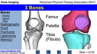
- 1. Bones Patella Femur Tibia Radiographs Fluid Fractures Ligaments Menisci © 2011 Ken L Schreibman, PhD/MD www.schreibman.info Knee Imaging Wisconsin Physical Therapy Association 4/8/11 Slide 1 of 92 3 Bones Femur Patella Tibia (Fibula)
- 2. Bones Patella Femur Tibia Radiographs Fluid Fractures Ligaments Menisci © 2011 Ken L Schreibman, PhD/MD www.schreibman.info Knee Imaging Wisconsin Physical Therapy Association 4/8/11 Slide 2 of 92 Patella: [L] “shallow pan” Largest sesamoid bone Sesamoid:“Bone embedded within a tendon” Many sesamoids throughout body, particularly in the foot 2 Great Toe sesamoids (100%) os peroneum (~30%)
- 3. Bones Patella Femur Tibia Radiographs Fluid Fractures Ligaments Menisci © 2011 Ken L Schreibman, PhD/MD www.schreibman.info Knee Imaging Wisconsin Physical Therapy Association 4/8/11 Slide 3 of 92 Patella: [L] “shallow pan” Largest sesamoid bone Sesamoid:“Bone embedded within a tendon” Quadriceps tendon From 4 quadriceps muscles Vastus Lateralis, Medialis, Intermedius Rectus Femoris To tibial tubercle (tuberosity) “Extensor Mechanism” Patellofemoral Compartment Patella: [L] “shallow pan”
- 4. Bones Patella Femur Tibia Radiographs Fluid Fractures Ligaments Menisci © 2011 Ken L Schreibman, PhD/MD www.schreibman.info Knee Imaging Wisconsin Physical Therapy Association 4/8/11 Slide 4 of 92 Articular Surfaces Trochlear groove trochlea: [L] “pulley” Patellofemoral compartment Femoral Condyles condyle: [Gk] “knuckle” round knob at end of a bone Medial/Lateralcompartments
- 5. Bones Patella Femur Tibia Radiographs Fluid Fractures Ligaments Menisci © 2011 Ken L Schreibman, PhD/MD www.schreibman.info Knee Imaging Wisconsin Physical Therapy Association 4/8/11 Slide 5 of 92 Femoral Condyles Anterior View Lateral View Posterior View Intercondylar Notch Lateral Compart Medial Compart
- 6. Bones Patella Femur Tibia Radiographs Fluid Fractures Ligaments Menisci © 2011 Ken L Schreibman, PhD/MD www.schreibman.info Knee Imaging Wisconsin Physical Therapy Association 4/8/11 Slide 6 of 92 Articular Surfaces As opposed to the round articular surfaces of the femoral condyles, the tibial articular surfaces are flat. Tibial plateau: [Fr] “plate” Anterior View Lateral View Posterior View Tibial Eminence Lateral Compart Medial Compart Eminence separate the lateral & medial compartments
- 7. Bones Patella Femur Tibia Radiographs Fluid Fractures Ligaments Menisci © 2011 Ken L Schreibman, PhD/MD www.schreibman.info Knee Imaging Wisconsin Physical Therapy Association 4/8/11 Slide 7 of 92 Tibial Eminence A,K 54yoF Tibial Eminence Jan: “Hurt knee skiing” Nov: “Knocked down by dog” Fracture tibial eminence Transverse fracture ↔ = Avulsion fracture Avulsion of ACL (Anterior Cruciate Ligament)
- 8. Bones Radiographs AP View Lateral View Sunrise View Notch View Fluid Fractures Ligaments Menisci © 2011 Ken L Schreibman, PhD/MD www.schreibman.info Knee Imaging Wisconsin Physical Therapy Association 4/8/11 Slide 8 of 92 Knee Imaging Radiographs (78%) Primary modality for knee pain Arthritis (joint narrowing, osteophytes) Fractures, loose bodies MR (21%) Internal derangement Ligament/meniscal tears, cartilage defects Occult fractures CT (1%) Surgical planning (of fxs seen on radiographs)
- 9. Bones Radiographs AP View Lateral View Sunrise View Notch View Fluid Fractures Ligaments Menisci © 2011 Ken L Schreibman, PhD/MD www.schreibman.info Knee Imaging Wisconsin Physical Therapy Association 4/8/11 Slide 9 of 92 AP View
- 10. Bones Radiographs AP View Lateral View Sunrise View Notch View Fluid Fractures Ligaments Menisci © 2011 Ken L Schreibman, PhD/MD www.schreibman.info Knee Imaging Wisconsin Physical Therapy Association 4/8/11 Slide 10 of 92 AP View Shows width of lateral, medial compartments Assess for arthritis Doesn’t show patello- femoral compartment Articular surfaces Weight-bearing portions femoral condyles Tibial plateau Assess for displaced fractures
- 11. Bones Radiographs AP View Lateral View Sunrise View Notch View Fluid Fractures Ligaments Menisci © 2011 Ken L Schreibman, PhD/MD www.schreibman.info Knee Imaging Wisconsin Physical Therapy Association 4/8/11 Slide 11 of 92 Lateral View
- 12. Bones Radiographs AP View Lateral View Sunrise View Notch View Fluid Fractures Ligaments Menisci © 2011 Ken L Schreibman, PhD/MD www.schreibman.info Knee Imaging Wisconsin Physical Therapy Association 4/8/11 Slide 12 of 92 Lateral View Shows lateral view of femoral condyles & tibial plateau Doesn’t show med/lat compartments Shows lateral view of patella Assess for patella fracture Doesn’t show patellofemoral compart Can show sizable joint effusions… physical exam is more sensitive!
- 13. Bones Radiographs AP View Lateral View Sunrise View Notch View Fluid Fractures Ligaments Menisci © 2011 Ken L Schreibman, PhD/MD www.schreibman.info Knee Imaging Wisconsin Physical Therapy Association 4/8/11 Slide 13 of 92 Sunrise View (Laurin View) Shows patellofemoral compart! Important view to assess arthritis Shows alignment between patella & trochlear groove. Patella www.ejbjs.org/cgi/reprint/60/1/55
- 14. Bones Radiographs AP View Lateral View Sunrise View Notch View Fluid Fractures Ligaments Menisci © 2011 Ken L Schreibman, PhD/MD www.schreibman.info Knee Imaging Wisconsin Physical Therapy Association 4/8/11 Slide 14 of 92 Notch View (Rosenberg) www.ejbjs.org/cgi/reprint/70/10/1479
- 15. Bones Radiographs AP View Lateral View Sunrise View Notch View Fluid Fractures Ligaments Menisci © 2011 Ken L Schreibman, PhD/MD www.schreibman.info Knee Imaging Wisconsin Physical Therapy Association 4/8/11 Slide 15 of 92 Notch View (Rosenberg) Shows width of posterior lateral, medial compartments Assess for arthritis Doesn’t show patellofemoral compartment Articular surfaces Posterior portions femoral condyles Tibial plateau Assess for displaced fractures
- 16. Bones Radiographs Fluid Suprapatellar Baker cyst Prepatellar Loose bodies Fabella Fractures Ligaments Menisci © 2011 Ken L Schreibman, PhD/MD www.schreibman.info Knee Imaging Wisconsin Physical Therapy Association 4/8/11 Slide 16 of 92 Fluid in Joint: Effusion Synovial fluid From inflamed synovium Arthritis Blood: “Hemarthrosis” From torn vascularized structure Anterior Cruciate Ligament tear Fat+Blood: “Lipohemarthrosis” Fat comes from fatty yellow bone marrow Indicates presence of intra-articular fracture
- 17. Bones Radiographs Fluid Suprapatellar Baker cyst Prepatellar Loose bodies Fabella Fractures Ligaments Menisci © 2011 Ken L Schreibman, PhD/MD www.schreibman.info Knee Imaging Wisconsin Physical Therapy Association 4/8/11 Slide 17 of 92 Suprapatellar Pouch The knee can hold a lot of fluid… not between articular surfaces. Joint effusions collect in the suprapatellar pouch. Synovial lined space Normal extension of the joint capsule Can easily hold > 40ml
- 18. Bones Radiographs Fluid Suprapatellar Baker cyst Prepatellar Loose bodies Fabella Fractures Ligaments Menisci © 2011 Ken L Schreibman, PhD/MD www.schreibman.info Knee Imaging Wisconsin Physical Therapy Association 4/8/11 Slide 18 of 92 H,B 15yoM Large effusion Effusion on Lateral View No effusion (radiographically) Radiographs can show sizable joint effusions… but physical exam is more sensitive! B,C 13yoF
- 19. Bones Radiographs Fluid Suprapatellar Baker cyst Prepatellar Loose bodies Fabella Fractures Ligaments Menisci © 2011 Ken L Schreibman, PhD/MD www.schreibman.info Knee Imaging Wisconsin Physical Therapy Association 4/8/11 Slide 19 of 92 Effusion on Lateral View Effusion MR: T2fs Sagittal Radiographs are relatively insensitive for fluid. M,C 54yoF T2 (fat suppressed) extremely sensitive for fluid. ? Effusion on MR
- 20. Bones Radiographs Fluid Suprapatellar Baker cyst Prepatellar Loose bodies Fabella Fractures Ligaments Menisci © 2011 Ken L Schreibman, PhD/MD www.schreibman.info Knee Imaging Wisconsin Physical Therapy Association 4/8/11 Slide 20 of 92 Effusion on MR M,C 54yoF Proton Density doesn’t show fluid very well. MR: PD Sagittal Effusion MR: T2fs Sagittal T2 (fat suppressed) extremely sensitive for fluid. MR: T2fs Axial Suprapatellar pouch wraps around femur
- 21. Bones Radiographs Fluid Suprapatellar Baker cyst Prepatellar Loose bodies Fabella Fractures Ligaments Menisci © 2011 Ken L Schreibman, PhD/MD www.schreibman.info Knee Imaging Wisconsin Physical Therapy Association 4/8/11 Slide 21 of 92 Effusion on MR M,C 54yoF MR: T2fs Sagittal thru Medial Compart MR: T2fs Sagittal thru Lateral Compart EffusionEffusion Suprapatellar pouch Baker cyst
- 22. Bones Radiographs Fluid Suprapatellar Baker cyst Prepatellar Loose bodies Fabella Fractures Ligaments Menisci © 2011 Ken L Schreibman, PhD/MD www.schreibman.info Knee Imaging Wisconsin Physical Therapy Association 4/8/11 Slide 22 of 92 Baker Cyst (Popliteal Cyst) Suprapatellar pouch Synovial lined space Normal place fluid collects Baker cyst Synovial lined space Abnormal place fluid collects Posterior extension joint capsule Medial, between: Medial gastrocnemius muscle One of the calf muscles Semi-Membranosis tendon One of the hamstring muscles
- 23. Bones Radiographs Fluid Suprapatellar Baker cyst Prepatellar Loose bodies Fabella Fractures Ligaments Menisci © 2011 Ken L Schreibman, PhD/MD www.schreibman.info Knee Imaging Wisconsin Physical Therapy Association 4/8/11 Slide 23 of 92 Baker Cyst (Popliteal Cyst) Baker cyst MR: T2fs Sagittal Medial Compart PD Sagittal Medial Medial Gastroc Muscle Semi- Mem Musc Origin: Above Femoral Condyle Insert: Below Tibial Plateau M,C 54yoF
- 24. Bones Radiographs Fluid Suprapatellar Baker cyst Prepatellar Loose bodies Fabella Fractures Ligaments Menisci © 2011 Ken L Schreibman, PhD/MD www.schreibman.info Knee Imaging Wisconsin Physical Therapy Association 4/8/11 Slide 24 of 92 Baker Cyst (Popliteal Cyst) P,T 51yoF PD Coronal Far Posterior T2fs Coronal Far Posterior Lateral Gastroc Muscle Medial Gastroc Muscle Lateral Gastroc Muscle Medial Gastroc Muscle Lateral Gastroc Muscle Medial Gastroc Muscle Semi- Mem Musc Semi- Mem Musc Baker cyst Semi- Mem Musc
- 25. Bones Radiographs Fluid Suprapatellar Baker cyst Prepatellar Loose bodies Fabella Fractures Ligaments Menisci © 2011 Ken L Schreibman, PhD/MD www.schreibman.info Knee Imaging Wisconsin Physical Therapy Association 4/8/11 Slide 25 of 92 Prepatellar Bursitis Prepatellar Bursa Synovial lined space Does not communicate with knee joint capsule Normally contains little/no fluid Irritation of bursa marked increase bursal fluid Prepatellar bursitis reported in people who kneel frequently Housemaid’s knee Preacher’s knee
- 26. Bones Radiographs Fluid Suprapatellar Baker cyst Prepatellar Loose bodies Fabella Fractures Ligaments Menisci © 2011 Ken L Schreibman, PhD/MD www.schreibman.info Knee Imaging Wisconsin Physical Therapy Association 4/8/11 Slide 26 of 92 Prepatellar Bursitis Often diagnosed by examOften diagnosed by exam http://en.wikipedia.org/wiki/Prepatellar_bursitis
- 27. Bones Radiographs Fluid Suprapatellar Baker cyst Prepatellar Loose bodies Fabella Fractures Ligaments Menisci © 2011 Ken L Schreibman, PhD/MD www.schreibman.info Knee Imaging Wisconsin Physical Therapy Association 4/8/11 Slide 27 of 92 Prepatellar Bursitis MR: T2fs Sagittal Prepatellar swelling S,R 61yoMH,K 61yoM
- 28. Bones Radiographs Fluid Suprapatellar Baker cyst Prepatellar Loose bodies Fabella Fractures Ligaments Menisci © 2011 Ken L Schreibman, PhD/MD www.schreibman.info Knee Imaging Wisconsin Physical Therapy Association 4/8/11 Slide 28 of 92 Loose Bodies Fragments of bone/cartilage in capsule Loose osteochondral fragment May be inconsequential if outside joint If remain in suprapatellar pouch, or in Baker cyst But if between articular surfaces… May cause locking, Destruction articular cartilage
- 29. Bones Radiographs Fluid Suprapatellar Baker cyst Prepatellar Loose bodies Fabella Fractures Ligaments Menisci © 2011 Ken L Schreibman, PhD/MD www.schreibman.info Knee Imaging Wisconsin Physical Therapy Association 4/8/11 Slide 29 of 92 Loose Bodies H,N 62yoF MR: T2fs SagittalMR: PD Sagittal Loose Bodies in Suprapatellar Pouch Loose Bodies in Baker Cyst ? Loose Bodies in Suprapatellar Pouch Baker Cyst Loose Bodies where? Osteophytes
- 30. Bones Radiographs Fluid Suprapatellar Baker cyst Prepatellar Loose bodies Fabella Fractures Ligaments Menisci © 2011 Ken L Schreibman, PhD/MD www.schreibman.info Knee Imaging Wisconsin Physical Therapy Association 4/8/11 Slide 30 of 92 Loose Bodies D,N 23yoM MR: PD Coronal …one week later Lateral View AP View Loose Body in Anterior Joint Loose Body in Medial Compart Loose Body in Lateral Compart Loose Body in Suprapatellar Pouch
- 31. Bones Radiographs Fluid Suprapatellar Baker cyst Prepatellar Loose bodies Fabella Fractures Ligaments Menisci © 2011 Ken L Schreibman, PhD/MD www.schreibman.info Knee Imaging Wisconsin Physical Therapy Association 4/8/11 Slide 31 of 92 Fabella: [L] little bean Sesamoid lateral gastrocnemius Present 10-30% people Bilateral 80% http://www.ajronline.org/cgi/reprint/183/2/352 Not a loose body
- 32. Bones Radiographs Fluid Fractures Patella Femur OCD Tibia Occult Fxs Ligaments Menisci © 2011 Ken L Schreibman, PhD/MD www.schreibman.info Knee Imaging Wisconsin Physical Therapy Association 4/8/11 Slide 32 of 92 Fractures Patella Uncommon Direct trauma, fall to patella Distraction – fragments pulled apart Femur Condyle fracture – rare Supra/Intra-condyle fx – more common Osteochondral defect – fairly common Tibia Plateau MVA – Very Common
- 33. Bones Radiographs Fluid Fractures Patella Femur OCD Tibia Occult Fxs Ligaments Menisci © 2011 Ken L Schreibman, PhD/MD www.schreibman.info Knee Imaging Wisconsin Physical Therapy Association 4/8/11 Slide 33 of 92 Bipartite Patella T,T 15yoF Not a fracture AP View Notch View Sunrise View
- 34. Bones Radiographs Fluid Fractures Patella Femur OCD Tibia Occult Fxs Ligaments Menisci © 2011 Ken L Schreibman, PhD/MD www.schreibman.info Knee Imaging Wisconsin Physical Therapy Association 4/8/11 Slide 34 of 92 Bipartite Patella Normal variant 2% of population 9x more common boys Bilateral 43% Asymptomatic 98% Usually superolateral www.radswiki.net/main/index.php?title=Bipartite_patella JP Lawson. Radiology 1985 157: 625-631. T,T 15yoF
- 35. Bones Radiographs Fluid Fractures Patella Femur OCD Tibia Occult Fxs Ligaments Menisci © 2011 Ken L Schreibman, PhD/MD www.schreibman.info Knee Imaging Wisconsin Physical Therapy Association 4/8/11 Slide 35 of 92 Patella Fractures (Uncommon) B,I 22yoM Direct Trauma (Fall onto knee) Comminuted, stellate pattern Typically not distracted
- 36. Bones Radiographs Fluid Fractures Patella Femur OCD Tibia Occult Fxs Ligaments Menisci © 2011 Ken L Schreibman, PhD/MD www.schreibman.info Knee Imaging Wisconsin Physical Therapy Association 4/8/11 Slide 36 of 92 Patella Fractures (Uncommon) Indirect distracting injury Unexpectedly rapid knee flexion against contracted quadriceps Distraction upper/lower poles M,M 64yoM
- 37. Bones Radiographs Fluid Fractures Patella Femur OCD Tibia Occult Fxs Ligaments Menisci © 2011 Ken L Schreibman, PhD/MD www.schreibman.info Knee Imaging Wisconsin Physical Therapy Association 4/8/11 Slide 37 of 92 Condyle fractures: rare Supra-condylar Inter-condylar Anterior View Lateral View Femur Fracture Posterior View more common
- 38. Bones Radiographs Fluid Fractures Patella Femur OCD Tibia Occult Fxs Ligaments Menisci © 2011 Ken L Schreibman, PhD/MD www.schreibman.info Knee Imaging Wisconsin Physical Therapy Association 4/8/11 Slide 38 of 92 Uncommon Fx Results from shearing of condyle as femur is dislocated across tibia Lateral View Femoral Condyle Fracture A,J 58yoM CT
- 39. Bones Radiographs Fluid Fractures Patella Femur OCD Tibia Occult Fxs Ligaments Menisci © 2011 Ken L Schreibman, PhD/MD www.schreibman.info Knee Imaging Wisconsin Physical Therapy Association 4/8/11 Slide 39 of 92 Femoral Supra/Inter Condylar Fractures More common From axial loading Fracturing between condyles Fracturing above condyles Increasingly common Older females (low energy) Younger males (high energy)
- 40. Bones Radiographs Fluid Fractures Patella Femur OCD Tibia Occult Fxs Ligaments Menisci © 2011 Ken L Schreibman, PhD/MD www.schreibman.info Knee Imaging Wisconsin Physical Therapy Association 4/8/11 Slide 40 of 92 Femoral Supra/Inter Condylar Fractures From axial loading L,L 49yoF with tractionFemoral shaft impacted into/ between condyles CT
- 41. Bones Radiographs Fluid Fractures Patella Femur OCD Tibia Occult Fxs Ligaments Menisci © 2011 Ken L Schreibman, PhD/MD www.schreibman.info Knee Imaging Wisconsin Physical Therapy Association 4/8/11 Slide 41 of 92 Osteochondral Defect (OCD) Fairly common M,A 24yoM
- 42. Bones Radiographs Fluid Fractures Patella Femur OCD Tibia Occult Fxs Ligaments Menisci © 2011 Ken L Schreibman, PhD/MD www.schreibman.info Knee Imaging Wisconsin Physical Therapy Association 4/8/11 Slide 42 of 92 Osteochondral Defect (OCD) Fairly common Fx of bone “osteo” & cartilage “chondral” Adults: traumatic Children: idiopathic “osteochondritis dissecans” Femoral condyles Medial 4x > Lateral M,A 24yoM
- 43. Bones Radiographs Fluid Fractures Patella Femur OCD Tibia Occult Fxs Ligaments Menisci © 2011 Ken L Schreibman, PhD/MD www.schreibman.info Knee Imaging Wisconsin Physical Therapy Association 4/8/11 Slide 43 of 92 Imaging Osteochondral Defects Start with radiographs… CT shows corticated margin MR shows “stability” These should be ordered by orthopedic surgeon MR: Coronal T1CT: Coronal Reformat M,A 24yoM
- 44. Bones Radiographs Fluid Fractures Patella Femur OCD Tibia Occult Fxs Ligaments Menisci © 2011 Ken L Schreibman, PhD/MD www.schreibman.info Knee Imaging Wisconsin Physical Therapy Association 4/8/11 Slide 44 of 92 Imaging Osteochondral Defects: MR Coronal T1 Post-op Coronal T2fs Sagittal T2fsArthroscopy Arthroscopy M,A 24yoM
- 45. Bones Radiographs Fluid Fractures Patella Femur OCD Tibia Occult Fxs Ligaments Menisci © 2011 Ken L Schreibman, PhD/MD www.schreibman.info Knee Imaging Wisconsin Physical Therapy Association 4/8/11 Slide 45 of 92 Tibial Plateau Fractures Very Common 80% MVA (the rest from falls, sports)* 60% involve lateral plateau** CT used for surgical planning Schatzker classification system* * http://emedicine.medscape.com/article/1249872-overview ** http://www.wheelessonline.com/ortho/tibial_plateau_fractures I II III IV V VI
- 46. Bones Radiographs Fluid Fractures Patella Femur OCD Tibia Occult Fxs Ligaments Menisci © 2011 Ken L Schreibman, PhD/MD www.schreibman.info Knee Imaging Wisconsin Physical Therapy Association 4/8/11 Slide 46 of 92 Tibial Plateau Fracture (SII) 46yo♂ physician injured knee skiing Walked around with knee pain 4 days When not improved went for imaging... MR: Coronal T2fs G,L 46yoM CT: Coronal Reformat
- 47. Bones Radiographs Fluid Fractures Patella Femur OCD Tibia Occult Fxs Ligaments Menisci © 2011 Ken L Schreibman, PhD/MD www.schreibman.info Knee Imaging Wisconsin Physical Therapy Association 4/8/11 Slide 47 of 92 Tibial Plateau Fracture (SII) G,L 46yoM CT: 3D Reformat CT: Coronal Reformat
- 48. Bones Radiographs Fluid Fractures Patella Femur OCD Tibia Occult Fxs Ligaments Menisci © 2011 Ken L Schreibman, PhD/MD www.schreibman.info Knee Imaging Wisconsin Physical Therapy Association 4/8/11 Slide 48 of 92 Tibial Plateau Fracture (SII) G,L 46yoM Intra-operative Post-operative
- 49. Bones Radiographs Fluid Fractures Patella Femur OCD Tibia Occult Fxs Ligaments Menisci © 2011 Ken L Schreibman, PhD/MD www.schreibman.info Knee Imaging Wisconsin Physical Therapy Association 4/8/11 Slide 49 of 92 Non-displaced (Occult) Fractures The way we diagnose fractures on radiographs is to see displaced fragments. Non-displaced Fx can be radiographically occult. So how do we detect occult knee fractures? CT often doesn’t help. Fxs non-displaced by radiographs are non-displaced on CT MR doesn’t miss non-displaced Fxs. Can’t send every ER patient with knee pain to MR There is a trick! Look for lipohemarthrosis on cross-table lateral!
- 50. Bones Radiographs Fluid Fractures Patella Femur OCD Tibia Occult Fxs Ligaments Menisci © 2011 Ken L Schreibman, PhD/MD www.schreibman.info Knee Imaging Wisconsin Physical Therapy Association 4/8/11 Slide 50 of 92 Radiographic Views: In Clinic AP & Lateral views shot with patient standing.
- 51. Bones Radiographs Fluid Fractures Patella Femur OCD Tibia Occult Fxs Ligaments Menisci © 2011 Ken L Schreibman, PhD/MD www.schreibman.info Knee Imaging Wisconsin Physical Therapy Association 4/8/11 Slide 51 of 92 Radiographic Views: In ER Shot with patient lying on a cart. AP view Vertical X-ray beam X-rays
- 52. Bones Radiographs Fluid Fractures Patella Femur OCD Tibia Occult Fxs Ligaments Menisci © 2011 Ken L Schreibman, PhD/MD www.schreibman.info Knee Imaging Wisconsin Physical Therapy Association 4/8/11 Slide 52 of 92 Radiographic Views: In ER Shot with patient lying on a cart. Lateral view Vertical X-ray beam Parallel to gravity X-rays
- 53. Bones Radiographs Fluid Fractures Patella Femur OCD Tibia Occult Fxs Ligaments Menisci © 2011 Ken L Schreibman, PhD/MD www.schreibman.info Knee Imaging Wisconsin Physical Therapy Association 4/8/11 Slide 53 of 92 Shot with patient lying on a cart. Cross-Table Lateral view Horizontal X-ray beam Perpendicular to gravity UW MSK Policy: All ER knees shot X-Table Lat. Radiographic Views: In ER
- 54. Bones Radiographs Fluid Fractures Patella Femur OCD Tibia Occult Fxs Ligaments Menisci © 2011 Ken L Schreibman, PhD/MD www.schreibman.info Knee Imaging Wisconsin Physical Therapy Association 4/8/11 Slide 54 of 92 Types of Joint Effusions Synovial fluid From inflamed synovium Arthritis Blood: “Hemarthrosis” From torn vascularized structure Anterior Cruciate Ligament tear Fat+Blood: “Lipohemarthrosis” Fat comes from fatty yellow bone marrow Indicates presence of intra-articular fracture
- 55. Bones Radiographs Fluid Fractures Patella Femur OCD Tibia Occult Fxs Ligaments Menisci © 2011 Ken L Schreibman, PhD/MD www.schreibman.info Knee Imaging Wisconsin Physical Therapy Association 4/8/11 Slide 55 of 92 Thought Experiment Lipo- hem- arthro- sis Whole Blood Cells Plasma Cells Plasma FatWhole Blood X-rays X-rays X-rays perpen- dicular to gravity
- 56. Bones Radiographs Fluid Fractures Patella Femur OCD Tibia Occult Fxs Ligaments Menisci © 2011 Ken L Schreibman, PhD/MD www.schreibman.info Knee Imaging Wisconsin Physical Therapy Association 4/8/11 Slide 56 of 92 Lipohemarthrosis on X-Table Lateral Fat Fluid Fat-Fluid Level = Lipohemarthrosis = Intra-Articular Fracture P,A 27yoF
- 57. Bones Radiographs Fluid Fractures Patella Femur OCD Tibia Occult Fxs Ligaments Menisci © 2011 Ken L Schreibman, PhD/MD www.schreibman.info Knee Imaging Wisconsin Physical Therapy Association 4/8/11 Slide 57 of 92 Lipohemarthrosis on X-Table Lateral CT: Coronal Reformat P,A 27yoF
- 58. Bones Radiographs Fluid Fractures Patella Femur OCD Tibia Occult Fxs Ligaments Menisci © 2011 Ken L Schreibman, PhD/MD www.schreibman.info Knee Imaging Wisconsin Physical Therapy Association 4/8/11 Slide 58 of 92 Impacted Tibial Plateau Fracture (SIII) CT: Coronal Reformat CT: Sagittal Reformat P,A 27yoF Intra-operative Fluoroscopy Post Operative Repaired with just: 2 Screws Bone Cement
- 59. Bones Radiographs Fluid Fractures Patella Femur OCD Tibia Occult Fxs Ligaments Menisci © 2011 Ken L Schreibman, PhD/MD www.schreibman.info Knee Imaging Wisconsin Physical Therapy Association 4/8/11 Slide 59 of 92 P,P 63yoF Occult Fracture AP View XTL View Lipohemarthrosis
- 60. Bones Radiographs Fluid Fractures Patella Femur OCD Tibia Occult Fxs Ligaments Menisci © 2011 Ken L Schreibman, PhD/MD www.schreibman.info Knee Imaging Wisconsin Physical Therapy Association 4/8/11 Slide 60 of 92 P,P 63yoF AP View CT: Coronal Reformat XTLView Occult Fracture ?
- 61. Bones Radiographs Fluid Fractures Patella Femur OCD Tibia Occult Fxs Ligaments Menisci © 2011 Ken L Schreibman, PhD/MD www.schreibman.info Knee Imaging Wisconsin Physical Therapy Association 4/8/11 Slide 61 of 92 P,P 63yoF MR Doesn’t Miss Fractures MR: Coronal T2fs AP View MR: Coronal T1CT: Coronal Reformat ? Bone Marrow Edema Black Fx Lines
- 62. Bones Radiographs Fluid Fractures Ligaments Quadriceps Collaterals Cruciates ACL Tears Menisci © 2011 Ken L Schreibman, PhD/MD www.schreibman.info Knee Imaging Wisconsin Physical Therapy Association 4/8/11 Slide 62 of 92 Ligaments & Tendons Tendons Connect muscles to bone Quadriceps Tendon Ligaments Connect bone to bone Patella Ligament Anterior Cruciate Ligament (ACL) Medial Collateral Ligament (MCL)
- 63. Bones Radiographs Fluid Fractures Ligaments Quadriceps Collaterals Cruciates ACL Tears Menisci © 2011 Ken L Schreibman, PhD/MD www.schreibman.info Knee Imaging Wisconsin Physical Therapy Association 4/8/11 Slide 63 of 92 Most commonly rupture >50yo Men 8x > Women Transversely, patella insertion Exam: Patella freely mobile Inability to actively extend knee Active flexion preserved Radiographs: May show low patella “patella baja” www.wheelessonline.com/ortho/rupture_of_the_quadriceps Quadriceps Tendon Rupture
- 64. Bones Radiographs Fluid Fractures Ligaments Quadriceps Collaterals Cruciates ACL Tears Menisci © 2011 Ken L Schreibman, PhD/MD www.schreibman.info Knee Imaging Wisconsin Physical Therapy Association 4/8/11 Slide 64 of 92 P,S 56yoM MR: Sagittal T2fsMR: Sagittal PD Quadriceps Tendon Rupture
- 65. Bones Radiographs Fluid Fractures Ligaments Quadriceps Collaterals Cruciates ACL Tears Menisci © 2011 Ken L Schreibman, PhD/MD www.schreibman.info Knee Imaging Wisconsin Physical Therapy Association 4/8/11 Slide 65 of 92 www.wheelessonline.com/ortho/patellar_tendon_avulsion Most commonly rupture <40yo More common in African descent Transversely, patella origin Exam: Inability to actively extend knee Radiographs: May show high patella “patella alta” Patella Ligament Avulsion
- 66. Bones Radiographs Fluid Fractures Ligaments Quadriceps Collaterals Cruciates ACL Tears Menisci © 2011 Ken L Schreibman, PhD/MD www.schreibman.info Knee Imaging Wisconsin Physical Therapy Association 4/8/11 Slide 66 of 92 Patella alta: Patella ligament >20% longer Patella length. Normally patella length = patella lig. length (+/- 20%) Courtesy of Kirkland Davis, MD Patella Ligament Avulsion
- 67. Bones Radiographs Fluid Fractures Ligaments Quadriceps Collaterals Cruciates ACL Tears Menisci © 2011 Ken L Schreibman, PhD/MD www.schreibman.info Knee Imaging Wisconsin Physical Therapy Association 4/8/11 Slide 67 of 92 Patella Ligament Avulsion T,D 42yoM MR: Sagittal T2fsMR: Sagittal PD
- 68. Bones Radiographs Fluid Fractures Ligaments Quadriceps Collaterals Cruciates ACL Tears Menisci © 2011 Ken L Schreibman, PhD/MD www.schreibman.info Knee Imaging Wisconsin Physical Therapy Association 4/8/11 Slide 68 of 92 Normal MCL MR: Coronal PD From above condyle to below plateau Frequently injured Lateral clipping injury Valgus stress on knee MR: Coronal PD MCL Avulsion off Tibial Insertion Medial Collateral Ligament (MCL) P,M 28yoM
- 69. Bones Radiographs Fluid Fractures Ligaments Quadriceps Collaterals Cruciates ACL Tears Menisci © 2011 Ken L Schreibman, PhD/MD www.schreibman.info Knee Imaging Wisconsin Physical Therapy Association 4/8/11 Slide 69 of 92 3 Structures Iliotibial Band Biceps Femoris Tendon Fibular Collateral Ligament Infrequently injured Lateral Collateral Complex MR: Coronal PD ITB MR: Coronal PD FCL BFT
- 70. Bones Radiographs Fluid Fractures Ligaments Quadriceps Collaterals Cruciates ACL Tears Menisci © 2011 Ken L Schreibman, PhD/MD www.schreibman.info Knee Imaging Wisconsin Physical Therapy Association 4/8/11 Slide 70 of 92 Cruciate Ligaments Cruciate: [L] “cross” Anterior Cruciate Ligament crosses anterior to the Posterior Cruciate Ligament ACL: Lat Fem Cond Med Tib Spine PCL: Med Fem Cond Lat Tib Spine
- 71. Bones Radiographs Fluid Fractures Ligaments Quadriceps Collaterals Cruciates ACL Tears Menisci © 2011 Ken L Schreibman, PhD/MD www.schreibman.info Knee Imaging Wisconsin Physical Therapy Association 4/8/11 Slide 71 of 92 Cruciates Cross in Both Planes MR: Sagittal PDCT: Sagittal Reformat ACL PCL ACL PCL
- 72. Bones Radiographs Fluid Fractures Ligaments Quadriceps Collaterals Cruciates ACL Tears Menisci © 2011 Ken L Schreibman, PhD/MD www.schreibman.info Knee Imaging Wisconsin Physical Therapy Association 4/8/11 Slide 72 of 92 Cruciate Ligament Tears PCL infrequently tears Surgical reconstruction of the PCL is controversial ACL frequently tears Requires surgical reconstruction Sports related twisting injury Torn ACL allows anterior displacement of the tibia relative to the femur. Anterior Draw Sign
- 73. Bones Radiographs Fluid Fractures Ligaments Quadriceps Collaterals Cruciates ACL Tears Menisci © 2011 Ken L Schreibman, PhD/MD www.schreibman.info Knee Imaging Wisconsin Physical Therapy Association 4/8/11 Slide 73 of 92 Anterior Draw Sign = ACL Tear S,E 58yoM Not usually seen on radiographs but… More common on MR N,G 16yoM MR: Sagittal PD
- 74. Bones Radiographs Fluid Fractures Ligaments Quadriceps Collaterals Cruciates ACL Tears Menisci © 2011 Ken L Schreibman, PhD/MD www.schreibman.info Knee Imaging Wisconsin Physical Therapy Association 4/8/11 Slide 74 of 92 Back of tibial plateau impacts upon front of femoral condyle Bone marrow contusions/edema Kissing Contusions = ACL Tear B,W19yoM MR: Sagittal T2fsMR: Sagittal PD BME BME Less sensitive for fluid Very sensitive for fluid Black Fx Lines
- 75. Bones Radiographs Fluid Fractures Ligaments Quadriceps Collaterals Cruciates ACL Tears Menisci © 2011 Ken L Schreibman, PhD/MD www.schreibman.info Knee Imaging Wisconsin Physical Therapy Association 4/8/11 Slide 75 of 92 Back of tibial plateau impacts upon front of femoral condyle Bone marrow contusions/edema Impaction fractures Anterior lateral femoral condyle Posterior lateral corner tibial plateau Impaction Fractures from ACL Tear MR: Sagittal T2fsMR: Sagittal PD W,S 45yoM BME BME
- 76. Bones Radiographs Fluid Fractures Ligaments Quadriceps Collaterals Cruciates ACL Tears Menisci © 2011 Ken L Schreibman, PhD/MD www.schreibman.info Knee Imaging Wisconsin Physical Therapy Association 4/8/11 Slide 76 of 92 ACL Tears on MRI Direct sign: Non-visualization of intact fibers Indirect signs: Anterior draw Kissing contusions Sagittal T2fsSagittal PD J,D 17yoF B,E 22yoF Sagittal T2fsSagittal PD
- 77. Bones Radiographs Fluid Fractures Ligaments Menisci Normal Anatomy Horizontal Tear Longitudinal Tear Radial Tear Flipped Fragment Bucket handle © 2011 Ken L Schreibman, PhD/MD www.schreibman.info Knee Imaging Wisconsin Physical Therapy Association 4/8/11 Slide 77 of 92 Menisci Looking at the menisci is the primary indication for ordering MRI of the knee To assess for fractures, arthritis Order radiographs Surgical planning fracture repair Order CT To assess for ACL tear At least start with physical exam… To assess for meniscal tear Radiographs, PE not helpful Order MRI!
- 78. Bones Radiographs Fluid Fractures Ligaments Menisci Normal Anatomy Horizontal Tear Longitudinal Tear Radial Tear Flipped Fragment Bucket handle © 2011 Ken L Schreibman, PhD/MD www.schreibman.info Knee Imaging Wisconsin Physical Therapy Association 4/8/11 Slide 78 of 92 Menisci: Function To improve contact between: Curved femoral condyles Flat tibial plateau Composed of fibrocartilage Triangular in cross-section Function like “O”-rings… “C”-shaped MM LM Function
- 79. Bones Radiographs Fluid Fractures Ligaments Menisci Normal Anatomy Horizontal Tear Longitudinal Tear Radial Tear Flipped Fragment Bucket handle © 2011 Ken L Schreibman, PhD/MD www.schreibman.info Knee Imaging Wisconsin Physical Therapy Association 4/8/11 Slide 79 of 92 Menisci: C-shaped A P A P MM LM A P MM LM Sagittal PHAH Sagittal PHAH Sagittal MB Mid Body Sagittal PD Sagittal PD Sagittal PD Coronal PD MB MM MB LM S,M 32yoM AH PH AH PH MB
- 80. Bones Radiographs Fluid Fractures Ligaments Menisci Normal Anatomy Horizontal Tear Longitudinal Tear Radial Tear Flipped Fragment Bucket handle © 2011 Ken L Schreibman, PhD/MD www.schreibman.info Knee Imaging Wisconsin Physical Therapy Association 4/8/11 Slide 80 of 92 Meniscal Tears Meniscal tears are seen on MR as increased signal that extends to an articular surface on at least 2 slices 1 slice only touches called “possible tear” 50% of these are torn at arthroscopy Articular surfaces: Superior Inferior Not an articular surface Free Edge Free Edge Arthroscopic View
- 81. Bones Radiographs Fluid Fractures Ligaments Menisci Normal Anatomy Horizontal Tear Longitudinal Tear Radial Tear Flipped Fragment Bucket handle © 2011 Ken L Schreibman, PhD/MD www.schreibman.info Knee Imaging Wisconsin Physical Therapy Association 4/8/11 Slide 81 of 92 Horizontal Tear “Cleavage tear” Cleaves the meniscus into upper/lower Sagittal PD Coronal PD PH MM PH MM A,R 50yoM
- 82. Bones Radiographs Fluid Fractures Ligaments Menisci Normal Anatomy Horizontal Tear Longitudinal Tear Radial Tear Flipped Fragment Bucket handle © 2011 Ken L Schreibman, PhD/MD www.schreibman.info Knee Imaging Wisconsin Physical Therapy Association 4/8/11 Slide 82 of 92 Longitudinal Tear “Vertical tear” Separates meniscus into inner/outer Sagittal PHAH Sagittal PD Coronal PD Axial T2fs PH MMAH MM MB MM P,B 18yoF
- 83. Bones Radiographs Fluid Fractures Ligaments Menisci Normal Anatomy Horizontal Tear Longitudinal Tear Radial Tear Flipped Fragment Bucket handle © 2011 Ken L Schreibman, PhD/MD www.schreibman.info Knee Imaging Wisconsin Physical Therapy Association 4/8/11 Slide 83 of 92 Radial Tear Radiates from free edge outward Separates meniscus into anterior/posterior MM LM Coronal MB LMMB MM MB MM MB MM Coronal T2fs Coronal PD E,S 57yoM
- 84. Bones Radiographs Fluid Fractures Ligaments Menisci Normal Anatomy Horizontal Tear Longitudinal Tear Radial Tear Flipped Fragment Bucket handle © 2011 Ken L Schreibman, PhD/MD www.schreibman.info Knee Imaging Wisconsin Physical Therapy Association 4/8/11 Slide 84 of 92 Flipped Meniscal Fragment Meniscus portion displaced within knee joint Can cause locking Decreased motion PH AHAH PH AH LM PH LM Sagittal PD N,D 46yoM Sagittal PH AH
- 85. Bones Radiographs Fluid Fractures Ligaments Menisci Normal Anatomy Horizontal Tear Longitudinal Tear Radial Tear Flipped Fragment Bucket handle © 2011 Ken L Schreibman, PhD/MD www.schreibman.info Knee Imaging Wisconsin Physical Therapy Association 4/8/11 Slide 85 of 92 Bucket Handle Tear Longitudinal tear where inner portion flips into the intercondylar notch, under PCL
- 86. Bones Radiographs Fluid Fractures Ligaments Menisci Normal Anatomy Horizontal Tear Longitudinal Tear Radial Tear Flipped Fragment Bucket handle © 2011 Ken L Schreibman, PhD/MD www.schreibman.info Knee Imaging Wisconsin Physical Therapy Association 4/8/11 Slide 86 of 92 Bucket Handle Tear Longitudinal tear where inner portion flips into the intercondylar notch, under PCL Coronal MB LMBucketed MB MM MB MM Coronal T2fs Coronal PD A,D 18yoM MB MM MB LM Bucketed MB MM PCL
- 87. Bones Radiographs Fluid Fractures Ligaments Menisci Normal Anatomy Horizontal Tear Longitudinal Tear Radial Tear Flipped Fragment Bucket handle © 2011 Ken L Schreibman, PhD/MD www.schreibman.info Knee Imaging Wisconsin Physical Therapy Association 4/8/11 Slide 87 of 92 Bucket Handle Tear Longitudinal tear where inner portion flips into the intercondylar notch, under PCL Sagittal PD PCL Bucketed MB MM H,D 15yoM Double PCL sign Coronal T2fs Coronal PD MB MM MB LM PCL Bucketed MB MM
- 88. Bones Radiographs Fluid Fractures Ligaments Menisci © 2011 Ken L Schreibman, PhD/MD www.schreibman.info Knee Imaging Wisconsin Physical Therapy Association 4/8/11 Slide 88 of 92 Knee: What to Order When (WOW) Always start with radiographs! Diagnose arthritis, assess progression Joint space narrowing, osteophytes Diagnose fractures, assess healing Fracture displacement, articular involvement Look for loose bodies See if bodies move over time Screen for unexpected findings Tumors Metal foreign bodies
- 89. Bones Radiographs Fluid Fractures Ligaments Menisci © 2011 Ken L Schreibman, PhD/MD www.schreibman.info Knee Imaging Wisconsin Physical Therapy Association 4/8/11 Slide 89 of 92 Knee: What to Order When (WOW) Radiographs: Need at least 3 views 1) AP Med & Lat: Condyles, Plateau, Compartments 2) Lateral Perpendicular to AP view Shows Patella ER: X-Table Lateral may show lipohemarthrosis 3) Sunrise Best view for patellofemoral compartment --------------------------------------------------------- 4) Notch (part of a 4-view series) Shows posterior condyles
- 90. Bones Radiographs Fluid Fractures Ligaments Menisci © 2011 Ken L Schreibman, PhD/MD www.schreibman.info Knee Imaging Wisconsin Physical Therapy Association 4/8/11 Slide 90 of 92 Knee: What to Order When (WOW) MRI: Frequently ordered for knee pain Internal derangement Meniscal tears Ligament tears (ACL, MCL) Cartilage defects Occult/non-displaced fractures MRI does not miss fractures BEST IF ORDERED BY KNEE SPECIALIST! They may prefer certain sequences They may prefer certain MR scanners
- 91. Bones Radiographs Fluid Fractures Ligaments Menisci © 2011 Ken L Schreibman, PhD/MD www.schreibman.info Knee Imaging Wisconsin Physical Therapy Association 4/8/11 Slide 91 of 92 Knee: What to Order When (WOW) CT: Infrequently ordered for knee pain Primary use is in the ER to assess the alignment of fracture fragments (shown on radiographs) to aid in surgical planning Should be ordered by the surgical team Occasionally, for patients who are not MR compatible, we will do an Arthrogram-CT to assess for internal derangement Should be ordered by the knee specialist
- 92. Bones Radiographs Fluid Fractures Ligaments Menisci © 2011 Ken L Schreibman, PhD/MD www.schreibman.info Knee Imaging Wisconsin Physical Therapy Association 4/8/11 Slide 92 of 92 Questions? P,D 59yoM MR: Coronal T2